C
a s eR
e p o Rt4 0 7 Arq Bras Oftalmol. 2016;79(6):407-10 http://dx.doi.org/10.5935/0004-2749.20160115
INTRODUCTION
Uveal metastasis of systemic carcinoma to the eye is estimated to occur in 8% of cases, according to the results of autopsy evaluations where microscopic detection rate is reported(1). The rate of intraocular metastasis varies according to the primary tumor site and the degree of systemic control. A previous report suggests that the frequency of macroscopic metastases has decreased over 12 years from 4.7% to
1% owing to improved cancer therapy(2).
Uveal metastases occur far more frequently in the choroid than in the iris, possibly due to the substantial blood supply to the posterior choroid by the posterior ciliary arteries(3).
Metastatic tumors to the iris are relatively uncommon; they may present as stromal nodules or ill-defined iris thickening, or they may be associated with atypical features such as pain, iridocyclitis, and hyphema(4,5). In 2014, Shields et al. reported their 40-year experience of 104 cases of iris metastases arising from systemic cancer, demons-trating the breast, lung, and skin as the most common primary sites for such lesions(4).
Many of these patients are never examined by an ophthalmologist because many have no visual complaints or have systemic advanced disease.
Here, we describe three patients with iris metastasis and with diffe-rent presentations and discuss the diagnostic challenges and unusual findings associated with these cases.
CASE REPORTS
C
ASE1
A 61-year-old man presented with a 3-month history of blurred vision affecting the right eye and the presence of iris lesion. His past ocular history was unremarkable, while his past medical history
in-ABSTRACT
Ocular metastasis is relatively uncommon, with a reported incidence of approxi-mately 8%, according to the results of autopsy evaluation.The majority of ocular metastases are located within the choroid, while metastatic tumors affecting the iris are rare. Metastatic tumors may manifest as stromal nodules or ill-defined iris thickening, or they may present with nonspecific features such as pain, iridocyclitis, and hyphema. Here, we describe three patients with iris metastasis and discuss the diagnostic challenges and unusual findings associated with these cases.
Keywords: Iris neoplasms; Iris metastasis; Case reports
RESUMO
A maioria das metástases oculares do câncer sistêmico são encontrados na coroide. As metástases para a íris são incomuns, podendo se manifestar como nódulo es tro mal, espessamento de íris de limites mal definidos ou como uma iridociclite ou hifema. Relatamos 3 pacientes com lesão de íris e história pregressa de câncer sistêmico. Enfatizamos a dificuldade no diagnóstico e raridade dessas lesões comparando com relatos anteriores.
Descritores: Neoplasias da íris/metástase neoplásica; Relatos de casos
cluded a renal adenocarcinoma treated by total nephrectomy 5 years previously. Follow-up with imaging and laboratory studies demons-trated no evidence of recurrence or metastatic disease.
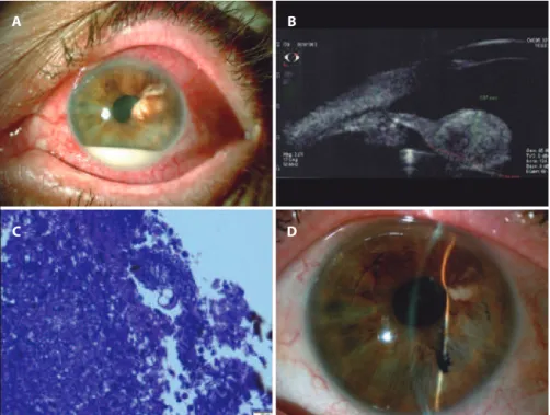
On admission, ocular examination revealed a best-corrected vi-sion of 20/30 in the right eye and 20/20 in the left eye. Full extraocular motion was observed. The pupils were 3 mm in diameter and reacti-ve to light. The right pupil had an area of irregularity inferiorly, and there were neovessels in the collarette (Figure 1 A). A 2+/4+ anterior chamber reaction with no keratic precipitates were also observed. Further, a 7 × 4 mm pinkish-yellow mass from 1 to 5 o’clock with intrinsic friable vessels was observed, in addition to a band of tissue from 7 to 9 o’clock in the anterior chamber. The intraocular pressure was 25 mmHg (right eye) and 12 mmHg (left eye), as determined by applanation tonometry. Fundoscopic examination was normal. Based on all these findings, a clinical diagnosis of an iris tumor was made. Ultrasound biomicroscopy (UBM) of the right eye revealed an irido-ciliary mass with homogeneous reflectivity, featuring areas of internal hyporeflective spaces within the tumor with clear margins (Figure 1 B). UBM of the left eye was normal.
Hypotensive agents and topical prednisolone 1% three times daily were initiated. Magnetic resonance imaging (MRI) revealed a renal mass, in addition to liver, brain, and pulmonary masses. A cli-nical diagnosis of iridociliary metastasis arising from a primary renal carcinoma was established. Treatment with oral anti-tyrosinase was initiated, leading to shrinkage of both the iris and the systemic lesions (Figures 1 C and 1 D). The patient remains alive at 18 months after the initial diagnosis.
C
ASE2
A 58-year-old female presented with pain and blurred vision in her left eye. She had breast cancer, which was treated 10 years earlier
Iris metastases from systemic cancer: a report of three cases
Metástase de íris de ĉncer sist̂mico: relato de tr̂s casos
Patrícia correade Mello1,2,3, oswaldo Ferreira Moura Brasil2, andré Vidoris3, Melina correia Morales3, ruBens n. BelFort3
Submitted for publication: September 25, 2015 Accepted for publication: March 6, 2016
1 Serviço de Oftalmologia, Hospital Federal dos Servidores do Estado do Rio de Janeiro, Rio de Janeiro, RJ, Brazil.
2 Setor de Retina e Vítreo, Instituto Brasileiro de Oftalmologia (IBOL), Rio de Janeiro, RJ, Brazil. 3 Setor de Tumores, Departamento de Oftalmologia e Ciências Visuais, Escola Paulista de Medicina
(EPM), Universidade Federal de São Paulo (UNIFESP), São Paulo, SP, Brazil.
Funding: No specific financial support was available for this study.
Disclosure of potential conflicts of interest: None of the authors have any potential conflict of interest to disclose.
Ir I sm e ta s ta s e sf r o ms y s t e m I cc a n c e r: ar e p o rto ft h r e ec a s e s
4 0 8 Arq Bras Oftalmol. 2016;79(6):407-10
with mastectomy, lymphadenectomy, and systemic chemotherapy. Ocular examination revealed a best corrected vision of 20/20 in the right eye and finger-counting only in the left eye. Full extraocular motion was observed. The left pupil was slowly responsive to light, with an area of irregularity at 2 o’clock and a 3 × 2-mm white pearl
mass in the temporal upper quadrant associated with bulging of the iris superiorly (Figure 2 A). Intense anterior chamber inflammation with 3+/4+ cells and an inferior white hypopyon without keratic precipitates was observed. UBM of the left eye revealed an iris lesion with heterogeneous reflectivity (Figure 2 B). Systemic work-up for
Figure 1. Case 1. A) Photography demonstrating a 7 × 4-mm pinkish-yellow mass from 1 to 5 o’clock with intrinsic friable vessels. A band of tissue was observed from 7 to 9 o’clock in the anterior chamber. B) Ultrasound biomicroscopy (UBM) image of an iridociliary mass with homogeneous relectivity and areas of internal hyporelective spaces within the tumor. C and D) Post-treatment images demonstrating shrinkage of the lesion.
A
C D
B
A B
D C
Me l l o PC, e ta l.
4 0 9
Arq Bras Oftalmol. 2016;79(6):407-10
A
C D
B
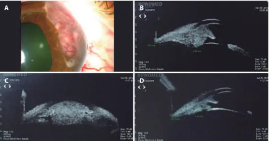
Figure 3. Case 3. A) Biomicroscopy image demonstrating a pink iris mass from 1 to 3 o’clock with intrinsic vessels. B and C) Ultrasound biomicroscopy (UBM) demonstrating a mass with homogeneous relectivity and areas of internal hyporelective spaces. D) UBM after treatment showing shrinkage of the lesion.
inflammatory and infectious uveitis was negative. Other systemic me -tastases were also ruled out. Findings of histological examination of an iris biopsy were highly suggestive of metastatic carcinoma (Figure 2 C). Immunohistochemical analyses were positive for CEA and CK7, and negative for estrogen. The patient was treated with external beam radiotherapy, with full regression of the lesion obser-ved (Figure 2 D). After 13 months follow-up, no evidence of disease was observed at the primary site or other sites of metastatic disease.
C
ASE3
A 25-year-old woman presented with an iris lesion affecting her right eye and a 3-month history of blurred vision. Her past ocular history was unremarkable. She had an adenocarcinoma 1 year pre viously, which was treated by a total parotidectomy and a man-dibulo-mastoidectomy with extraction of her cervical lymph nodes; however, surgical margins were not clear. Adjuvant external beam radiotherapy was performed at the surgical site. No evidence of local recurrence was observed on follow-up. Ocular examination revea-led a best corrected vision of 20/100 in the right eye and 20/20 in the left eye. Full extraocular motion was observed. The right pupil was irregular and slowly responsive to light, with no signs of uveal inflammation. A pink iris mass was observed from 1 to 3 o’clock with the presence of intrinsic vessels (Figure 3 A). UBM of the right eye re-vealed an iridociliary mass with homogeneous reflectivity and areas of internal hyporeflective spaces within the tumor (Figure 3 B and 3 C). A clinical diagnosis of metastatic disease was made. Full work-up, including positron emission tomography (PET), findings were nega-tive. Plaque brachytherapy was performed to treat the focal iris me-tastasis, with consequent lesion improvement observed (Figure 3 D). However, the patient died 10 months later due to the progression of metastatic disease.
DISCUSSION
In a clinical survey(6) of 3,680 iris tumors, 21% were reported as
cystic and 79% as solid. In the solid group, 68% were melanocytic and 11% were non-melanocytic. Non-melanocytic iris tumors included a range of diagnoses, including choristomatous, vascular, fibrous, neu-ral, myogenic, epithelial, xanthomatous, metastatic, lymphoid, leukemic, and non-neoplastic conditions masquerading as iris masses. Only 67 (2%) tumors were metastatic.
In the largest published series of 107 eyes with metastatic tumors
of the iris, Shields et al.(4) reported primary tumors were more likely
to originate from the breast (33%), lung (27%), skin (12%), kidney (7%), esophagus (3%), and other regions. The kidney and parotid are
considered very rare primary tumor sites. Shields et al.(4) found that
the majority of iris metastases were inferior (35%) and superior (28%), whereas the majority of cases were nasal (28%) or temporal (28%) in
a series reported Ferry and Font(2). All three of our patients reported
blurred vision, with the presence of non-melanocytic iris lesions on the nasal side in cases 1 and 3 and in the upper temporal region in
case 2. Shields et al.(4) reported pain and blurred vision as the most
common complaints, as observed in cases 1 and 2. Pain was related to secondary glaucoma in case 1 and to inflammation in case 2. All cases had a history of previously treated malignancy, as did the majority
of patients in previous studies(4,5). Melanoma is considered the main
differential diagnosis of iris metastasis, but it is clinically challenging
to diagnose due to a lack of specific pathognomonic features(7).
UBM has been used to study and monitor iris tumors in previous
studies(8); however, no pathognomic features have demonstrated
utility in distinguishing melanomas from other metastases. In a series
of UBM studies(8) of iris melanomas, two distinct patterns of tumor
vascularity were observed by histological analysis with UBM: small hyperechoic dots relating to small-caliber vessels in the iris stroma; and hypoechoic spaces relating to larger vascular spaces. Cases 1 and 3 exhibited the latter pattern, which may have been attributable to intrinsic vessels visible on slit lamp examination. In case 2, UBM de monstrated irregular reflectivity, with no visible intrinsic tumoral vessels observed on biomicroscopy.
The majority of iris tumors can be diagnosed using clinical his-torical criteria without the need for cytological or pathologic biopsy. In the largest series of fine-needle biopsies for iris tumors, the most
common indications were suspected iris melanomas or metastases(9).
In these cases, cytological evaluation of fine-needle aspirates was extremely helpful in distinguishing melanoma from metastatic
neo-plasm(9). We decided to perform a biopsy of the iris lesion in case 2
Ir I sm e ta s ta s e sf r o ms y s t e m I cc a n c e r: ar e p o rto ft h r e ec a s e s
4 1 0 Arq Bras Oftalmol. 2016;79(6):407-10
involvement of the regional lymph nodes in addition to an iris lesion. Adenocarcinoma of the parotid develops from the secretory element of the gland and is considered an aggressive lesion with potential for
both local lymphatic and distant metastasis(10). The patient in case 3
died shortly after treatment of the iris lesion as she developed distant metastatic disease a few months later.
Unfortunately, the median time interval between the detection
of iris metastasis and death is reportedly 24 months(4). It should be
noted that cases 1 and 2 remain alive after follow-up durations of 18 and 13 months, respectively.
The treatment of uveal metastases, including iris metastases, is palliative. Patients with other systemic involvement should be treated with systemic chemotherapy, while external radiotherapy or brachy-therapy are recommended for the treatment of focal metastases.
REFERENCES
1. Bloch RS, Gartner S. The incidence of ocular metastatic carcinoma. Arch Ophthalmol. 1971;85(6):673-5.
2. Shields CL, Shields JA, Gross NE, Schwartz GP, Lally SE. Survey of 520 eyes with uveal metastases. Ophthalmology. 1997;104(8):1265-76.
3. Shields CL, Kaliki S, Crabtree GS, Peshatani A, Morton S, Anand RA, et al. Iris metastasis from systemic cancer in 104 patients: The 2014 Jerry A. shields Lecture. Cornea. 2015; 34(1):42-8.
4. Ferry AP, Font RL. Carcinoma metastatic to the eye and orbit. I. a clinic-pathological study of 227 cases. Arch Ophthalmol. 1974;92(4):276-86.
5. Shields CL, Kancherla S, Patel J, Vijayvargiva P, Suriano MM, Kolbus E, et al. Clinical Survey of 3680 iris tumor based on the age at presentation. Opththalmology. 2012; 119(2):407-14.
6. Ferry AP, Font RL. Carcinoma metastatic to the eye and orbit. II a clinic-pathological study of 26 patients with carcinoma metastatic to the anterior segment of the eye. Arch Ophthalmol. 1975;93(7):472-82.
7. Hood CT, Schoenfield LR, Torres V, Singh AD. Iris melanoma. Ophthalmology. 2011; 118(1):221-2.
8. Giuliari GP, McGowan HD, Pavlin CJ, Heathcote JG, Simpson ER. Ultrasound biomicros-copic imaging of iris melanoma: a clinicopathologic study. Am J Ophthalmol. 2011; 151(4):579-85.
9. Shields CL, Manquez ME, Ehya H, Mashayekhi A, Danzig CJ, Shields JA. Fine-needle aspiration biopsy of iris tumors in 100 consecutive cases: technique and complications. Ophthalmology. 2006;113(11):2080-6.
10. Ho K, Lin H, Ann DK, Chu PG, Yen Y. An overview of the rare parotid gland cancer. Head & Neck Oncol. 2011;3:40.