INTRODUCTION
Full-field electroretinogram (ERG) measures the diffuse response of retina cells to a light stimulation. The response is the consequence of light-induced changes in the transretinal movements of ions in the ex-tracellular space, particularly sodium and potassium(1). The elec trical
activity recorded represents the combination of positive and negative components originated from different stages of retinal processing.
The ERG response is generally expressed as a waveform with ne-gative and positive deflections. The nene-gative deflection, called a-wave, represents the activity of photoreceptors. The positive deflection, called b-wave, represents the ON-bipolar cell function(1). ERG has been
used as a diagnostic method for inherited retinal degenerative
disea-ses such as retinitis pigmentosa, congenital stationary night blindness, cone dystrophy, and other conditions(2-4). At times, ERG abnor malities
can precede fundus changes(5).
Different types of electrodes can be used for ERG recording, and those that contact the cornea or nearby bulbar conjunctiva during the procedure are most strongly suggested for full-field ERG recor-ding. These electrodes include conductive fibers and foils, bipolar contact lenses, conjunctival loop electrodes, and corneal wicks(6). ERG
recording in uncooperative patients and/or patients with palpebral alterations is a challenge because of the difficulty of appropriate pla-cement of the conventional active electrodes. Alternative recording methods such as the use of skin electrodes have been tested to allow ERG recording in this specific group of patients.
Full-field electroretinogram recorded with skin electrodes in normal adults
Eletrorretinograma de campo total registrado com eletrodos de pele em adultos normais
Arthur GustAvo FernAndes1, solAnGe rios sAlomão1, Josenilson mArtins PereirA1, AdriAnA Berezovsky1
Submitted for publication: February 25, 2016 Accepted for publication: May 27, 2016
1 Laboratório de Eletrofisiologia Visual Clínica, Departamento de Oftalmologia e Ciências Visuais,
Escola Paulista de Medicina (EPM), Universidade Federal de São Paulo (UNIFESP), São Paulo, SP, Brazil.
Funding: No specific financial support was available for this study.
Disclosure of potential conflicts of interest: None of the authors have any potential conflict of interest to disclose.
Corresponding author: Adriana Berezovsky. Depto. de Oftalmologia e Ciências Visuais. Rua Bo tu catu, 821 São Paulo, SP 04023062 Brazil Email: [email protected]
Approved by the following research ethics committee: Universidade Federal de São Paulo UNIFESP (#0174/04).
ABSTRACT
Purpose: Alternative recording methods have been tested to allow the electro-retinogram (ERG) recording in uncooperative patients and/or patients with pal-pebral alterations, including recordings with skin electrodes. The purpose of this study was to compare ERG recorded with skin electrodes and well-established microfiber electrodes and to determine normative values of ERG parameters for recording with skin electrodes.
Methods: Fifty healthy volunteers (17-26 years; mean 20.63 ± 2.01 years) partici-pated in the study. A gold disk skin electrode was placed on the lower orbital rim of a randomly chosen eye. On the contralateral eye, a microfiber electrode was positioned in the lower conjunctival sac. Gold disc electrodes were positioned at the ipsilateral outer canthus of both eyes acting as reference electrodes for the creation of a potential difference. Two ground electrodes were placed on the lobe of each ear. ERGs were recorded according to the International Society of Clinical Electrophysiology Visual (ISCEV) protocol using the VERIS 5.1.9 system for data acquisition and analysis.
Results: Both types of electrodes showed similar wave response morphologies. The implicit time of responses between the two electrodes was comparable. On peak-to-peak amplitude, skin electrode recordings showed an amplitude reduction of 61.4% for rod responses, 61.5% for maximal responses, 46.2% for oscillatory potentials, 57.4% for cone responses, and 54.4% for 30Hz-flicker responses, when compared with microfiber electrode recordings. Based on these findings, normative values for peak-to-peak amplitude and implicit time to be used as a reference for ERGs recorded with skin electrodes were determined.
Conclusions: ERGs recorded with skin electrodes presented lower peak-to-peak amplitude compared with microfiber electrodes. However, using appropriate nor-mative values, skin electrodes may be useful for specific target populations such as uncooperative infants and/or patients with ocular surface alterations.
Keywords: Electroretinography; Electrodes; Skin; Eyelids; Comparative study
RESUMO
Objetivo: O eletrorretinograma de campo total (ERG) em pacientes não colabo-rativos e/ou com alterações palpebrais pode ser registrado com diferentes métodos de captação de respostas, dentre eles os eletrodos de pele. O objetivo deste estudo foi comparar o eletroretinograma obtido com eletrodos de pele e eletrodos de mi-crofibra, determinando valores normativos para os parâmetros registrados com eletrodos de pele.
Métodos: Cinquenta voluntários saudáveis (17-26 anos; média 20,63 ± 2,01) par-ticiparam deste estudo. Um eletrodo de cúpula de ouro foi aderido à pele na margem orbital inferior de um dos olhos escolhido ao acaso. No olho contralateral, um eletrodo de microfibra foi posicionado no saco conjuntival inferior. Eletrodos de cúpula de ouro foram posicionados na região lateral de cada olho para a formação da diferença de potencial com o eletrodo ativo. Dois eletrodos terra foram posicionados no lobo de cada orelha. O eletrorretinograma foi registrado de acordo com o protocolo da ISCEV (Sociedade Internacional de Eletrofisiologia Visual), com o sistema VERIS 5.1.9 para aquisição e análise dos registros.
Resultados: Os dois tipos de eletrodos apresentaram morfologias de onda similares. O tempo implícito das respostas foi comparável entre os dois tipos de eletrodo. Os registros feitos com eletrodo de pele quando comparados aos registros com eletrodos de mi crofibra mostraram reduções na amplitude das ondas, de 61,4% para resposta de bastonetes, 61,5% para resposta máxima, 46,2% para potenciais oscilatórios, 57,4% para resposta de cones, e 54,4% para flicker 30Hz. Baseado nestes resultados, foram determinados valores normativos para amplitude e tempo de implícito para ERGs obtidos com eletrodos de pele.
Conclusões: ERGs registrados com eletrodos de pele apresentam respostas com am-plitudes menores quando comparados aos registrados com eletrodos de microfibra. No entanto, usando valores normativos apropriados, os eletrodos de pele podem ser uma alternativa útil para populações especificas como pacientes não colaborativos e/ou com alterações palpebrais.
The objective of this study was to compare the ERG recorded with skin electrodes and well-established microfiber active electrodes. In addition, normative values for ERG parameters recorded with skin elec-trodes were determined.
METHODS
This study protocol was evaluated by the Committee of Ethics in Research of the Universidade Federal de São Paulo and was approved in accordance with the principles of the Declaration of Helsinki.
P
ARTICIPANTSHealthy volunteers were recruited from university students and employees of Federal University of Sao Paulo and were invited for the full-field ERG test after a full explanation of the procedure, its benefits and risks, followed by written informed consent. The inclusion criteria consisted of normal visual acuity for distance 0.0 logMAR (20/20) or better, lack of visual disorders, normal fundoscopy, and acceptance of the informed consent form. Exclusion criteria consisted of high ametropia (myopia= -4.00 spherical equivalent; hyperopia= +4.00 spherical equivalent), history of previous ocular surgeries, or heredi-tary eye diseases.
V
ISUALACUITYPresenting visual acuity (with spectacles if used) was measured for each eye using a logMAR chart at 4 m.
F
ULL-
FIELDERG
Full-field ERG was performed in a completely dark room, after maximal pupil dilatation (minimum pupil diameter of 6 mm) with one drop each of 1% tropicamide and 10% phenylephrine hydrochloride, followed by 30 min of dark adaptation.
The Veris electrodiagnostic system (Veris 5.1.9; Electro-Diagnostic Imaging, Sao Mateo, CA, EUA) was used for the ERG recording. Peak-to-peak amplitudes were measured in microvolts (µV), and the implicit time was measured in milliseconds (ms).
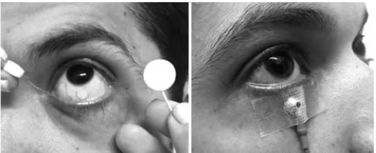
A skin electrode (Grass Gold Disc Electrodes-E5GH) was placed on the lower orbital rim of a randomly chosen eye. On the contralateral eye, an active microfiber electrode was positioned in the lower con-junctival sac (Figure 1). Gold disc electrodes were positioned at the ipsilateral outer canthus of both eyes acting as reference electrodes for the creation of a potential difference. Two ground electrodes were placed on the lobe of each ear.
ERGs were recorded according to the International Society of Cli-nical Electrophysiology Visual (ISCEV) protocol(7), which is considered
the minimum requisite for comparison of different tests performed in different centers(8).
The participants were adapted to complete dark condition for 30 min, and the protocol of ERG recording followed this sequential order: (a) Rod response (a maximum intensity white light with a 2.4 log neutral density filter attenuation); (b) Maximal response (a maximum intensity white light to obtain a combined response of cones and rods after dark-adaptation); (c) Oscillatory potentials (a maximum intensity white light with a 75-Hz low-cut and 500-Hz high-cut filter); and a maximum intensity white light in a 30 cd/m2
background presented at (d) 1 Hz and (e) 30 Hz to obtain cone res-ponses after a 10 min adaptation to a 30 cd/m2 background light (d) single-flash cone response (maximum intensity light at 1 Hz) and (e) 30 Hz flicker response (maximum intensity light at 30 Hz) were recorded.
Figure 2 shows a representative full-field ERG recording from a 22-year-old male participant. Right eye responses were recorded with the microfiber electrode, and left eye responses were recorded with the skin electrode.
S
TATISTICALANALYSISPeak-to-peak amplitude and implicit time from each step of the ISCEV standard ERG protocol were compared between the microfiber electrode and the skin electrode using a statistical package (Jandel Sigmastat-Statistical Software Version 2.0, USA). The significance level used was 0.05. All the parameters were compared between the electrodes using the Mann-Whitney rank sum test. Normative values for skin electrode full-field ERG recordings were calculated as 2.5 and 97.5 percentiles of peak-to-peak amplitude and implicit time.
RESULTS
A total of 50 normal subjects (17-26 years; mean 20.63 ± 2.01 years; 19 males) were recruited for the study based on the inclusion criteria.
Mean amplitudes and implicit times for the five ERG responses are shown in table 1; figure 3 shows the comparison of the recordings of the microfiber electrodes and the skin electrodes. All responses showed similar waveforms for both the electrodes. There was no statistical difference in the implicit time of responses between the two types of electrodes. On amplitude, skin electrode responses showed a significant amplitude reduction (p<0.001) of 61.4% for rod responses, 61.5% for maximal responses, 46.2% for oscillatory potentials, 57.4% for cone responses, and 54.4% for flicker responses, when compared with microfiber electrode responses.
Based on these results, normative values for amplitude and implicit time to be used as a reference for full-field ERG recorded with skin electrodes were determined as shown in table 2.
DISCUSSION
ERG recordings using skin electrodes produced lower amplitudes and higher electrical noise. This technique is not recommended by ISCEV; however, it can be adopted for specific clinical conditions(7).
In this study, it was observed that when using skin electrodes as an alternative method, the responses present similar wave morphology and had no significant difference regarding implicit times for all five ISCEV protocol responses.
The reduction found in peak-to-peak amplitudes recorded with skin electrodes in our study is consistent with previous reports of re-ductions of 75% and 80%(9), 43%-73%(10), 50%-70%(11), 50%(12), 23%-62%(13),
and 42%-57%(14). Our data showed reductions between 46% and 62%
on the peak-to-peak amplitudes. Physical factors are involved in this re-duction given their influence on the current flow through the electro-des. Such factors include the resistive and capacitative characteristics of the materials from which the electrodes are made and coated with and the area of the recording surface. In addition, the amplitude re-corded with skin electrodes is noisier, mainly as a result of electromyo-graphic activity intervention(15). However, it has been shown that even
with reduced amplitudes, recordings with skin electrodes can be useful to differentiate between normal and abnormal responses, as long as an appropriate normative reference is used(16). The combination of clinical
findings and ERG results demonstrated that ERGs with skin electrodes are accurate for the diagnosis of retinal dystrophies(16).
For pediatric patients, ERG recorded with skin electrodes presents an important advantage because there is no need for sedation. In patients with ocular surface alterations, this technique is suggested as a unique alternative for recording ERG.
Consideration of pediatric age groups could have provided better evaluation of the electrode performance; however, several studies report that peak-to-peak amplitudes of ERG responses are similar in children and adults, irrespective of whether recording is performed with conventional or skin electrodes(17-21). ERG responses are said to
achieve adult levels by the fourth year of life; therefore, separate pe-diatric normative values evaluation would be necessary for children aged <4 years(6).
Previous studies with skin electrodes have demonstrated that peak-to-peak amplitude can show variability as the skin electrode can move during the exam, reducing the amplitudes on the final steps as the electrodes lose adherence to the skin(11). However, comparative
studies demonstrated that the variability found with skin electrodes is similar to the variability found with corneal contact electrodes, and examiners need to ensure that the electrode is correctly placed close to the lower orbital rim during the entire exam(9,22).
Figure 2. ERG recording from a 22-year-old male (OD: microiber electrode/OS: skin electrode).
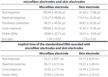
Table 1. Amplitude and implicit time measurements for microiber electrodes and skin electrodes
Peak-to-peak amplitudes of the standardized ERG recorded with microfiber electro des and skin electrodes
Microiber electrode Skin electrode
Rod response 198.49 ± 40.30 µV 081.60 ± 17.88 µV Maximal response 276.27 ± 48.86 µV 114.14 ± 22.30 µV
Oscillatory potentials 090.31 ± 49.56 µV 049.61 ± 32.56 µV
Cone response 108.66 ± 32.02 µV 047.42 ± 13.36 µV Flicker 30Hz 050.86 ± 20.73 µV 024.27 ± 09.00 µV
b/a ratio 1.95 ± 0.42 1.76 ± 0.26
Implicit time of the standardized ERG recorded with microiber electrodes and skin electrodes
Microiber electrode Skin electrode
Rod response 55.27 ± 8.97 ms 53.77 ± 8.54 ms
Maximal response 36.17 ± 6.15 ms 33.25 ± 5.48 ms
Table 2. Normative values (2.5 and 97.5 percentiles) for full-ield ERGs obtained with skin electrodes
Normative values for peak-to-peak amplitude for responses recorded with skin electrodes
Rod response 45.70-112.31 µV
Maximal response 68.82-154.39 µV
Oscillatory potentials 15.05-137.51 µV
Cone response 19.35-074.19 µV
Flicker 30 Hz 11.19-045.56 µV
b/a ratio 1.30-2.31
Normative values for implicit time for responses recorded with skin elec tro des
Rod response 43.84-75.38 ms
Maximal response 25.50-44.98 ms
Cone response 22.00-30.65 ms
Flicker 30 Hz 24.00-33.69 ms
Figure 3. Mean amplitude and implicit time comparison between microiber electrodes and skin electrodes (*p<0.001). Error bars represent standard deviation of the mean.
The ISCEV protocol and the recommendation for each laboratory to determine its own normative values are extremely important for a high quality test. Normative values from different laboratories may differ for several reasons including different equipment, different elec-trodes, and different settings on the Ganzfeld(23).
Testing using skin electrodes for full-field ERG recording in pa-tients previously diagnosed with retina dystrophy using ERG with con ventional electrodes is recommended to allow an evaluation of the validity of the results and accuracy of the skin electrodes as an alter native method.
Our findings are comparable to previous studies, indicating a re duction in amplitude of electroretinography responses recorded with skin electrodes. However, skin electrodes are useful for specific target populations such as uncooperative infants and/or patients with ocular surface alterations. The normative values calculated in this study can be used as a reference for full-field ERG recorded with skin electrodes.
REFERENCES
Available from: http://www.intechopen.com/books/electroretinograms/electroreti-nogram-in-hereditary-retinal-disorders.
2. Birch DG. Clinical electroretinography. In: Fuller DG, Birch DG, editors. Assessment of visual function for the clinician. Ophthalmol Clin North Am. 1989;2:469-98.
3. Paranhos FR, Avila MP, Paranhos A, Cialdini AP. Estudo estatístico de valores normais do eletrorretinograma: contribuição à padronizaçäo do exame. Arq Bras Oftalmol. 1997; 60(3):278-84.
4. Birch DG, Anderson JL. Standardized full-field electroretinography: normal values and their variation with age. Arch Ophthalmol. 1992;110(11):1571-6.
5. Pereira JM, Mendieta L, Sacai PY, Salomão SR, Berezovsky A. Estudo normativo do eletror-retinograma de campo total em adultos jovens. Arq Bras Oftalmol. 2003;66(2):137-44. 6. Papathanasiou ES, Papacostas SS. Flash electroretinography: normative values with
surface skin electrodes and no pupil dilation using a standard stimulation protocol. Doc Ophthalmol. 2008;116(1):61-73.
7. McCulloch DL, Marmor MF, Brigell MG, Hamilton R, Holder GE, Tzekov R, et al. ISCEV Standard for full-field clinical electroretinography (2015 update). Doc Ophthalmol. 2015; 130(1):1-12.
8. Berezovsky A, Pereira JM, Salomão SR, Santos VR, Schor P. Validation of a new fiber electrode prototype for clinical electroretinography. Arq Bras Oftalmol. 2008;71(3): 316-20.
9. Bradshaw K, Hansen R, Fulton A. Comparison of ERGs recorded with skin and cor neal-contact electrodes in normal children and adults. Doc Ophthalmol. 2004;109(1):43-55. 10. Stevens T. Electrophysiologic testing in disorders of the retina, optic nerve, and visual pathway. (Ophthalmology Monographs, No. 2). Arch Ophthalmol. 2001;119(11):1734. 11. Kriss A. Skin ERGs: their effectiveness in paediatric visual assessment, confounding
factors, and comparison with ERGs recorded using various types of corneal electrode. Int J Psychophysiol. 1994;16(2):137-46.
12. Hawlina M. Pattern electroretinography with the new HK-loop electrode. Chibret Int J Ophthalmol. 1993;9:51-8.
13. Mortlock KE, Binns AM, Aldebasi YH, North RV. Inter-subject, inter-ocular and inter-session repeatability of the photopic negative response of the electroretinogram recorded using DTL and skin electrodes. Doc Ophthalmol. 2010;121(2):123-34.
14. Hamilton R, Al Abdlseaed A, Healey J, Neveu M, Brown L, Keating D, et al. Multi-centre variability of ISCEV standard ERGs in two normal adults. Doc Ophthalmol. 2015;130(2): 83-101.
15. Esakowitz L, Kriss A, Shawkat F. A comparison of flash electroretinograms recorded from Burian Allen, JET, C-glide, gold foil, DTL and skin electrodes. Eye (Lond). 1993;7 (Pt 1):169-71.
16. Meredith S, Reddy M, Allen L, Moore A, Bradshaw K. Full-field ERG responses recorded with skin electrodes in pediatric patients with retinal dystrophy. Doc Ophthalmol. 2004; 109(1):57-66.
17. Fulton AB, Hansen RM, Westall CA. Development of ERG responses: the ISCEV rod, maximal and cone responses in normal subjects. Doc Ophthalmol. 2003;107(3):235-41. 18. Westall CA, Panton CM, Levin AV. Time courses for maturation of electroretinogram
responses from infancy to adulthood. Doc Ophthalmol. 1998;96(4):355-79. 19. Fulton AB, Hansen RM. Electroretinography: application to clinical studies of infants.
J Pediatr Ophthalmol Strabismus. 1985;22(6):251-5.
20. Breton ME, Quinn GE, Schueller AW. Development of electroretinogram and rod pho-totransduction response in human infants. Invest Ophthalmol Visual Sci. 1995;36(8): 1588-602.
21. Fulton AB, Hansen RM. The development of scotopic sensitivity. Invest Ophthalmol Visual Sci. 2000;41(6):1588-96.
22. Wali N, Leguire LE. Dark-adapted luminance-response functions with skin and corneal electrodes. Doc Ophthalmol. 1991;76(4):367-75.
23. Jacobi PC, Miliczek K, Zrenner E. Experiences with the international standard for clinical electroretinography: normative values for clinical practice, interindividual and intraindi-vidual variations and possible extensions. Doc Ophthalmol. 1993;85(2):95-114.