Original Article
2 3 Arq Bras Oftalmol. 2015;78(1):23-6 http://dx.doi.org/10.5935/0004-2749.20150007
INTRODUCTION
Thinning and loss of the choroidal vasculature may lead to pho-toreceptor damage-related diseases(1), including central serous chorio-retinopathy (CSC)(2), age-related macular degeneration (AMD)(3), choroidal melanoma(4), Vogt-Koyanagi-Harada (VKH) syndrome(5), and others(6).
Choroidal thickness (CT) can be measured using diferent techniques such as ultrasonography and histology. However, the location of the choroid behind the pigmented cells of the retina attenuates incident light and reduces the reliability of these methods. In recent years, spectral-domain optical coherence tomography (SD-OCT) has been used to assess choroidal tissue in vivo and in real time with an accepta-ble reproducibility and sensitivity(7-10).Previous studies in a healthy adult population report a range of central CT from 270 to 350 μm(11). The eyes of healthy children difer from those of adults, and the in -luen ce of a normal developmental process on the choroidal struc tu re
RESUMO
Objetivo: Investigar a associação entre a espessura central da coroide e o comprimento axial (AL), idade, sexo e erros de refração em uma população pediátrica saudável por meio da tomografia de coerência óptica (OCT ).
Métodos: Estudo institucional envolvendo 137 crianças saudáveis (57 meninos, 80
meninas), com idades entre 4 e 18 anos. Cada criança foi submetida a um exame de fundo de olho, refração sob cicloplegia e medida do comprimento axial usando o biométrico óptico Nidek AL-Scan (Nidek CO, LTD.). A espessura foveal central (CFT ) e espessura da coroide (CT ) foram medidas utilizando o Cirrus HD-OCT (Carl Zeiss Meditec). O olho direito de cada sujeito foi selecionado para análise.
Resultados: A idade média das crianças foi de 10,0 ± 4,7 anos (variação, 4 a 18 anos). O equivalente esférico médio (SE) foi -0,24 ± 1,24 dioptrias (D) (variação de -2,00 D a +2,25 D). A média do comprimento axial foi de 23,1 ± 1,2 mm (variação, 20 a 27 mm). A espessura da coroide central média foi de 388,2 ± 50,0 mm e não se correlacionou com a idade, sexo, comprimento axial ou erro refrativo.
Conclusões: Os resultados proporcionam uma base de dados pediátrica normativa
da espessura da coroide usando tomografia de coerência óptica com profundidade de imagem aprimorada. Esta informação pode ser útil no diagnóstico e acompanhamento de doenças de retina e coroide em crianças.
Descritores: Coroide; Doenças da coroide/patologia; Comprimento axial do olho; Erros de refração; Tomografia de coerência óptica
can be evaluated by determining CT in children of various ages. There are limited studies evaluating CT in healthy children using SD-OCT. In these studies, CT measurements were performed using the Heidelberg Spectralis (Heidelberg Engineering, Heidelberg, Germany) OCT device, and the correlation with age and other factors such as axial length (AL) and refractive error was reported to be contrary(12-15).
In this study, we aimed to determine CT in a healthy pediatric population aged between 4 and 18 years using Cirrus HD-OCT (Carl Zeiss Meditec Inc., Dublin, CA, USA). To the best of our knowledge, this is the irst study using Cirrus HD-OCT to measure CT in a pediatric po pulation.
METHODS
This prospective study was approved by the institutional medical ethics committee and conducted in accordance with the Declaration of Helsinki for research in human subjects. Written informed consent
Choroidal thickness measurement in healthy pediatric population using
Cirrus HD optical coherence tomography
Medida da espessura da coroide na população pediátrica saudável usando tomograia
de coerência óptica Cirrus HD
Aylin Tenlik1, Fatma B. Gürağaç1, emre Güler2, mehmet Serdar dervişoğullari1, YükSel totan1
Submitted for publication: July 31, 2014 Accepted for publication: November 11, 2014
1 Department of Ophthalmology, Medical School, Turgut Özal University, Ankara, Turkey. 2 Department of Ophthalmology, Erciş State Hospital, Van, Turkey.
Funding: No specific financial support was available for this study.
Disclosure of potential conflicts of interest: None of the authors have any potential conflicts of interest to disclose.
Corresponding author: Emre Güler. Van Yolu Cad. No 57 - Erciş, Van, 65400 - Turkey
E-mail: [email protected] ABSTRACT
Purpose: To investigate the association between central choroidal thickness (CT ), axial length (AL), age, gender, and refractive error in a healthy pediatric population using optical coherence tomography (OCT ).
Methods: This institutional study involved 137 healthy children (57 boys, 80 girls) aged between 4 and 18 years. Each child underwent a dilated eye examination, cycloplegic refraction, and AL measurement using a Nidek AL-Scan optical biometer. The central foveal thickness (CFT ) and CT were measured using Cirrus high definition (HD)-OCT. The right eye of each subject was selected for analysis.
Results: The mean age of the children was 10.0 ± 4.7 years (range, 4-18 years). The mean spherical equivalent (SE) was -0.24 ± 1.24 diopters (D) (range, -2.00 D to +2.25 D). The mean AL was 23.1 ± 1.2 mm (range, 20-27 mm). The mean central CT was 388.2 ± 50.0 µm and was not correlated with age, gender, AL, or refractive error.
Conclusions: The data provide a pediatric normative database of CT using enhanced depth imaging OCT. This information may be useful in the diagnosis and monitoring of retino-choroidal diseases in children.
Choroidal thickness measurement in healthy pediatric population using Cirrus HD optical coherence tomography
2 4 Arq Bras Oftalmol. 2015;78(1):23-6
was obtained from the parents of the children after explaining the imaging modality to them and to the child.
In this study, we evaluated 137 healthy Turkish children who visited our clinic for refractive error examinations between March 2013 and September 2013. Children aged between 4 and 18 years of age with no known current or previous ocular disease or ocular medication were included. We divided the subjects into 3 groups on the basis of age: 4-6 years, 7-12 years, and 13-18 years. Children aged younger than 4 years were excluded because they were considered unable to cooperate with OCT. We also excluded all patients with ma-culopathy, myopia of more than 2 diopters (D), congenital glaucoma, past history of prematurity, previous eye trauma or eye surgery, and previous ocular abnormalities such as strabismus, amblyopia, conge-nital cataract, and vitreo-retinal disorders. We also excluded children who could not cooperate with OCT examination.
Before OCT, each child underwent a complete ophthalmic exami-nation, including best-corrected visual acuity (BCVA) as the logarithm of the minimum angle of resolution (logMAR), cycloplegic refraction, evaluation of ocular alignment, slit lamp biomicroscopy, and fundus examination. Cycloplegic refraction was performed with an autore-fractometer (Topcon KR 8800), after applying 3 drops, 5 minutes apart, of cyclopentolate 0.5%. Spherical equivalent (SE) refraction was cal-culated as the sum of the value of the spherical value and half of the cylindrical value. AL was determined using a Nidek AL-Scan optical biometer (Nidek Co, Ltd.) before cycloplegia. All examinations were performed between 13:00 to 15:00 to avoid diurnal variations. However, these can be diferent for each individual.
O
PTICALCOHERENCETOMOGRAPHYIMAGINGThe SD Cirrus high deinition (HD)-OCT light source is centered on a wavelength of 800 nm, achieving 5 µm axial resolutions in tissue. Previously, the full thickness of the choroid could not be seen in most eyes because of scattering and insuicient light penetration beyond the retinal pigment epithelium (RPE), as well as signal strength roll-of distal to the zero-delay line. Selective pixel proiling available in the newest software (Version 6) generates a HD 5-line raster image from 20 B-scans taken at a single location. This processing software is compa-rable to frame averaging, but is unique in that images are generated by evaluating all of the pixel data to reduce noise and to construct the best possible image. Decreased signal strength posterior to the RPE is compensated by this image enhancement software, which enables visualization of the border where choroidal tissue meets sclera and allows CT measurements to be performed.
The images were taken in the usual manner and were not in-verted to bring the choroid into closer proximity of the zero-delay line, because image inversion using the Cirrus software results in a low-resolution, pixelated image. The protocol of HD 5 Line Raster spaced at 0.25 mm was performed centering on the fovea and consis-ted of 6-mm parallel lines with 1024 A-scans/B-scans and averaging 4 B-scans per image. In this study, images had to be at least 6 out of 10 in intensity and taken as close to the fovea as possible, imaging the thinnest point of the macula with the understanding that slight diferences in positioning could afect the measured thicknesses. The right eye of each subject was selected for analysis.
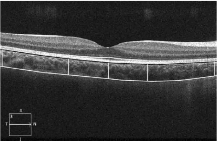
The choroid was visualized by Cirrus HD-OCT with the enhanced depth imaging system (EDI) according to a previously described method. (16) The choroid was imaged by positioning an OCT camera close enough to the eye to obtain an inverted image. Using the Cirrus linear measurement tool, 1 experienced examiner measured CT thickness perpendicularly from the outer edge of the hyper-relective RPE to the inner sclera at 1000 µm intervals temporal and nasal to the fovea, up to 3000 µm (temporal and nasal CT, respectively; Figure 1). The same examiner performed 2 additional measurements and the average of the 3 measurements was used for the statistical analyses.
S
TATISTICALANALYSESStatistical analysis was performed using SPSS 21.0 for Windows (SPSS Inc., Chicago, IL, USA). The data were normally distributed on the Kolmogorov-Smirnov test (p>0.05). A paired t-test and 1-way ANOVA was used to analyze the diferences in CT between the age groups. Pearson’s correlation coeicients were used to evaluate the efect of SE, AL, central foveal thickness (CFT), age, and sex on the CT. A p value of <0.05 was considered to be statistically signiicant.
RESULTS
In total, 137 children (57 boys, 80 girls) were included in this study. The mean age of the children was 10.0 ± 4.7 years (range, 4-18 years). The mean SE was -0.24 ± 1.24 D (range, -2.00 D to +2.25 D). The AL was 23.3 ± 1.2 mm (range, 20-25.5 mm). Table 1 shows the characteristics of the children and the right eye for each age group.
The mean central CT was 388.2 ± 50.0 µm (range, 241-464 µm). The CT at 1 and 3 mm nasal to the fovea was 334.1 ± 49.7 µm (range, 244-433 µm) and 306.1 ± 44.9 µm (range, 220-421 µm), respectively. The CT at 1 and 3 mm temporal to the fovea was 347.6 ± 49.4 µm (range, 249-469 µm) and 365.1 ± 44.9 µm (range, 273-457 µm), res-pectively. The CT at 1 and 3 mm nasal and temporal to the fovea was signiicantly thinner than the central CT (p<0.001). The CT at 1 and 3 mm temporal to the fovea was signiicantly thicker than the CT at 1 and 3 mm nasal to the fovea (p<0.001). Table 2 shows the mean central CT and the mean for 1 mm and 3 mm nasal and temporal quadrants by age.
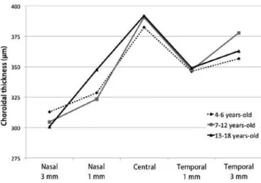
There was no statistically signiicant diference in central CT among subgroups. There was also no signiicant diference in 1 mm temporal and 3 mm nasal and temporal CT among the age groups. The only signiicant diference was found for 1 mm nasal CT, which was thicker in the group aged 13-18 years than group aged 7-12 years (p=0.02; Figures 2 and 3).
The diference in central CT between males and females was not statistically signiicant after adjustment for age and AL (p=0.49). There was no signiicant correlation between central CT and age, SE, or AL (p=0.18, p=0.41, p=0.31, respectively). Correlation analysis showed that the 3 mm nasal CT was thicker in males than in females (r=0.15, p=0.03) and the 3 mm nasal CT was positively correlated with SE (r=0.23, p=0.003; Table 3).
The mean CFT was 242.1 ± 21.3 µm (range 188-297 µm). Central CT was not correlated with mean CFT on Pearson’s correlation analysis (r=0.001, p=0.49).
Tenlik A, et al.
25 Arq Bras Oftalmol. 2015;78(1):23-6 DISCUSSION
Few studies have evaluated CT in the pediatric population and the factors that inluence CT remain unclear. In this study we aimed to evaluate the mean values and determinants of CT among healthy children.
The previous studies evaluating CT in childhood were performed with SD-OCT devices, which use EDI technology. EDI-OCT measures the distance between the RPE and the chorio-scleral interface to obtain the CT. We evaluated the choroid features using another commercially available EDI-OCT system, Cirrus HD OCT (Carl Zeiss Meditec Inc.).
In the present study, the central CT was higher (mean, 388.2 ± 50.0 µm) compared with previous studies of CT in children(12-15,17). This result may be the result of diferences in the measuring software, OCT light source, ethnicity, or characteristics such as age, SE, or AL. The mean value of central CT in children in the present study was also higher than that determined in healthy adults in previous studies(18-22).
The relationship between age and CT is controversial. Some re-ports have suggested that CT decreases progressively with age(18-21,23,24). Margolis(18) reported a 1.5 µm thinning in CT for each year of life. Park and Oh(13) found a signiicant negative correlation between central CT and age among 48 healthy Korean children aged between 4 and 10 years. The thinning of CT with age in those previous studies was related to degenerative vascular changes(18,19) and stretching of tissue secondary to eyeball elongation(13).
Conversely, Bidaut-Garnier et al.(12) reported that central CT showed a signiicant positive relationship with age in 348 eyes from 174 children. Similarly, Read et al.(14) determined a signiicantly thinner CT value in early childhood (4-6 years) compared with older children. These indings may relect the natural growth of the eye during childhood. However, some studies did not ind a correlation between CT and age. Agawa et al.(25) and Li et al.(22) did not identify any correlation between CT and age in eyes with AL <25 mm. Ruiz-Moreno et al.(17) compared a group of healthy children aged 3-17 years with a group of healthy adults aged 25-85 years. They found that no signiicant dife-rence in central CT between the pediatric population and the adults.
In the present study, there was no correlation between central CT and age in healthy children, which was compatible with indings of Agawa et al.(25). However, we did observe that the average CT 1 mm nasal to the fovea was signiicantly thicker in children aged 13-18 years than in those aged 7-12 years.
Many of the previous studies reported a negative correlation between central CT and AL or SE(12,14,26-28). However, Park and Oh(13) did
Figure 2. The relationship between age and choroidal thickness.
Figure 3. Scatter plot showing the relationship between age and choroidal thickness 1 µm nasal to the fovea (Pearson correlation analysis).
Table 1. Characteristics of the children and the right eye by age group
Age group No subjects Percent (%) Mean SE (SD) Diopters Range diopters Mean AL (SD)mm Range mm
4-18 years 137 100.0 -0.24 (1.24) -2.00 to +2.25 23.1 (1.2) 20-27
4-6 years 048 035.0 0.64 (0.43) -0.50 to +1.25 22.5 (0.8) 21-24
7-12 years 039 028.4 -0.33 (1.10) -2.00 to +1.25 23.1 (1.0) 21-26
13-18 years 050 036.4 -0.97 (1.30) -2.00 to +1.25 23.8 (1.1) 22-27
No= number; SE= spherical equivalent; AL= axial length; SD= standard deviation.
Table 2. Distribution of choroidal thickness values using Cirrus HD-OCT
Mean CCT (SD) Mean CTN1 (SD) Mean CTT1 (SD) Mean CTN3 (SD) Mean CTT3 (SD)
4-18 years 388.2 (50.01) 334.1 (49.7) 347.6 (49.4) 306.1 (44.9) 365.1 (44.9)
4-6 years 382.4 (41.10) 328.6 (51.0) 346.3 (47.3) 312.8 (51.1) 356.7 (46.4)
7-12 years 390.6 (49.40) 323.5 (41.4) 347.3 (45.4) 304.6 (42.5) 377.9 (39.6)
13-18 years 392.0 (58.00) 347.7 (52.1) 349.0 (54.9) 300.8 (40.3) 363.1 (45.9)
Choroidal thickness measurement in healthy pediatric population using Cirrus HD optical coherence tomography
26 Arq Bras Oftalmol. 2015;78(1):23-6
not ind any correlation between those parameters. In the present study, central CT was not correlated with AL or SE, which agrees with the indings of Park and Oh(13).
Li et al.(22) found a thicker subfoveal choroid in men compared with women among 93 healthy Danish university students with the same AL. Their inding of sex-associated diferences in CT could be the result of diferences in hormonal exposure. Recently, the same authors reported that CT in girls increased with body height and sexual maturation, whereas it did not change in boys. The results suggest that puberty promotes choroidal thickening in girls, an efect that may be mediated by the pubertal growth spurt. The lack of a pubertal efect in boys may be related to a smaller proportion of boys in that study having entered puberty(29). However, CT was not correlated with gender in the present study, which is consistent with the indings of Bidaut-Garnier et al.(12) and Park and Oh(13). Therefore, we suggest that the efect of hormones does not have a signiicant inluence on CT in children, although we acknowledge that the hormonal hypothesis is partially valid. Further studies of pediatric po pulations are needed to better understand the efect of hormones.
According to our indings, in children the central CT was highest, followed by the temporal and the nasal CT, similar to reports on healthy adults(9,18,20,21,24). However, some previous studies of pediatric populations reported that CT was the highest in the temporal cho-roid(13,17). The diferences in CT between adults and children may be a consequence of choroidal vascular remodeling and maturation. The fovea has higher metabolic needs than the surrounding retina, and this may lead to a decrease in the temporal CT, while the subfoveal choroid is spared.
To the best of our knowledge, this is the irst study of CT measure-ments among Turkish children using Cirrus HD-OCT. The choroid was the thickest in the subfoveal area in this study population, followed by the temporal and nasal sectors at 1 mm and 3 mm from the fovea. In addition, AL, SE, age, and gender did not signiicantly afect the CT. We believe that these indings may provide useful background data to diagnose and monitor retino-choroidal diseases in children. However,a variety of factors, for example, the topographic features of the retina, the impression of other ocular anatomic tissues, and diferences in the optics of the eye may lead to focal changes in CT. Therefore, larger, long-term prospective studies are needed to reveal age-related changes in CT and the inluencing factors.
REFERENCES
1. Huang W, Wang W, Zhou M, Chen S, Gao X, Fan Q, et al. Peripapillary choroidal thickness in healthy chinese subjects. BMC Ophthalmology. 2013;13:23.
2. Gemenetzi M, De Salvo G, Lotery AJ. Central serous chorioretinopathy: an update on pathogenesis and treatment. Eye (Lond). 2010;24(12):1743-56.
3. Spaide RF. Age-related choroidal atrophy. Am J Ophthalmol. 2009;147(5):801-10. 4. Torres VL, Brugnoni N, Kaiser PK, Singh AD. Optical coherence tomography enhanced
depth imaging of choroidal tumors. Am J Ophthalmol. 2011;151(4):586-93.e.2. 5. Maruko I, Iida T, Sugano Y, Saito M, Sekiryu T. Subfoveal retinal and choroidal thickness
after verteporin photodynamic therapy for polypoidal choroidal vasculopathy. Am J Ophthalmol. 2011;151(4):594-603.
6. Regatieri CV, Branchini L, Fujimoto JG, Duker JS. Choroidal imaging using spectral-domain optical coherence tomography. Retina. 2012;32(5):865-76.
7. Nassif N, Cense B, Park BH, Yun SH, Chen TC, Bouma BE, et al. In vivo human retinal imaging by ultrahigh-speed spectral domain optical coherence tomography. Opt Lett. 2004;29(5):480-2.
8. Wojtkowski M. In vivo human retinal imaging by ultrahigh-speed spectral domain optical coherence tomography. J Biomed Opt. 2002;7(3):457-63.
9. Branchini L, Regatieri CV, Flores-Moreno I, Baumann B, Fujimoto JG, Duker JS. Repro-ducibility of choroidal thickness measurements across three spectral domain optical coherence tomography systems. Ophthalmology. 2012;119(1):119-23. Comment in: Ophthalmology. 2012;119(6):1286; author reply 1286-7.
10. Lee SW, Yu SY, Seo KH, Kim ES, Kwak HW. Diurnal variation in choroidal thickness in relation to sex, axial length, and baseline choroidal thickness in healthy Korean subjects. Retina. 2014;34(2):385-93.
11. Shin JW, Shin YU, Cho HY, Lee BR. Measurement of choroidal thickness in normal eyes using 3D OCT-1000 spectral domain optical coherence tomography. Korean J Ophthalmol. 2012;26(4):255-9.
12. Bidaut-Garnier M, Schwartz C, Puyraveau M, Montard M, Delbosc B, Saleh M. Choroi-dal thickness measurement in children using optical coherence tomography. Retina. 2014;34(4):768-74.
13. Park KA, Oh SY. Choroidal thickness in healthy children. Retina. 2013;33(9):1971-6. 14. Read SA, Collins MJ, Vincent SJ, Alonso-Caneiro D. Choroidal thickness in childhood.
Invest Ophthalmol Vis Sci. 2013;54(5):3586-93.
15. Mapelli C, Dell’Arti L, Barteselli G, Osnaghi S, Tabacc hi, Clerici M, et al. Choroidal volume variations during childhood. Invest Ophthalmol Vis Sci. 2013;54(10):6841-5. 16. Manjunath V, Fujimoto JG, Duker JS. Cirrus HD-OCT high deinition imaging is another
tool available for visualization of the choroid and provides agreement with the in-ding that the choroidal thickness is increased in central serous chorioretinopathy in comparison to normal eyes. Retina. 2010;30(8):1320-1; author reply 1321-2. 17. Ruiz-Moreno JM, Flores-Moreno I, Lugo F, Ruiz-Medrano J, Montero JA, Akiba M. Macular
choroidal thickness in normal pediatric population measured by swept-source opti-cal coherence tomography. Invest Ophthalmol Vis Sci. 2013;54(1):353-9.
18. Margolis R, Spaide RF. A pilot study of enhanced depth imaging optical coherence tomography of the choroid in normal eyes. Am J Ophthalmol. 2009;147(5):811-5. 19. Ding X, Li J, Zeng J, Ma W, Liu R, Li T, et al. Choroidal thickness in healthy Chinese
subjects. Invest Ophthalmol Vis Sci. 2011;52(13):9555-60.
20. Ikuno Y, Kawaguchi K, Nouchi T, Yasuno Y. Choroidal thickness in healthy Japanese subjects. Invest Ophthalmol Vis Sci. 2010;51(4):2173-6.
21. Manjunath V, Taha M, Fujimoto JG, Duker JS. Choroidal thickness in normal eyes measured using Cirrus HD optical coherence tomography. Am J Ophthalmol. 2010; 150(3):325-9.e.1.
22. Li XQ, Larsen M, Munch IC. Subfoveal choroidal thickness in relation to sex and axial length in 93 danish university students. Invest Ophthalmol Vis Sci. 2011;52(11):8438-41. 23. Ho J, Branchini L, Regatieri C, Krishnan C, Fujimoto JG, Duker JS. Analysis of normal
peripapillary choroidal thickness via spectral domain optical coherence tomography. Ophthalmology. 2011;118(10):2001-7.
24. Ouyang Y, Heussen FM, Mokwa N, Walsh AC, Durbin MK, Keane PA, et al. Spatial distri-bution of posterior pole choroidal thickness by spectral domain optical coherence tomography. Invest Ophthalmol Vis Sci. 2011;52(9):7019-26.
25. Agawa T, Miura M, Ikuno Y, Makita S, Fabritius T, Iwasaki T, et al. Choroidal thickness measurement in healthy japanese subjects by three-dimensional high-penetration optical coherence tomography. Graefes Arch Clin Exp Ophthalmol. 2011;249(10):1485-92. 26. Fujiwara T, Imamura Y, Margolis R, Slakter JS, Spaide RF. Enhanced depth imaging
op-tical coherence tomography of the choroid in highly myopic eyes. Am J Ophthalmol. 2009;148(3):445-50.
27. Flores-Moreno I, Lugo F, Duker JS, Ruiz-Moreno JM. The relationship between axial length and choroidal thickness in eyes with high myopia. Am J Ophthalmol. 2013; 155(2):314-9.e1.
28. Wei WB, Xu L, Jonas JB, Shao L, Du KF, Wang S. et al. Subfoveal choroidal thickness: the Beijing Eye Study. Ophthalmology. 2013;120(1):175-80.
29. Li XQ, Jeppesen P, Larsen M, Munch IC. Subfoveal choroidal thickness in 1323 children aged 11 to 12 years and association with puberty: the Copenhagen Child Cohort 2000 Eye Study. Invest Ophthalmol Vis Sci. 2014;55(1):550-5.
Table 3. Pearson’s correlation of choroidal thickness quadrants
CCT CTN1 CTN3 CTT1 CTT3
r P r P r P r P r P
Age 0.079 0.181 0.129 0.066 -0.126 0.072 -0.030 0.362 0.029 0.369
Gender -0.111 0.099 -0.081 0.174 0.158 0.033 -0.019 0.411 0.014 0.436
SE -0.056 0.259 0.060 0.244 0.238 0.003 0.141 0.050 0.099 0.125
AL -0.001 0.494 -0.006 0.471 -0.119 0.083 -0.044 0.305 -0.025 0.384