O
r i g i n a la
rt i c l e3 0 8 Arq Bras Oftalmol. 2016;79(5):308-11 http://dx.doi.org/10.5935/0004-2749.20160088
INTRODUCTION
Central serous chorioretinopathy (CSC) is an idiopathic condition characterized by the development of a well-circumscribed, serous detachment of the sensory retina in the posterior pole. Although the pathogenesis of CSC has been extensively studied, it still remains
controversial(1-3). The primary pathology may involve dysfunction of
the retina pigment epithelium (RPE) or choriocapillaris hyperpermea-bility or both. Gass postulated that choriocapillaris hyperpermeabi-lity was the cause of CSC. Later, studies using indocyanine green an giography (ICGA) showed multiple areas of choroidal vascular hyperpermeability, vascular dilatation, illing delay in the choroidal
arteries and choriocapillaris, and punctate hyperluorescent spots(4-8).
These angiographic indings strongly support the theory of Gass, which suggests that the primary pathology underlying CSC involves choroidal vascular changes. Recent studies using enhanced depth imaging spectral-domain optical coherence tomography (EDI-OCT)
have showed that the choroid is very thick in patients with CSC, which
might indicate the crucial role of the choroid in CSC(9-11).
Although most cases with CSC may resolve spontaneously without any intervention, treatment may be required in chronic and recurrent cases and in acute cases who need prompt restoration of vision due to occupational or other needs. Several treatment options are now available, including pharmacological treatment with aceta-zolamide, nonsteroidal anti-inlammatory drugs, argon laser photo-coagulation of the leaking site, and photodynamic therapy with verte-porin. It is well known that vascular endothelial growth factor (VEGF) is
related to vascular permeability(12). Anti-VEGF agents may reduce
cho-roidal hyperpermeability. Recently, several reports have demonstrated acceptable outcomes after intravitreal anti-VEGF agent (ranibizumab
and bevacizumab) injection in patients with CSC(12-18).
Conventional OCT devices have limited ability for choroidal ima-ging due to low penetration and high light scattering at the level of
Subfoveal choroidal thickness changes after intravitreal bevacizumab therapy
for central serous chorioretinopathy
Alterações da espessura subfoveal coroide após terapia com bevacizumab para
coriorretinopatia serosa central
Cihan ÜnlÜ1, Gurkan ErdoGan1, TuGba aydoGan GEzGinaslan1, bETul ilkay sEzGin akCay1, Esra kardEs1, Tahir kansu bozkurT1
Submitted for publication: January 27, 2016 Accepted for publication: April 14, 2016
1 Opthalmology Department, Ümraniye Training and Research Hospital, İstanbul, Turkey.
Funding: No specific financial support was available for this study.
Disclosure of potential conflicts of interest: None of the authors have any potential conflicts of
interest to disclose.
Corresponding author: Cihan Ünlü. Bengisu evleri sitesi D2/2 Bengisu cad. Aşağı Dudullu mah. Ümraniye, İstanbul, Turkey - E-mail: [email protected]
Approved by the following research ethics committee: Ümraniye Training and Research Hospital,
İstanbul, Turkey.
ABSTRACT
Purpose: To evaluate subfoveal choroidal thickness (SFCT ) changes after in-travitreal bevacizumab (IVB) therapy for central serous chorioretinopathy (CSC) using enhanced depth imaging spectral-domain optical coherence tomography (EDI-OCT ).
Methods: In this retrospective study, we assessed the medical records of patients with CSC who received IVB (IVB group) or who were observed without intervention (control group). SFCT was measured using EDI-OCT. The main outcome measure was the change in SFCT.
Results: Twenty-one eyes were included in the IVB group and 16 eyes were included in the control group. All patients showed resolution of neurosensory detachment and improvement in vision. In the IVB group, the mean SFCT was 315 µm at baseline, which decreased to 296 µm at the most recent visit. In the control group, the mean SFCT was 307 µm at baseline, which decreased to 266 µm at the most recent visit. Although there was a significant decrease in the mean SFCT for the control group, the decrease was not significant for the IVB group (41 vs 19 µm, p=0.003 vs p=0.071).
Conclusions: SFCT decreased in both groups with remission of the disease. However, the decrease was significantly greater in the control group. In terms of anatomic and functional outcomes, IVB injection is not promising.
Keywords: Intravitreal injection; Bevacizumab; Central serous chorioretinopathy; Choroid; Optical coherence tomography
RESUMO
Objetivo: Avaliar as alterações da espessura da coroide subfoveal (SFCT ) após te ra pia com bevacizumab (IVB) para coriorretinopatia serosa central (CSC) usando tomografia de coerência óptica de domínio espectral com profundidade apri -morada (EDI-OCT ).
Métodos: Neste estudo retrospectivo, foram avaliados prontuários de pacientes com CSC que receberam IVB (grupo IVB) ou que foram apenas observados, sem intervenção (grupo controle). SFCT foi medido por meio de EDI-OCT. O desfecho principal avaliado foi a mudança na SFCT.
Resultados: Houve 21 olhos no grupo IVB e 16 olhos no grupo de controle. Todos os pacientes apresentaram resolução de descolamento neurossensorial e melhora na visão. No grupo IVB, a SFCT media foi 315 µm no início e diminuiu para 296 µm na visita mais recente. No grupo controle, a SFCT média foi 307 µm no início e diminuiu para 266 µm na visita mais recente. Embora tenha havido uma diminuição significativa na SFCT média para o grupo controle, a diminuição não foi significativa para o grupo IVB (41 µm contra 19 µm, p=0.003 vs p=0.071).
Conclusões: A SFCT diminuiu em ambos os grupos após a remissão da doença. Contudo, a diminuição foi significativamente maior no grupo de controle. Em termos de resultados anatômicos e funcionais, a injeção de IVB não foi promissora.
Ün l Ü C, e t a l.
3 0 9
Arq Bras Oftalmol. 2016;79(5):308-11 the RPE. Spaide introduced the EDI-OCT technique, which improved
imaging of the choroid. Since this landmark study, an increasing number of investigators have studied choroidal thickness in several
diseases of the eye(9,19-21). In this study, we aimed to evaluate the
cho-roidal thickness changes after intravitreal bevacizumab (IVB) injection in patients with CSC and compare the results with those of control patients who did not receive any treatment for CSC. This inding may help to elucidate the pathophysiology of CSC.
METHODS
This study followed the tenets of the Declaration of Helsinki. This retrospective comparative study was approved by the local ethics committee (Ümraniye Training and Research Hospital, İstanbul, Turkey). Written informed consent, explaining all potential risks and possible beneits of the IVB injection and the of-label nature of this therapy, was obtained from all patients who received the IVB injection. The charts of patients who were diagnosed with unilateral CSC were reviewed. The diagnosis of CSC was established by the presence of serous macular detachment on dilated fundus examination, luo-rescein leakage on luoluo-rescein angiography (FA), and corresponding subretinal luid accumulation as evidenced by OCT. Each of the pa-tients had a history of comprehensive ocular examinations, including best-corrected visual acuity (BCVA) on a Snellen chart, intraocular pressure measurement, biomicroscopic examination, dilated retinal examination, and OCT examinations. All eyes were examined with the RTVue-100 OCT device (Optovue Inc., Fremont, CA, USA). The charts of the patients were excluded from this study if the patients had a history of previous chorioretinal disease, history of intraocular surgery, previous photodynamic therapy, or intravitreal anti-VEGF therapy, glaucoma, high myopia with a refractive error <-6.0, or other eye diseases that could compromise visual acuity. Patients with sys-temic diseases that could have afected the choroidal thickness such as diabetes mellitus or malignant hypertension were also excluded. Patients who were included in this study were followed up for at least 3 months.
Patients who were treated with an intravitreal injection of 0.05 ml
(1.25 mg) of bevacizumab (Avastin®) were grouped as the IVB group,
and patients who were only observed without any treatment were grouped as the control group. We administered IVB injections to the acute patients with CSC who were eager to receive this treatment due to occupational needs or excessive discomfort because of the decreased vision. IVB injection was performed through the pars plana into the vitreous cavity under strict aseptic conditions. Eyes were injected less than 2 weeks after diagnosis in our clinic. The control group was observed without any treatment.
Subfoveal choroidal thickness (SFCT) measurements were obtai ned
by the EDI-OCT technique previously described by Spaide(18). SFCT
was deined as the vertical distance from the hyperrelective line of Bruch’s membrane to the inner surface of the observed sclera under the center of the fovea. Baseline and inal SFCT measurements were obtained for the IVB and control groups. Final SFCT measurements for comparison between the groups were performed at the inal visit at the end of the follow-up period.
At all follow-up visits, patients were examined with slit-lamp exa mination and OCT. FA was performed at the discretion of the examiner. The BCVA was obtained in all patients. Re-injection was performed in some of the patients if sustained, and reaccumulated subretinal luid was associated with moderate-to-severe vision loss. Re-injections were performed at least 2 months following the pri-mary injection.
S
TATISTICALANALYSISStatistical analyses were performed using Number Cruncher Statistical System 2007 and Power Analysis and Sample Size 2008 Statistical Software (Utah, USA). Visual acuity measurements were converted into the logarithm of the minimum angle of resolution
(logMAR) for analysis. The primary outcome measures were changes in SFCT and BCVA. Descriptive statistics methods such as the mean, standard deviation, and median were used to analyze the data.
Nor-mally distributed data were analyzed using Student’s t-test between
groups and paired sample t-tests within each group. Non-normally
distributed data were analyzed using the Mann-Whitney U-test
between groups and the Wilcoxon signed-rank test within each group. Fisher’s exact test was used to compare percentages. For all tests,
p<0.05 was considered to be statistically signiicant.
RESULTS
The study included 37 eyes of 37 patients. The mean age of the patients was 44.7 ± 9.4 years (range, 24-64 years). Twenty-six patients (70%) were male, and 11 patients (30%) were female. The mean follow-up time was 9.4 ± 8.4 months (range, 3-30 months). There were 21 patients in the IVB group and 16 patients in the control group. The demographic characteristics of the groups are summarized in table 1. No signiicant diferences were detected between the two groups.
The mean SFCT and the mean BCVA at baseline and the inal visits for both groups are summarized in table 2. Figures 1 and 2 show a
Table 1. Demographic characteristics of the groups
IVB (n=21) Control (n=16)
p value mean ± SD mean ± SD
Patient age (years) 46.2 ± 8.2 42.70 ± 10.60 a0.269
Duration of CSC episode (weeks) 11.3 ± 5.3 10.94 ± 06.02 b0.540 Follow-up time (months) (median) 10.2 ± 9.5 (6.0) 08.40 ± 07.00 (3.5) b0.404
n (%) n (%)
Gender
Female 04 (19.0%) 7 (43.8%) c0.151
Male 17 (81.0%) 9 (56.2%)
CSC= central serous chorioretinopathy; IVB=intravitreal bevacizumab. a= Student’s t-test; b= Mann-Whitney U-test; c= Fisher’s exact test.
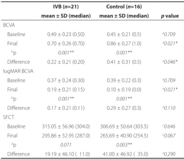
Table 2. The mean subfoveal choroidal thickness and the mean BCVA at baseline and at the inal visit for both groups
IVB (n=21) Control (n=16)
p value mean ± SD (median) mean ± SD (median)
BCVA
Baseline 0.49 ± 0.23 (0.50) 0.45 ± 0.21 (0.5) a0.709*
Final 0.70 ± 0.26 (0.70) 0.86 ± 0.27 (1.0) a0.021*
bp 0.001** 0.001**
Diference 0.22 ± 0.21 (0.20) 0.41 ± 0.31 (0.5) a0.046* logMAR BCVA
Baseline 0.37 ± 0.24 (0.30) 0.39 ± 0.22 (0.3) a0.709*
Final 0.19 ± 0.21 (0.15) 0.10 ± 0.19 (0.0) a0.021*
bp 0.001** 0.001**
Diference 0.17 ± 0.21 (0.11) 0.29 ± 0.27 (0.3) a0.110* SFCT
Baseline 315.05 ± 56.96 (304.0) 306.69 ± 50.64 (303.5) c0.646* Final 295.86 ± 52.95 (287.0) 265.69 ± 40.90 (254.5) c0.067*
dp 0.071 0.003**
Diference 019.19 ± 46.10 (011.0) 041.00 ± 46.92 (035.0) a0.290* BCVA= best-corrected visual acuity; IVB= intravitreal bevacizumab; logMAR= logarithm of the minimum angle of resolution; SFCT= subfoveal choroidal thickness.
*= p<0.05; **= p<0.01.
Su b f o v e a lc h o r o i d a lt h i c k n e S Sc h a n g e Sa f t e ri n t r av i t r e a l b e va c i z u m a bt h e r a p yf o rc e n t r a lS e r o u S c h o r i o r e t i n o pat h y
3 1 0 Arq Bras Oftalmol. 2016;79(5):308-11
representative case at the baseline and inal visits, respectively. All patients showed complete resolution of neurosensory detachment and improvement in vision at the inal visit after the follow-up period. In the IVB group, the mean SFCT was 315 ± 57 µm at the baseline visit, which decreased to 296 ± 53 µm at the inal visit after a mean follow-up period of 10.2 months. In the control group, the mean SFCT was 307 ± 51 µm at the baseline visit, which decreased to 266 ± 41 µm at the inal visit after a mean follow-up period of 8.4 months. The mean SFCT at the baseline and inal visits did not signiicantly difer between the groups. Although there was a signiicant decrease in the mean SFCT for the control group, no signiicant decrease was
found for the IVB group (41 vs 19 µm, p=0.003 vs p=0.071). The mean
BCVA levels in the IVB group at the baseline and inal visits were 0.49 (logMAR 0.37) and 0.70 (logMAR 0.19), respectively. In contrast, the mean BCVA levels in the control group at the baseline and inal visits were 0.45 (logMAR 0.39) and 0.86 (logMAR 0.10), respectively. The mean BCVA at the baseline visit was similar for both groups, and
the mean BCVA increased signiicantly in both groups (p=0.001).
However, the increase in the mean BCVA was signiicantly greater for the control group (0.41; logMAR 0.29) than for the IVB group (0.22;
logMAR 0.17; p<0.05 for BCVA), and the mean inal BCVA of the
con-trol group was signiicantly greater than that of the IVB group [0.86
(logMAR 0.10) vs 0.70 (logMAR 0.19 p<0.05].
Twenty-eight patients (76%) had total and nine patients (24%) had near-total resolution of subretinal luid at the end of the acute CSC episode; only 12 of 16 patients (75%) in the observation group had complete resolution, and only 16 of 21 patients (76%) in the IVB group had complete resolution. The resolution rates were similar in both groups, and re-injection was performed on ive patients in the
IVB group (n=21). No treatment complications were observed during
the follow-up period.
DISCUSSION
No established treatment modalities exist for CSC. Various treat-ments, including topical or systemic carbonic anhydrase inhibitors, laser photocoagulation to the leaking site, photodynamic therapy
with verteporin, and intravitreal injection of anti-VEGF agents, have been applied with variable success rates. In this study, we aimed to demonstrate the efect of IVB therapy on the choroid, which plays a vital role in the pathogenesis of CSC.
There have been many theories concerning the pathogenesis of CSC. RPE dysfunction or defect has been blamed in the development
of serous retinal detachment in CSC(22,23). Gass proposed that CSC was
the result of choroidal vascular hyperpermeability(1). Later studies
using ICGA supported the theory of Gass and demonstrated
evi-dence of hyperpermeability from the choriocapillaries(24,25). Increased
hydrostatic pressure from choroidal vascular hyperpermeability may cause leaks from the level of the RPE and subsequent serous retinal
detachment(25). Based on this knowledge, choroidal vascular
abnor-malities seem to play a key role in the pathogenesis of CSC as the underlying mechanism. Using EDI-OCT, Imamura et al.,
demonstra-ted the presence of a thick choroid in patients with CSC(9). Increased
choroidal thickness in patients with unilateral CSC has been shown
not only in the afected eyes but also in the unafected fellow eyes(10).
In the present study, the baseline mean SFCTs were 315 ± 57 µm in the IVB group and 307 ± 51 µm in the control group, which are greater than that of the normal population [unpublished data: we had previously evaluated 412 eyes of 206 normal Turkish subjects with a mean age of 45 years and found a mean SFCT of 254.4 ± 43.1 µm (range, 122-426 µm)], which supports the presence of a thick choroid in patients with CSC. This inding is consistent with the results from a previous study by Kim et al., in which the SFCT of normal
in-dividuals was found to be 266 ± 55 µm(10). In the present study, after
a mean follow-up period of 10 months, the mean SFCT decreased to 296 ± 53 and 266 ± 41 µm in the IVB and control groups, respectively, and the mean BCVA levels increased to 0.70 and 0.86 in the IVB and control groups, respectively. According to these results, better visual acuity is associated with thinner SFCT. This decrease can be interpre-ted as an approach toward the normal physiological state for SFCT, which was associated with better visual acuity in the control group.
Bevacizumab is a full-length monoclonal antibody that selecti-vely bonds with VEGF. Since 2005, it has been used in ophthalmology to treat various conditions, including neovascular age-related macu-lar degeneration, diabetic macumacu-lar edema, retinal vein occlusions,
and neovascular glaucoma(26). Variable outcomes have been reported
on the use of bevacizumab in CSC(13,16), and the mechanism of action
of bevacizumab in CSC is unknown. Choroidal ischemia may cause an increase in the concentration of VEGF and subsequent choroidal hyperpermeability, which results in a thickened choroid in CSC. At this point, some beneits of an anti-VEGF agent may be proposed on the basis of choroidal ischemia and hyperpermeability as the
pathogenesis of CSC(12-14). However, no studies have demonstrated
an increased level of VEGF in CSC until now. Conversely, Lim et al., re ported no signiicant diference in the VEGF levels in the aqueous
humor of patients with CSC compared with a control group(27). In
addition, the optimal dosage of IVB for CSC has not been formally evaluated. We have used a dosage of 1.25 mg in 0.05 ml, which is the typical dosage of bevacizumab used in other well-studied disease
states(28-29). Furthermore, there are still some controversies about the
nature of the disease. It is not yet understood whether the resolution of subretinal luid is due to IVB or the natural history of the disease. In the present study, the efect of bevacizumab injection was not promising compared with observation in terms of the functional and structural outcomes. This inding supports the literature, which
indi-cates no beneicial efect of anti-VEGF agents in CSC(16).
Since the introduction of EDI-OCT by Spaide et al., this technique has been used by many authors to evaluate the choroidal thickness in various diseases and conditions such as age-related macular
dege-neration, high myopia, and CSC(9,18). Increased choroidal thickness
was demonstrated in both eyes of patients with unilateral active
CSC(10), and changes in choroidal thickness have been shown to be
closely related to choroidal vasculature(30). However, it remains unclear
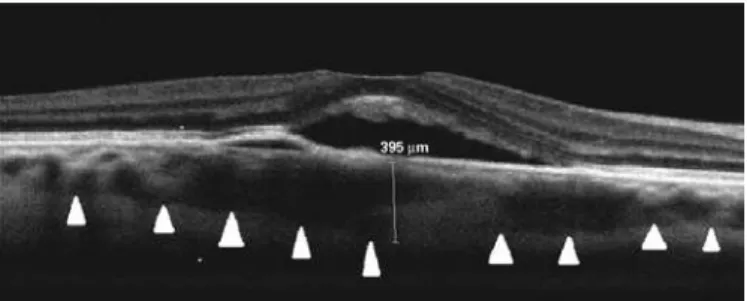
Figure 1. Baseline enhanced depth imaging spectral-domain optical coherence tomo-graphy imaging of a patient with acute central serous chorioretinopathy. The subfoveal choroidal thickness was 395 µm.
Ün l Ü C, e t a l.
3 1 1
Arq Bras Oftalmol. 2016;79(5):308-11 whether or not choroidal thickening on OCT might represent the
disease activity in CSC. In the present study, decreased SFCT was associated with better visual acuity in both groups. Further studies with more patients and a more objective measurement method for the choroidal thickness may reveal a relationship between choroidal topographic changes and disease activity.
This study had some limitations, including its retrospective nature, small sample size, and manual measurement of SFCT. Because our OCT device does not include software for the automated measure-ment of SFCT, we manually measured the distance from the hyperre-lective line of Bruch’s membrane to the inner surface of the observed sclera under the fovea.
In the present study, we demonstrated that IVB injection in CSC is not superior to observation in terms of the anatomic and functional outcomes, and that SFCT decreased with remission of the disease. Further investigation is needed to demonstrate the role of the cho-roid in CSC pathophysiology and the place of IVB as a useful treatment option.
REFERENCES
1. Gass JD. Pathogenesis of disciform detachment of neuroepithelium. Am J Ophthalmol. 1967;63(3):1-139.
2. Spaide RF, Goldbaum M, Wong DW, Tang KC, Iida T. Serous detachment of the retina. Retina. 2003;23(6):820-46; quiz 895-6.
3. Marmor MF. New hypotheses on the pathogenesis and treatment of serous retinal detachment. Graefes Arch Clin Exp Ophthalmol. 1988;226(6):548-52.
4. Prünte C. Indocyanine green angiographic indings in central serous chorioretinopa-thy. Int Ophthalmol. 1995;19(2):77-82.
5. Prünte C, Flammer J. Choroidal capillary and venous congestion in central serous cho-rioretinopathy. Am J Ophthalmol. 1996;121(1):26-34.
6. Iida T, Kishi S, Hagimura N, Shimizu K. Persistent and bilateral choroidal vascular abnor-malities in central serous chorioretinopathy. Retina 1999;19(6):508-12.
7. Kitaya N, Nagaoka T, Hikichi T, Sugawara R, Fukui K, Ishiko S, et al. Features of abnormal choroidal circulation in central serous chorioretinopathy. Br J Ophthalmol. 2003;87(6): 709-12.
8. Tsujikawa A, Ojima Y, Yamashiro K, Ooto S, Tamura H, Nakagawa S, et al. Punctate hy perluorescent spots associated with central serous chorioretinopathy as seen on indocyanine green angiography. Retina. 2010;30(5):801-9.
9. Imamura Y, Fujiwara T, Margolis R, Spaide RF. Enhanced depth imaging optical cohe-rence tomography of the choroid in central serous chorioretinopathy. Retina. 2009; 29(10):1469-73. Comment in: Retina. 2010;30(8):1320-1; author reply 1321-2. 10. Kim YT, Kang SW, Bai KH. Choroidal thickness in both eyes of patients with unilaterally
active central serous chorioretinopathy. Eye (Lond). 2011;25(12):1635-40.
11. Jirarattanasopa P, Ooto S, Tsujikawa A, Yamashiro K, Hangai M, Hirata M, et al. Assessment of macular choroidal thickness by optical coherence tomography and angiogra-phic changes in central serous chorioretinopathy. Ophthalmology. 2012;119(8): 1666-78.
12. Inoue M, Kadonosono K, Watanabe Y, Kobayashi S, Yamane S, Arakawa A. Results of one-year follow-up examinations after intravitreal bevacizumab administration for chronic central serous chorioretinopathy. Ophthalmologica. 2011;225(1):37-40.
13. Artunay O, Yuzbasioglu E, Rasier R, Sengul A, Bahcecioglu H. Intravitreal bevacizumab in treatment of idiopathic persistent central serous chorioretinopathy: a prospective, controlled clinical study. Curr Eye Res. 2010;35(2):91-8.
14. Lim SJ, Roh MI, Kwon OW. Intravitreal bevacizumab injection for central serous chorio-retinopathy. Retina. 2010;30(1):100-6. Comment in: Retina. 2010;30(8):1322-3; author reply 1323; Retina. 2011;31(1):198-9; author reply 199. Retina. 2010;30(8):1323-24; author reply 1324.
15. Torres-Soriano ME, García-Aguirre G, Kon-Jara V, Ustariz-Gonzáles O, Abraham-Marín M, Ober MD, et al. A pilot study of intravitreal bevacizumab for the treatment of central serous chorioretinopathy (case reports). Graefes Arch Clin Exp Ophthalmol. 2008; 246(9):1235-9.
16. Lim JW, Ryu SJ, Shin MC. The efect of intravitreal bevacizumab in patients with acute central serous chorioretinopathy. Korean J Ophthalmol. 2010;24(3):155-8. 17. Seong HK, Bae JH, Kim ES, Han JR, Nam WH, Kim HK. Intravitreal bevacizumab to treat
acute central serous chorioretinopathy: short-term efect. Ophthalmologica. 2009; 223(5):343-7.
18. Spaide RF, Koizumi H, Pozzoni MC. Enhanced depth imaging spectral-domain optical coherence tomography. Am J Ophthalmol. 2008;146(4):496-500. Erratum in: Am J Ophthalmol. 2009;148(2):325.
19. Reibaldi M, Boscia F, Avitabile T, Uva MG, Russo V, Zagari M, et al. Enhanced depth imaging optical coherence tomography of the choroid in idiopathic macular hole: A cross-sectional prospective study. Am J Ophthalmol. 2011;151(1):112-7. Comment in: Am J Ophthalmol. 2011;151(3):560-1; author reply 561.
20. Chung SE, Kang SW, Lee JH, Kim YT. Choroidal thickness in polypoidal choroidal vas-culopathy and exudative age-related macular degeneration. Ophthalmology. 2011; 118(5):840-5.
21. Maruko I, Iida T, Sugano Y, Oyamada H, Sekiryu T, Fujiwara T, et al. Subfoveal choroidal thickness after treatment of Vogt-Koyanagi-Harada disease. Retina. 2011;31(3):510-7. 22. Maruko I, Iida T, Sugano Y, Ojima A, Ogasawara M, Spaide RF. Subfoveal choroidal
thickness after treatment of central serous chorioretinopathy. Ophthalmology. 2010; 117(9):1792-9.
23. Bae SH, Heo JW, Kim C, Kim TW, Lee JY, Song SJ, et al. A randomized pilot study of low-luence photodynamic therapy versus intravitreal ranibizumab for chronic central serous chorioretinopathy. Am J Ophthalmol. 2011;152(5):784-92.
24. Scheider A, Nasemann JE, Lund OE. Fluorescein and indocyanine green angiographies of central serous choroidopathy by scanning laser ophthalmoscopy. Am J Ophthal-mol. 1993;115(1):50-6.
25. Guyer DR, Yannuzzi LA, Slakter JS, Sorenson JA, Hope-Ross M, Orlock DR. Digital indocyanine-green videoangiography of occult choroidal neovascularization. Ophthal-mology. 1994;101(10):1727-35; discussion 1735-7.
26. Stewart MW. The expanding role of vascular endothelial growth factor inhibitors in ophthalmology. Mayo Clin Proc. 2012;87(1):77-88.
27. Lim JW, Kim MU, Shin MC. Aqueous humor and plasma levels of vascular endothelial growth factor and interleukin-8 in patients with central serous chorioretinopathy. Retina. 2010;30(9):1465-71.
28. Avery RL, Pieramici DJ, Rabena MD, Castellarin AA, Nasir MA, Giust MJ. Intravitreal bevacizumab (Avastin) for neovascular age-related macular degeneration. Ophthal-mology. 2006;113(3):363-72. Comment in: OphthalOphthal-mology. 2007;114(2):400; author reply 400-1.
29. Spaide RF, Laud K, Fine HF, Klancnik JM Jr, Meyerle CB, Yannuzzi LA, et al. Intravitreal bevacizumab treatment of choroidal neovascularization secondary to age-related macular degeneration. Retina. 2006;26(4):383-90.