https://doi.org/10.1590/0004-282X20170151
ARTICLE
The relative frequency of common
neuromuscular diagnoses in a reference center
Frequência relativa de diagnósticos neuromusculares comuns em um serviço de referência
Ana Cotta1, Júlia Filardi Paim1, Elmano Carvalho2, Antonio Lopes da-Cunha-Júnior3, Monica M. Navarro4,Jaquelin Valicek2, Miriam Melo Menezes5, Simone Vilela Nunes5, Rafael Xavier-Neto5, Sidney Baptista
Junior1, Luciano Romero Lima6, Reinaldo Issao Takata7, Antonio Pedro Vargas5
he diagnostic investigation of neuromuscular patients is a complex procedure that involves the participation of several professionals. Due to the high diversity of nosologic entities, it is necessary to use diferent kinds of examinations to conirm the clinical diagnostic hypotheses. It is useful to know the relative
frequencies of neuromuscular disorders in the investigated population in order to utilize the most appropriate diagnostic tests. Some neuromuscular reference centers have previously reported on several Brazilian series on the diagnosis of myopa -thies and these are included on the discussion below1,2,3,4,5.
1Rede SARAH de Hospitais de Reabilitação, Departamento de Patologia, Belo Horizonte MG, Brasil; 2Rede SARAH de Hospitais de Reabilitação, Departamento de Neuroisiologia, Belo Horizonte MG, Brasil; 3Rede SARAH de Hospitais de Reabilitação, Departamento de Radiologia, Belo Horizonte MG, Brasil; 4Rede SARAH de Hospitais de Reabilitação, Departamento de Pediatria, Belo Horizonte MG, Brasil; 5Rede SARAH de Hospitais de Reabilitação, Departamento de Neurologia, Belo Horizonte MG, Brasil; 6Rede SARAH de Hospitais de Reabilitação, Departamento de Informática, Belo Horizonte MG, Brasil; 7Rede SARAH de Hospitais de Reabilitação, Departamento de Biologia Molecular, Brasília DF, Brasil;
Correspondence: Ana Cotta; Rede SARAH de Hospitais de Reabilitação; Av. Amazonas, 5953; 30510-000 Belo Horizonte MG, Brasil; E-mail: [email protected]
Conflict of interest: There is no conlict of interest to declare.
Received 08 August 2016; Received in inal form 05 December 2016; Accepted 11 August 2017.
ABSTRACT
The diagnostic procedure in neuromuscular patients is complex. Knowledge of the relative frequency of neuromuscular diseases within the investigated population is important to allow the neurologist to perform the most appropriate diagnostic tests. Objective: To report the relative frequency of common neuromuscular diagnoses in a reference center. Methods: A 17-year chart review of patients with suspicion of myopathy. Results: Among 3,412 examinations, 1,603 (46.98%) yielded conirmatory results: 782 (48.78%) underwent molecular studies, and 821 (51.21%) had muscle biopsies. The most frequent diagnoses were: dystrophinopathy 460 (28.70%), mitochondriopathy 330 (20.59%), spinal muscular atrophy 158 (9.86%), limb girdle muscular dystrophy 157 (9.79%), Steinert myotonic dystrophy 138 (8.61%), facioscapulohumeral muscular dystrophy 99 (6.17%), and other diagnoses 261 (16.28%). Conclusion: Using the presently-available diagnostic techniques in this service, a speciic limb girdle muscular dystrophy subtype diagnosis was reached in 61% of the patients. A neuromuscular-appropriate diagnosis is important for genetic counseling, rehabilitation orientation, and early treatment of respiratory and cardiac complications.
Keywords: diagnosis; neuromuscular diseases; biopsy; epidemiology.
RESUMO
O procedimento diagnóstico neuromuscular é complexo. O conhecimento da frequência relativa das doenças neuromusculares em uma população é importante para utilização dos testes diagnósticos mais apropriados. Objetivo: Relatar a frequência relativa de doenças neuromusculares em um centro de referência. Métodos: Revisão de prontuários de pacientes com suspeita de miopatia em 17 anos. Resultados: Dentre 3412 exames, 1603 (46,98%) foram conirmatórios: 782 (48,78%) estudos moleculares e 821 (51,21%) biópsias musculares. Os diagnósticos mais frequentes foram: distroinopatia 460 (28,70%), mitocondriopatia 330 (20.59%), atroia muscular espinhal 158 (9,86%), distroia muscular cintura-membros 157 (9,79%), distroia miotônica de Steinert 138 (8,61%), distroia muscular face-escápulo-umeral 99 (6,17%) e outros diagnósticos 261 (16,28%). Conclusão: Utilizando as técnicas diagnósticas atualmente disponíveis em nosso serviço, o diagnóstico especíico do subtipo de distroia muscular cintura-membros foi obtido em 61% dos pacientes. O diagnóstico neuromuscular apropriado é importante para o aconselhamento genético, orientações de reabilitação e tratamento precoce de complicações respiratórias e cardíacas.
he aim of this study was to describe the relative frequency of neuromuscular diseases in a neuromuscular reference center.
METHODS
his was a retrospective, descriptive study of consecu-tive molecular examinations and muscle biopsies. Molecular examinations performed between April 19, 1999 and January 18, 2016, and muscle biopsies performed from October 3, 2000 to January 18, 2016 were included.
This study was approved by the Ethics and Research Committee.
Over 17 years, 3,412 patients, admitted to the neuromus -cular outpatient clinic, were investigated for diseases in 16 diagnostic categories: 1,200 muscle biopsies and 2,212 molec -ular exams were performed.
he diagnostic procedure included the correlation between clinical data and laboratory tests, neurophysiol-ogy, and image results, with conirmation through molecular examinations, muscle biopsy, and speciic enzymatic studies (e.g. alpha-glucosidase) in the patients or their relatives with a conirmed diagnosis. Inclusion criteria were molecular or muscle biopsy diagnoses in 16 categories: 1) Steinert myo -tonic dystrophy; 2) dystrophinopathy (Duchenne muscular
dystrophy, Becker muscular dystrophy, intermediate pheno-type dystrophinopathy, and female dystrophinopathy car -riers); 3) facioscapulohumeral muscular dystrophy; 4) limb girdle muscular dystrophy, 5) spinal muscular atrophy; 6) con -genital muscular dystrophy (except collagen VI disorders); 7) collagen VI neuromuscular disorders (Bethlem myopathy and Ullrich congenital muscular dystrophy); 8) congenital myop-athies (subtypes: central core congenital myopathy, nemaline congenital myopathy, centronuclear/myotubular congenital myopathy, cap disease congenital myopathy, and congeni -tal iber type disproportion); 9) congeni-tal myasthenic syn-dromes; 10) myoibrillar myopathy; 11) glycogen-storage dis -ease type V (McArdle dis-ease); 12) glycogen-storage dis-ease type II (Pompe disease); 13) dermatomyositis; 14) sporadic inclusion body myositis; 15) other inlammatory myopathies (polymyositis, nonspeciic myositis, and necrotizing immune myopathy); and 16) mitochondriopathy.
Exclusion criteria were: 1) myopathic abnormalities with -out other speciications; 2) neurogenic muscular abnormali-ties; 3) rare genetic syndromes and other neuromuscular dis -orders not included in the 16 investigated categories; and 4) duplicated examinations of patients submitted to both mus -cle biopsy and conirmatory molecular examinations ( for duplicated examinations, the results were reported only once on the molecular examination tables) (Table 1).
Table 1. Diagnostic categories: inclusion and exclusion criteria.
Inclusion criteria - 16 diagnostic categories
1) Steinert myotonic dystrophy
2) dystrophinopathy: Duchenne, Becker, intermediate phenotype, and female carriers
3) facioscapulohumeral muscular dystrophy
4) limb girdle muscular dystrophy
5) spinal muscular atrophy
6) congential muscular dystrophy (except collagen VI disorders)
7) Bethlem myopathy/ Ullrich congenital muscular dystrophy (type VI collagenopathy)
8) congenital myopathies (subtypes “central core” congenital myopathy, nemaline congenital myopathy, centronuclear/myotubular congenital myopathy, congenital iber type disproportion, cap disease congenital myopathy)
9) congenital myasthenic syndrome
10) myoibrillar myopathy
11) glycogen-storage disease type V (McArdle disease)
12) glycogen-storage disease type II (Pompe disease)
13) dermatomyositis
14) sporadic inclusion body myositis
15) other inlammatory myopathies (polymyositis, nonspeciic myositis, and immune mediated necrotizing myopathy) 16) mitochondrial myopathy
Exclusion criteria
1) myopathic abnormalities without other speciication
2) neurogenic muscular abnormalities
3) rare genetic syndromes and other neuromuscular disorders not included in the 16 inclusion criteria diagnostic categories
Molecular examinations for dystrophinopathies included the investigation of dystrophin gene deletion through poly -merase chain reaction (PCR), and deletion and duplication of the dystrophin gene through multiplex ligation-dependent probe ampliication.
Molecular investigation of spinal muscular atrophy con-sisted of the detection of exons 7 and 8 deletion of the sur -vival motor neuron (SMN) gene through PCR.
Facioscapulohumeral muscular dystrophy investiga -tion consisted of the search of dele-tions of repeated tan -dem 3.3kb units within the D4Z4 locus at the 4q35 region through restriction fragments EcoRI/ HindIII, EcoRI/ AvrII and ApoI through Southern blotting with pulsed ield elec -trophoresis (CHEF-DRIII - Biorad) followed by p13E-11 probe hybridization.
Steinert myotonic dystrophy molecular investigation was performed through the study of the dystrophia myotonica protein kinase (DMPK) gene CTG expansion with analysis of EcoRI and BamHI through Southern blotting, followed by pM10M-6 probe hybridization.
Calpainopathy (limb girdle muscular dystrophy type 2A [LGMD2A]) molecular investigation was performed with the search for 24 exons calpain gene point mutations (NM_000070.2) through denaturing high performance liq-uid chromatography and altered amplicon direct sequencing with the ABI 3130XL Genetic Analyzer (Applied Biosystems Sequencing (v. 5.2) software), and SeqScape (v. 2.5) analysis software, as well as a search for mutation on the Leiden data -base (www.dmd.nl/index.html).
Sarcoglycanopathy (LGMD2C, LGMD2D, LGMD2E, and LGMD2F) molecular investigation was performed with the study of alpha, beta, gamma, and delta sarcoglycan genes (respectively SGCD, SGCE, SGCC, and SGCF genes) through denaturing high performance liquid chromatog -raphy followed by altered amplicon direct sequencing with the ABI 3130XL Genetic Analyzer (Applied Biosystems Sequencing (v. 5.2) software), and SeqScape (v. 2.5) analysis software. he exons analyzed were alpha-sarcoglycan exon 3, beta-sarcoglycan exons 3 and 4, delta-sarcoglycan exon 8, and gamma-sarcoglycan exon 6, these being the most fre-quent mutations in Brazil6.
Fukutin-related proteinopathy (LGMD2I) molecular investigation was performed with the fukutin-related protein (FKRP) investigation through denaturing high performance liquid chromatography followed by altered amplicon direct sequencing with the ABI 3130XL Genetic Analyzer (Applied Biosystems Sequencing (v. 5.2) software), and SeqScape (v. 2.5) analysis software.
The review of muscle biopsy diagnoses was performed through a Systematized Nomenclature of Medicine search in the fields “results” and “observations”. The stan -dard procedure for muscle biopsy consisted of histo -chemical studies and, whenever necessary, immunohisto-chemical and electron microscopy studies. Histoimmunohisto-chemical
investigations were performed on liquid nitrogen-frozen sections with: hematoxylin and eosin, Gomori modified trichrome, periodic acid-Schiff with and without diastase, Oil-red-O, myosin ATPase (pH9.4, pH4.6, and pH4.3), suc -cinate dehydrogenase, cytochrome-c-oxidase, nicotin -amide adenine dinucleotide, acid phosphatase, and non-specific esterase. For patients under investigation for metabolic myopathy, tests for myophosphorylase, phos -phofructokinase, and myoadenylate deaminase deficien -cies were performed.
Diagnosis of a mitochondrial disorder included dei-nite, probable and possible diagnostic criteria, codiied by the Systematized Nomenclature of Medicine, considering histochemical, ultrastructural, molecular ( from December 11, 2008), and respiratory chain enzymatic studies ( from August 6, 2013).
A dysferlinopathy (LGMD2B) diagnosis was suspected for patients with a characteristic clinical presentation and phe -notypic conirmation through immunohistochemistry for dysferlin deiciency.
A congenital myopathy diagnosis followed the International Standard of Care Committee for Congenital Myopathies7 criteria. Congenital myopathy subtypes
included: “central core” congenital myopathy, nemaline con -genital myopathy, centronuclear/myotubular con-genital myopathy, congenital iber type disproportion, and cap dis -ease congenital myopathy. A congenital muscular dystro-phy diagnosis followed the International Standard of Care Committee for Congenital Muscular Dystrophies criteria8.
he congenital myasthenic syndrome diagnosis was made by considering anamnesis, clinical examination, and neurophysiological studies with signiicant repetitive nerve stimulation decrement with or without abnormal ultrastruc -tural neuromuscular junctions9.
RESULTS
he 2,212 molecular examinations corresponded to diag -nostic categories 1 to 5. Category 1: 297 patients with clini -cal suspicion of Steinert myotonic dystrophy; category 2: 583 patients with clinical suspicion of dystrophinopathy; cat-egory 3: 352 patients with clinical suspicion of facioscapulo -humeral muscular dystrophy; category 4: 299 patients with clinical suspicion of limb girdle muscular dystrophy, among them 139 patients with clinical suspicion of calpainopathy (LGMD2A), 58 with clinical suspicion of sarcoglycanopa-thy (LGMD2C/ LGMD2D/ LGMD2E/ LGMD2F), and 102 with clinical suspicion of fukutin-related proteinopathy (LGMD2I); and category 5: 681 patients with clinical suspi-cion of spinal muscular atrophy.
18, 2016, and one muscle biopsy had a review of the diagnosis during the study period, based on the muscle biopsy slides on material that had been previously col-lected on September 21, 1997 (one patient with a diagno -sis of Pompe disease).
After the application of inclusion and exclusion criteria (Table 1) to the group of 3,412 patients, 1,603 (47%) patients were selected with conirmed diagnoses in the 16 diagnos -tic categories, 782 (48.8%) molecular examinations, and 821 (51.2%) muscle biopsies (Figure 1). he most frequent diagnoses were, in descending order: dystrophinopathy 460 (28.70%); deinite, probable or possible mitochondriopathy 330 (20.59%); spinal muscular atrophy 158 (9.86%); limb gir-dle muscular dystrophy 157 (9.79%); Steinert myotonic dys -trophy 138 (8.61%); facioscapulohumeral muscular dystro-phy 99 (6.17%); congenital muscular dystrodystro-phy 79 (4.93%); congenital myopathy 58 (3.62%); polymyositis 35 (2.18%); dermatomyositis 34 (2.12%); sporadic inclusion body myosi -tis 15 (0.94%); type VI collagenopathy 13 (0,81%); myoibril-lar myopathy 11 (0.68%); congenital myasthenic syndrome 7 (0.44%); glycogen-storage disease type V/McArdle disease 5 (0.31%), and glycogen-storage disease type II/Pompe dis -ease 4 (0.25%) (Figure 2) (Table 2).
here were 157 patients with limb girdle muscular dys -trophy diagnoses. Among them, 34 were excluded from the muscle biopsy diagnosis table because they were submitted to further conirmatory molecular examinations. herefore, 14 of the 24 patients with calpainopathy (LGMD2A) were excluded from the muscle biopsy table. Of the 26 patients with a sarcoglycanopathy diagnosis, 17 had the diagno -sis made through muscle biopsy with immunohistochem-istry and nine through molecular examinations; six were excluded from the muscle biopsy table as they had been
submitted to both muscle biopsy and molecular examina-tions. Among the eight patients with a diagnosis of fukutin-related proteinopathy, three were excluded from the muscle biopsy table as they were submitted to molecular examina -tions after the muscle biopsy. hree patients from the same family received the diagnosis of laminopathy (LGMD1B); three patients from another family received the diagnosis of caveolinopathy (LGMD1C) with the index case submit -ted to muscle biopsy, and three patients received the diag-nosis of telethoninopathy (LGMD2G), conirmed through muscle biopsy with immunohistochemical deiciency on telethonin expression. With the presently-available tech -niques in our service, a conirmatory diagnosis of limb gir-dle muscular dystrophy subtype was made in 61% of the patients (Table 3)5,10,11,12,13,14.
here were 344 male patients with a diagnosis of dys -trophinopathy. Among the 287 patients with a diagnosis of Duchenne muscular dystrophy, 184 were conirmed by molecular examination and 103 by muscle biopsy. Among 45 patients with Becker muscular dystrophy, 33 were con -irmed by molecular examination and 12 by muscle biopsy. Among the 12 patients with intermediate dystrophinopa -thy phenotype, 10 were conirmed by molecular examina-tion and two by muscle biopsy; four patients were excluded from the muscle biopsy table as they were later submitted to conirmatory molecular examination.
he initial date of the examinations varied according to the speciic technique implementation: Steinert myo -tonic dystrophy molecular examinations started on April 19, 1999, dystrophinopathy on June 4, 2001, spinal muscu -lar atrophy on May 29, 2000, facioscapulohumeral muscu-lar dystrophy on February 12, 2003, calpainopathy (LGMD2A) on May 24, 2011, sarcoglycanopathy (LGMD2C, LGMD2D,
Figure 2. Relative frequencies of neuromuscular diagnoses. Dystrophinopathy
n = 1603
460 (28.7%)
330 (20.6%)
158 (9.9%) 157
(9.8%) 138 (8.6%)
99 (6.2%)
84 (5.2%)
79 (4.9%)
58 (3.6%)
40 (2.5%)
Mitochondriopathy
Spinal muscular
atrophy Limb girdle
muscular dystrophy Steinert myotonic dystrophy FSH muscular
dystrophy
Inflammatory myopathy
Congenital muscular dystrophy
Congenital myopathy
Other
Figure 1. Muscle biopsy-molecular neuromuscular examination ratio.
N = 1603
821 (51.2%) Muscle biopsies
LGMD2E, and LGMD2F) on May 26, 2011, fukutin-related proteinopathy on May 18, 2011. Muscle biopsies started in 1997 and the informatized diagnostic system, codi-ied through the Systematized Nomenclature of Medicine, started on October 3, 2000.
After April 5, 2013, the diagnosis of dysferlinopathy was made through muscle biopsy with immunohistochemi -cal deiciency in dysferlin expression after the conirmation of normal caveolin immunohistochemical expression and absent calpain mutation15,16,17.
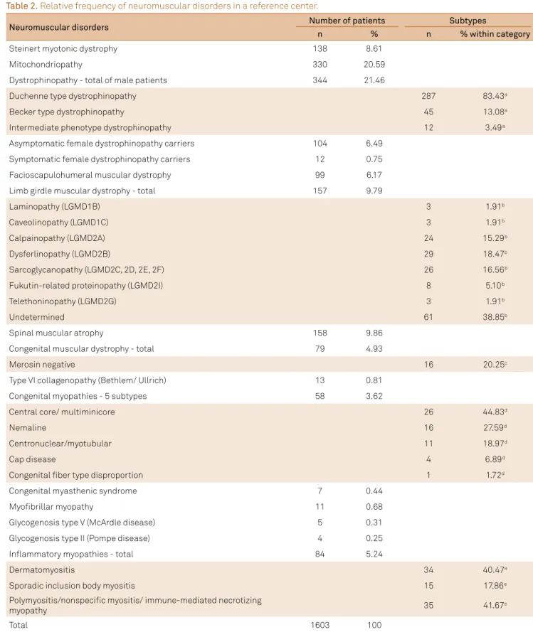
Table 2. Relative frequency of neuromuscular disorders in a reference center.
Neuromuscular disorders Number of patients Subtypes
n % n % within category
Steinert myotonic dystrophy 138 8.61
Mitochondriopathy 330 20.59
Dystrophinopathy - total of male patients 344 21.46
Duchenne type dystrophinopathy
287 83.43a
Becker type dystrophinopathy 45 13.08a
Intermediate phenotype dystrophinopathy 12 3.49a
Asymptomatic female dystrophinopathy carriers 104 6.49
Symptomatic female dystrophinopathy carriers 12 0.75
Facioscapulohumeral muscular dystrophy 99 6.17
Limb girdle muscular dystrophy - total 157 9.79
Laminopathy (LGMD1B)
3 1.91b
Caveolinopathy (LGMD1C) 3 1.91b
Calpainopathy (LGMD2A) 24 15.29b
Dysferlinopathy (LGMD2B) 29 18.47b
Sarcoglycanopathy (LGMD2C, 2D, 2E, 2F) 26 16.56b
Fukutin-related proteinopathy (LGMD2I) 8 5.10b
Telethoninopathy (LGMD2G) 3 1.91b
Undetermined 61 38.85b
Spinal muscular atrophy 158 9.86
Congenital muscular dystrophy - total 79 4.93
Merosin negative 16 20.25c
Type VI collagenopathy (Bethlem/ Ullrich) 13 0.81
Congenital myopathies - 5 subtypes 58 3.62
Central core/ multiminicore
26 44.83d
Nemaline 16 27.59d
Centronuclear/myotubular 11 18.97d
Cap disease 4 6.89d
Congenital iber type disproportion 1 1.72d
Congenital myasthenic syndrome 7 0.44
Myoibrillar myopathy 11 0.68
Glycogenosis type V (McArdle disease) 5 0.31
Glycogenosis type II (Pompe disease) 4 0.25
Inlammatory myopathies - total 84 5.24
Dermatomyositis
34 40.47e
Sporadic inclusion body myositis 15 17.86e
Polymyositis/nonspeciic myositis/ immune-mediated necrotizing
myopathy 35 41.67e
Total 1603 100
DISCUSSION
his study provided an estimated relative frequency of speciic diagnoses (16 diagnostic categories) through molecular and muscle biopsy examinations in a neuromus -cular reference clinic, over the last 17 years. About half the patients received a diagnosis, within one of the 16 catego -ries, by molecular studies, and half by muscle biopsy. he relative frequency of neuromuscular diseases showed some similarities to the neuromuscular disease prevalence in a study in northern England14. In that study, the most frequent
diagnoses, in descending order, were: myotonic dystrophy, mitochondriopathy, dystrophinopathy, facioscapulohumeral muscular dystrophy, limb girdle muscular dystrophy, spinal muscular atrophy, congenital muscular dystrophy, Bethlem type VI collagenopathy, and congenital myopathies (central core and nemaline)14. On the other hand, in the present study,
the most frequent diagnoses, in descending order were: dys-trophinopathy, mitochondriopathy, spinal muscular atrophy, limb girdle muscular dystrophy, Steinert myotonic dystro-phy, and facioscapulohumeral muscular dystrophy. Due to methodological diferences between both studies, it is not possible to conclude that the diferent relative frequencies of myotonic dystrophy in both studies should be attributed to diferences in prevalences. Besides, only patients with myo -tonic dystrophy type 1 (Steinert myo-tonic dystrophy) were included in the present study, and type 2 myotonic dystrophy patients were not included in our study.
One limitation of this study is related to the diagnosis of dysferlinopathy, as the diagnosis was immunophenotypical and not genotypical. Considering the possibility of immuno -histochemical secondary deiciencies, after April 5, 2013, the diagnosis was made taking into consideration the absence of calpain gene mutations, detection of calpain expression on Western blot, and detection of immunophenotypical caveo -lin expression15,16,17 (Table 3)5,10,11,12,13,14.
Considering the diagnostic techniques available over the last 17 years, a conclusive, deinite limb girdle muscular dystrophy subtype diagnosis was achieved in about 61% of the patients. In the previously-mentioned study in northern England, using muscle biopsy, immunohistochemistry, Western blot, and genetic sequencing, a conclusive limb girdle muscular dystro-phy subtype diagnosis was achieved in 75% of the patients18,
whereas in another study in Italy, the percentage of conirmed limb girdle muscular dystrophy subtype diagnoses was 60%10,18.
Even after the utilization of molecular examinations dur-ing the diagnostic procedure at the neuromuscular outpa -tient clinic, muscle biopsies represented 51.2% (821/1,603) of the examinations used to conirm the diagnosis. In a French study, muscle biopsies resulted in 43.6% of conclusive diagno-ses19. In that study, the most frequent muscle biopsy diagnoses,
in descending order were: congenital myopathy (for the most part corresponding to nonstructural congenital myopathy), progressive muscular dystrophy (for the most part correspond-ing to dystrophinopathy), mitochondrial encephalomyopathy, other metabolic myopathies, congenital muscular dystrophy,
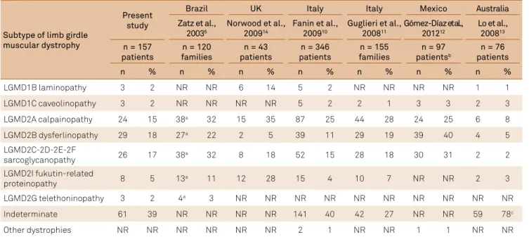
Table 3. Relative frequency of limb girdle muscular dystrophies.
Subtype of limb girdle muscular dystrophy
Present study
Brazil UK Italy Italy Mexico Australia
Zatz et al., 20035
Norwood et al., 200914
Fanin et al., 200910
Guglieri et al., 200811
Gómez-Díaz et al., 201212
Lo et al., 200813
n = 157 patients
n = 120 families
n = 43 patients
n = 346 patients
n = 155 families
n = 97 patientsb
n = 76 patients
n % n % n % n % n % n % n %
LGMD1B laminopathy 3 2 NR NR 6 14 5 2 NR NR NR NR 1 1
LGMD1C caveolinopathy 3 2 NR NR NR NR 5 2 2 1 3 3 2 3
LGMD2A calpainopathy 24 15 38a 32 15 35 87 25 44 28 24 25 6 8
LGMD2B dysferlinopathy 29 18 27a 22 2 5 39 11 29 19 39 40 4 5
LGMD2C-2D-2E-2F
sarcoglycanopathy 26 17 38a 32 8 18 52 15 28 18 30 31 2 2 LGMD2I fukutin-related
proteinopathy 8 5 13
a 11 12 28 15 4 10 7 NR NR 2 3
LGMD2G telethoninopathy 3 2 4a 3 NR NR NR NR NR NR NR NR NR NR
Indeterminate 61 39 NR NR NR NR 141 40 42 27 NR NR 59 78c
Other dystrophies NR NR NR NR NR NR 2 1 NR NR 1 1 NR NR
NR: not reported; a: absolute number of patients calculated based on the published percentages; b: initial number of 290 biopsies (dystrophin and merosin deiciencies were excluded); c: 78 indeterminate, of whom 58 have normal dysferlin expression and 20 have defective dysferlin. The frequencies of sarcoglycanopathy were calculated with the total of patients of each subtype: Fanin 200910, LGMD2C-2F = 11+30+10+1 = 52 (15%); Guglieri 200811,
and dermatomyositis19. In the present study, the most frequent
muscle biopsy diagnoses were, in descending order: mitochon -driopathy, dystrophinopathy, limb girdle muscular dystrophy, inlammatory myopathy (polymyositis, dermatomyositis, and others), congenital muscular dystrophy, and congenital myop-athy. he diference in the frequency of congenital myopathy between these studies may be due to the inclusion criteria as, in the present study, only ive structural congenital myopathy subtypes were included (central core congenital myopathy, nemaline congenital myopathy, centronuclear/myotubular congenital myopathy, congenital iber type disproportion, and cap disease congenital myopathy).
An analysis of 4,500 muscle biopsies performed from 1979 to 2012 in another Brazilian reference center identiied 19 patients with Pompe disease1; the same reference center
reported 106 patients submitted to dystrophin gene DNA analysis between 1999 and 2005 and at least one deletion was detected in 76 cases2, as well as 56 patients with limb girdle
muscular dystrophy submitted to muscle biopsy from 1976 to 20013. From a group of 3,802 patients submitted to muscle
biopsy between 1989 and 2001, at another Brazilian neuro-muscular reference center, 86 patients were found to have mitochondriopathy of the chronic progressive external oph-thalmoplegia subtype4. Yet another Brazilian group reported
120 unrelated families with limb girdle muscular dystrophy, submitted to molecular investigation until 20035.
With the advent of novel technologies, such as next gen-eration sequencing, there is hope that the resolution rate of molecular studies may increase with consequent reduction in the number of undetermined diagnoses. his would provide the patients with: 1) adequate genetic counseling; 2) early clinical, respiratory and cardiac management; 3) rehabilita -tion orienta-tion concerning gait prognosis and daily activi-ties, according to the subtype of neuromuscular disorder.
Acknowledgments
We thank Cleides Campos de Oliveira and Simone Ferreira do Nascimento for muscle biopsy technical assistance.
References
1. Werneck LC, Lorenzoni PJ, Kay CS, Scola RH. Muscle biopsy in Pompe disease. Arq Neuropsiquiatr. 2013;71(5):284-9. https://doi.org/10.1590/0004-282X20130022
2. Freund AA, Scola RH, Arndt RC, Lorenzoni PJ, Kay CK,
Werneck LC. Duchenne and Becker muscular dystrophy: a molecular and immunohistochemical approach. Arq Neuropsiquiatr.
2007;65(1):73-6. https://doi.org/10.1590/S0004-282X2007000100016
3. Comerlato EA, Scola RH, Werneck LC. Limb-girdle muscular dystrophy: an immunohistochemical diagnostic approach. Arq Neuropsiquiatr. 2005;63(2A):235-45. https://doi.org/10.1590/S0004-282X2005000200009
4. Kiyomoto BH, Tengan CH, Costa CK, Oliveira AS, Schmidt B, Gabbai AA. Frequency of dystrophic muscle abnormalities in chronic progressive external ophthalmoplegia: analysis of 86 patients. J Neurol Neurosurg Psychiatry. 2006;77(4):541-3. https://doi.org/10.1136/jnnp.2005.079954
5. Zatz M, de Paula F, Starling A, Vainzof M. The 10 autosomal recessive limb-girdle muscular dystrophies. Neuromuscul Disord. 2003;13(7-8):532-44. https://doi.org/10.1016/S0960-8966(03)00100-7
6. Gouveia TL, Paim JF, Pavanello RC, Zatz M, Vainzof M. Sarcoglycanopathies: a multiplex molecular analysis for the most common mutations. Diagn Mol Pathol. 2006;15(2)95-100. https://doi.org/10.1097/00019606-200606000-00006
7. North KN, Wang CH, Clarke N, Jungbluth H, Vainzof M, Dowling JJ et al. Approach to the diagnosis of congenital myopathies. Neuromuscul Disord. 2014;24(2):97-116. https://doi.org/10.1016/j.nmd.2013.11.003
8. Bönnemann CG, Wang CH, Quijano-Roy S, Deconinck N, Bertini E, Ferreiro A et al. Diagnostic approach to the congenital muscular dystrophies. Neuromuscul Disord. 2014;24(4):289-311. https://doi.org/10.1016/j.nmd.2013.12.011
9. Engel AG. Current status of the congenital myasthenic syndromes. Neuromuscul Disord. 2012;22(2):99-111. https://doi.org/10.1016/j.nmd.2011.10.009
10. Fanin M, Nascimbeni AC, Aurino S, Tasca E, Pegoraro E,
Nigro V et al. Frequency of LGMD gene mutations in Italian patients with distinct clinical phenotypes. Neurology. 2009;72(16):1432-5. https://doi.org/10.1212/WNL.0b013e3181a1885e
11. Guglieri M, Magri F, D’Angelo MG, Prelle A, Morandi L, Rodolico C et al. Clinical, molecular, and protein correlations in a large sample of genetically diagnosed Italian limb girdle muscular dystrophy patients. Hum Mutat. 2008;29(2):258-66. https://doi.org/10.1002/humu.20642
12. Gómez-Díaz B, Rosas-Vargas H, Roque-Ramírez B, Meza-Espinoza P, Ruano-Calderón LA, Fernández-Valverde F et al. Immunodetection analysis of muscular dystrophies in Mexico. Muscle Nerve 2012;45(3):338-345. https://doi.org/10.1002/mus.22314
13. Lo HP, Cooper ST, Evesson FJ, Seto JT, Chiotis M, Tay V et al. Limb-girdle muscular dystrophy: diagnostic evaluation, frequency and clues to pathogenesis. Neuromuscul Disord. 2008;18(1):34-44. https://doi.org/10.1016/j.nmd.2007.08.009
14. Norwood FL, Harling C, Chinnery PF, Eagle M, Bushby K, Straub V. Prevalence of genetic muscle disease in Northern England: in-depth analysis of a muscle clinic population. Brain. 2009;132(11):3175-86. https://doi.org/10.1093/brain/awp236
15. Nguyen K, Bassez G, Krahn M, Bernard R, Laforêt P, Labelle V et al. Phenotypic study in 40 patients with dysferlin gene mutations: high frequency of atypical phenotypes. Arch Neurol. 2007;64(8):1176-82. https://doi.org/10.1001/archneur.64.8.1176
16. Groen EJ, Charlton R, Barresi R, Anderson LV, Eagle M, Hudson J et al. Analysis of the UK diagnostic strategy for limb girdle muscular dystrophy 2A. Brain. 2007;130(12):3237-49. https://doi.org/10.1093/brain/awm259
17. Müller JS, Piko H, Schoser BG, Schlotter-Weigel B, Reilich P, Gürster S et al. Novel splice site mutation in the caveolin-3 gene leading to autosomal recessive limb girdle muscular dystrophy. Neuromuscul Disord. 2006;16(7):432-6. https://doi.org/10.1016/j.nmd.2006.04.006
18. Bushby K. Diagnosis and management of the limb girdle muscular dystrophies. Pract Neurol. 2009;9(6):314-23. https://doi.org/10.1136/jnnp.2009.193938
19. Cuisset JM, Maurage CA, Carpentier A, Briand G,