Arq Bras Cardiol 2003; 81: 97-100.
Bortolotto et al Malignant hypertension and hypertensive encephalopathy in primary aldosteronism
9 7 9 79 7 9 79 7
Instituto do Coração do Hospital das Clínicas – FMUSP
Mailing address: Luiz Aparecido Bortolotto – InCor Unidade de Hipertensão -Av. Dr. Enéas C. Aguiar, 44 - 05403-000 – São Paulo, SP, Brazil – E-mail: [email protected]
English version by Stela Maris C. e Gandour
Arq Bras Cardiol, volume 81 (nº 1), 97-100, 2003
Luiz Aparecido Bortolotto, Fernando Henpin Yue Cesena, Fabio Biscegli Jatene, Hélio Bernardes Silva
São Paulo, SP - Brazil
Malignant Hypertension and Hypertensive Encephalopathy in
Primary Aldosteronism Caused by Adrenal Adenoma
Brief Report
Two cases are reported as follows: 1) 1 female patient with accelerated-malignant hypertension secondary to an aldosterone-producing adrenal adenoma; and 2) 1 female patient with adrenal adenoma, severe hyperten-sion, and hypertensive encephalopathy. This association is a rare clinical finding, and malignant hypertension may modify the hormonal characteristic of primary aldoste-ronism, making its diagnosis more difficult. The diagnosis of primary aldosteronism should be considered in patients with malignant hypertension or hypertensive encephalo-pathy if persistent hypokalemia occurs. Identification of primary aldosteronism is of paramount importance for the patient’s evolution, because the surgical treatment makes the prognosis more favorable.
Primary aldosteronism (Conn’s syndrome) due to adrenal adenoma is a very rare and potentially curable cause of arterial hypertension 1,2. Contrary to what Conn et al
re-ported in 1964, we currently know that hypertension caused by adrenal adenoma may behave in a malignant way 1,2.
We report the cases of 2 female patients with severe arterial hypertension, 1 with accelerated malignant hyper-tension and the other with hypertensive encephalopathy secondary to primary aldosteronism due to an aldosterone-producing adrenal adenoma. The major characteristics of primary aldosteronism and the particularities of its associa-tion with accelerated malignant hypertension are reviewed. In addition, the important role played by the renin-angio-tensin-aldosterone system in the development of accelera-ted malignant hypertension is discussed, as is its behavior when associated with primary aldosteronism.
9 8 9 8 9 8 9 8 9 8
Bortolotto et al
Malignant hypertension and hypertensive encephalopathy in primary aldosteronism
Arq Bras Cardiol 2003; 81: 97-100.
her serum potassium level was then 4.9 mEq/L, her plasma renin activity was 0.8 ng/mL/h, and her plasma aldosterone level was 8.4 ng/100mL. On that occasion, funduscopy re-vealed the absence of exudates and hemorrhages. One year after adrenalectomy, the patient remains without hyperten-sive crises; her blood pressure is 140/90 mm Hg, and she is taking fewer medications than she did before the surgery (0.4 mg/day of clonidine, 50 mg/day of atenolol, and 10 mg/ day of amlodipine). Her serum potassium is 5.0 mEq/L, her plasma renin activity is 1.8 ng/mL/h, and her plasma aldoste-rone level is 13.6 ng/100mL.
Case 2- A 34-year-old female patient was admitted to the Hypertension Unit at InCor complaining of headache and blurred vision. She reported having systemic arterial hypertension, which had been diagnosed during pregnan-cy 9 years before and was difficult to control in the last 4 years. During this period, she required several hospitaliza-tions because of hypertensive crises. The patient was ta-king 50 mg/day of hydrochlorothiazide, 60 mg/day of pro-pranolol, 150 mg/day of captopril, and 75 mg/day of hydra-lazine. On admission, her physical examination revealed a
good general condition, heart rate of 70 bpm, blood pressu-re of 240/140 mmHg, and no other alterations. Funduscopy revealed bilateral papilledema. The electrocardiogram sho-wed signs of hypertrophy of the left chambers. The echo-cardiogram revealed left ventricular concentric hypertrophy (mass index of 180 g/m2, posterior wall of 12 mm, diastolic
diameter of 50 mm) and ejection fraction of 0.71. The com-plementary laboratory tests showed normal renal function (serum urea of 25 mg/dL, serum creatinine of 1.1 mg/dL) and normal urinary sediment. The abdominal ultrasonography showed kidneys with size and morphology within the normal range. The renal arteriography showed no vascular obstructive lesion. The measurements of vanillylmandelic acid and metanephrines in the 24-hour urine were normal. The initial measurement of plasma electrolytes showed K of 2.3 mEq/L and Na of 144 mEq/L. The measurements of the electrolytes in the 24-hour urine (total volume of 920 mL) a few days later showed K of 13 mEq and Na of 33 mEq, con-comitantly with plasma K of 3.3 mEq/mL and plasma Na of 142 mEq/L. The plasma renin activity was suppressed: 0.25 ng/mL/h in the horizontal position and 0.90 ng/mL/h after a 2-hour walk (normal value: 5.0±1.8 ng/mL/h). At the same time, a significant increase in the plasma levels of aldosterone was observed: 56.9 ng/100mL in the horizontal position (normal range: 1 to 16 ng/100mL) and 50.2 ng/ 100mL after a 2-hour walk (normal range: 4 to 31 ng/100mL). Abdominal computerized tomography detected a hypoden-se nodule of 1.2 cm of diameter in the right adrenal gland. Blood pressure control during hospitalization was initially unsatisfactory, when the patient maintained high blood pressure levels despite the use of 160 mg/day of proprano-lol, 150 mg/day of captopril, and 150 mg/day hydralazine. The introduction of 300 mg/day of spironolactone for 10 days did not reduce blood pressure. An appropriate control of blood pressure was only obtained after the combination of minoxidil (5 mg every other day), which also significantly improved headache. The patient underwent right adrenalec-tomy, and a yellow nodule of 1.5 cm of diameter was found in the cortical zone. The anatomicopathological examination diagnosed adenoma. During the postoperative period, the plasma K levels normalized and the patient was discharged from the hospital asymptomatic. She maintained blood pres-sure levels of 130/90 mmHg taking 50 mg/day of hydrochlo-rothiazide and 80 mg/day of propranolol. One year later, the patient was readmitted for reassessment. The patient was taking fewer drugs than before the surgery, and her blood pressure remained 140/90 mmHg. Funduscopy revealed no exudates, hemorrhages, or papilledema. A new laboratory assessment revealed plasma potassium levels of 4.6 mEq/L, plasma renin activity at rest of 2 ng/mL/h, and plasma aldos-terone of 10 ng/100mL. Ventricular hypertrophy also de-creased (mass index of 110 g/m2).
Discussion
We report the cases of 2 patients with severe arterial hypertension, 1 with an alteration in funduscopy diagnostic
Fig. 1 – Histologic section at the level of the adrenal nodule. The diagnosis of adenoma was established based on the presence of relatively large cells with abundant cytoplasm, scarcity of atypia, and absence of mitoses. Magnification: 10 X.
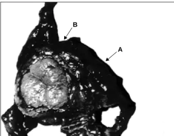
Fig. 2 – Histologic section of the adrenal cortex showing an area of normal appearance (A) and the nodule corresponding to the adenoma (B). Magnification: 1.25 X.
B
Arq Bras Cardiol 2003; 81: 97-100.
Bortolotto et al Malignant hypertension and hypertensive encephalopathy in primary aldosteronism
9 9 9 99 9 9 99 9
of accelerated malignant hypertension (grade 3 in the Keith-Wagener classification), and another with bilateral papille-dema, characteristic of hypertensive encephalopathy, but also present in patients with malignant hypertension, even in the absence of hemorrhages and exudates, which may disappear in a short period of time, as happened with the pa-tient with confirmed accelerated malignant hypertension. Both accelerated malignant hypertension and hypertensive encephalopathy are rare complications of primary aldoste-ronism, and, therefore, deserve to be discussed. To better discuss the most important aspects of this association from the pathophysiologic point of view, we can consider both patients as having severe arterial hypertension with accele-rated or malignant behavior.
Primary aldosteronism due to adrenal adenoma is a rare disease, which affects approximately 1% of all hyperten-sive patients 3. Its prevalence is higher in young females (30
to 50 years), and it is usually accompanied by hypokalemia and orthostatic hypotension 3. Its identification is
extreme-ly important, because tumor resection invariabextreme-ly improves blood pressure levels, and, many times completely reverts the hypertensive condition. In addition, accelerated malig-nant hypertension is an extremely severe condition that causes a great reduction in survival.
The coexistence of primary aldosteronism and accele-rated malignant hypertension, although extremely rare, makes the prognosis more favorable, because surgical re-section of the tumor eliminates the triggering cause of hy-pertension. This association should be differentiated from secondary aldosteronism, which is frequently observed in patients with accelerated malignant hypertension due to adrenal hyperactivity 2. In the initial description by Conn 4,
the association of accelerated malignant hypertension and primary aldosteronism was not observed. Kaplan 4 reported
this association for the first time in 1963.
Since then, several attempts to explain this reduced incidence of malignant transformation of primary aldostero-nism have occurred. One of them postulated that renin would be an important factor for the development of the mi-crovascular lesions of malignant hypertension, and renin suppression, in most cases of primary aldosteronism, would prevent malignant transformation 2. In accelerated
malig-nant hypertension, stimulation of the renin-angiotensin-al-dosterone system is known to occur, which may be implica-ted in the process of malignant transformation 5,6. In these
patients, the initial increase in blood pressure in the afferent glomerular arteriole would lead to a reflex constriction and elevation in the renin levels, followed by an elevation in blood pressure levels, resulting in a vicious cycle with ma-lignant transformation 5,6. Contrary to this explanation,
some authors found elevated plasma renin activity in only 36% of the patients with accelerated malignant hyperten-sion 1. Therefore, some researchers have postulated that the
rare association of aldosterone-producing adenoma and accelerated malignant hypertension would occur due to the mere infrequent prevalence of each of these diseases sepa-rately 1.
Screening of this secondary form of hypertension is performed through plasma potassium measurement, which is reduced in 70 to 90% of the patients 3. The excessive
uri-nary loss of K is confirmed through potassium level measu-rement in the 24-hour urine. The patient with accelerated malignant hypertension had an elevated urinary excretion of K in the presence of reduced plasma potassium levels, while the patient with hypertensive encephalopathy did not have an elevated urinary K level, a fact that may be explai-ned by the low-sodium diet and the relatively small urinary volume (920 mL).
Laboratory confirmation of primary aldosteronism is achieved through the measurement of plasma renin activity and aldosterone level. Plasma renin activity is suppressed in most cases 3, although it may be normal or elevated in up
to 23% of the patients 2. In accelerated malignant
hyperten-sion, on the other hand, an increased plasma renin activity is frequently observed 2. When the 2 diagnoses are
associa-ted, the behavior of plasma renin activity is variable. In this context, according to the proposal by Aloia and Beutow 1 in 1974, patients with primary aldosteronism
and accelerated malignant hypertension may be divided into 3 different groups according to their pathophysiologi-cal characteristics. In a first group, the patients would have primary aldosteronism and malignant hypertension with low plasma renin activity. In the second group, primary al-dosteronism would lead to benign nephrosclerosis and an increase in the plasma renin activity with consequent malig-nant transformation. And, in a third group, with the possibi-lity of the so-called tertiary aldosteronism, a continuous sti-mulation of the adrenal gland by elevated levels of renin would occur, due to the malignant transformation of hyper-tension, leading to the appearance of a nodule with autono-mous production of aldosterone and subsequent renin sup-pression 1.
These pathophysiological explanations are difficult to demonstrate, because only a few cases of the association of accelerated malignant hypertension and primary aldostero-nism have been reported. However, in these reports, the fre-quent association of increased plasma renin activity with a worse clinical evolution and a lower response to surgery is worth noting, maybe because of the occurrence of a more advanced degree of nephrosclerosis 1. Another pertinent
observation is the relative lack of reports of malignant hypertension associated with idiopathic hyperplasia, prac-tically all cases being due to adrenal adenoma. In the cases reported, suppression of plasma renin activity and the good response to surgical treatment, which confirmed the diagno-sis of adenoma, emphasize these observations.
1 0 0 1 0 0 1 0 0 1 0 0 1 0 0
Bortolotto et al
Malignant hypertension and hypertensive encephalopathy in primary aldosteronism
Arq Bras Cardiol 2003; 81: 97-100.
hypertension, a normal or elevated plasma renin activity does not eliminate the hypothesis of aldosterone-produ-cing adrenal adenoma, and, when this diagnosis is strongly
References
1. Aloia JF, Beutow G. Malignant hypertension with aldosterone-producing ade-noma. Am J Med Sci 1974; 268: 241-5.
2. Murphy BF, Whitworth JA, Kincaid-Smith P. Malignant hypertension due to an aldosterone-producing adrenal adenoma. Clin Exper Theory Practice 1985; A7: 939-50.
3. Biglieri EG, Irony I, Kater CE. Adrenocortical forms of human hypertension. In: Laragh JH, Brenner, BM, eds. Hypertension Pathophysiology, Diagnosis, and Management. New York: Raven Press Ltd., 1990: 1609-23.
4. Kaplan, NM. Primary aldosteronism with malignant hypertension. N Engl J Med 1963; 269: 1282-6.
5. Samani NJ, Ruprai AK, Brammar WJ, Swales JD. The renin gene in patients with malignant hypertension and raised plasma renin activity. Clin Sci 1989; 76: 151-5.
6. Kawazoe N, Eto T, Abe I, et al. Pathophysiology in malignant hypertension: with special reference to the renin-angiotensin system. Clin Cardiol 1987; 10: 513-8.