From INCOR, University of São Paulo School of Medicine, São Paulo - Brazil and the Unit of Thoracic Illness - Beneficiência Portuguesa Hospital, São Paulo - Brazil.
CASE REPORTS
UNUSUAL ABDOMINAL TUMORS WITH
INTRACARDIAC EXTENSION. TWO CASES WITH
SUCCESSFUL SURGICAL RESECTION
Noedir A. G. Stolf, Gilmar Geraldo dos Santos and Victor L. S. Haddad
RHCFAP/2984
STOLF, NAG et al. – Unusual abdominal tumors with intracardiac extension. Two cases with successful surgical resection. Rev Hosp. Clín. Fac. Med. S. Paulo 54 (5):159-164;1999.
SUMMARY: Abdominal tumors that can grow through vascular lumen and spread to the right heart are rare. Although these tumors have different histologic aspects, they may cause similar abdominal and cardiac symptoms and are a serious risk factor for pulmonary embolism and sudden death when they reach the right atrium and tricuspid valve. The best treatment is radical surgical resection of the entire tumor using cardiopulmonary bypass with or without deep hypothermia and total circulatory arrest. We report the cases of two patients, the first with leiomyosarcoma of the inferior vena cava and the other with intravenous leiomyomatosis of the uterus that showed intravascular growth up to right atrium and ventricle, who underwent successful radical resection in a one-stage procedure with the use of cardiopulmonary bypass. We discuss the clinical and histologic aspects and imaging diagnosis and review the literature.
DESCRIPTORS: Intracardiac tumor. Vena cava neoplasms. Heart neoplasms. Surgical procedure.
Abdominal tumors with intravas-cular growth and cardiac involvement are rare. Of abdominal tumors that can grow in the lumen of the inferior vena cava (IVC) up to the right heart the most frequent are renal carcinomas 1,2,
intravenous leiomyomatosis of the uterus (IVL) 3,4, and leiomyosarcoma
of the IVC 5.
Leiomyosarcomas (LMS) are rare malignant mesenchymal tumors origi-nating in the smooth muscles of the vessels and were first reported by Perl6
in 1871. It has been more often described in the last 15 years and, up to now, 215 cases have been reported. Intravascular growth spreading to the right heart chambers was found in 40 patients, about 18.6% of all cases, and of those, only nine underwent surgical treatment.
Intravenous leiomyomatosis is a rare uterine neoplasm, reported by Birsh-Hirshfeld in 18967 and Knauer in
19038, and is characterized by nodular
masses of histologically benign smooth muscle and intraluminal growth that may extend variable distances.
We herein report the case of two patients successfully treated through radical surgical resection of intra-cardiac tumors of abdominal origin using cardiopulmonary bypass (CPB)—normothermic in the first case and total circulatory arrest and deep hypothermia in the other— with com-plete removal of the primary site and its extension in a one-stage procedure.
CASE REPORTS
Case 1.: A 41-year-old White man
was admitted with progressive dyspnea, orthopnea and paroxysmal
nocturnal dyspnea, and palpitations of four months duration. A previous history of venous thromboses of the lower limbs was found, diagnosed one year previously. Recently, edema of the lower limbs and mild peripheral cya-noses was noted. He was receiving Digoxin and Furosemide. Physical examination showed severe congestive heart failure, mild peripheral cyanoses and jaundice. No heart murmur was found. The abdomen was globus, with remarkable ascites. The liver was enlarged about 12 cm. and quite painful. No abdominal mass was found. Functionally, he was in class IV of NYHA. Echocardiogram, Compute-rized tomography and magnetic reso-nance imaging showed a tumor in the IVC, which was partially occluded, and the tumor was propagating into the right atrium. The patient underwent surgery on December 9th 1992 through
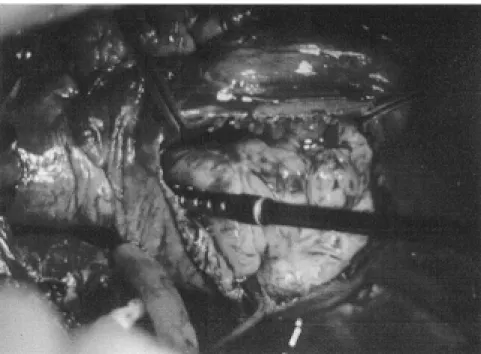
cardiopulmonary bypass (CPB) was accomplished with venous cannulas in the superior vena cava and left femoral vein and the arterial line in the ascen-ding aorta, which was left unclamped. A large tumoral mass was found in the right atrium extending into the right ventricle (Fig. 1), IVC, and supra-hepatic veins, which were partially occluded. The intracardiac and upper segments of the tumor were resected. The IVC was opened below the liver, and the tumor origin was found in the middle segment of this vein, extending towards the renal veins. The tumor was completely removed (Fig. 2). The IVC was closed with a direct running suture. Diffuse bleeding remained and was controlled with difficulty. The tumor was a multi-lobulated gray-reddish mass, 36 cm. in length and five to eight cm. in diameter. The patient had a problematic immediate postope-rative period with low cardiac output, respiratory and renal failure, rises in hepatic enzymes, and jaundice. Subse-quently, he had a good recovery and was discharged in the 28th
postopera-tive day, functionally NYHA class I. He remained well, but in spite of receiving chemotherapy, he died of diffuse abdominal metastases 24 months after surgery.
Case 2.: A.M., a 43 year-old white
woman, with three previous pregnan-cies and deliveries, was admitted with progressive dyspnea, orthopnea, par-oxysmal nocturnal dyspnea and palpi-tations. She denied weight loss and gynecologic symptoms. Physical ex-amination revealed jugular stasis, si-nus rhythm, and a grade 3/6 systolic murmur (at the third and fourth inter-costal space on the left sternal border. There was neither palpable abdominal mass nor peripheral edema. The chest x-ray showed moderate right atrial and ventricular enlargement. No abnormality was found in the elec-trocardiogram. An echocardiogram
revealed during diastole an intracar-diac tumor arising from the IVC and moving from the right atrium into right ventricle through the tricuspid valve. Computed tomography and magnetic resonance confirmed the presence of a tumor in the IVC and right atrium. The diagnosis of a tumor of the IVC was made.
The patient underwent surgery on December 7th, 1994. A median
thoraco-abdominal incision was made exposing the heart and infra-hepatic vena cava, and a CPB was performed with a single venous cannula, hypothermia to 16oC,
and total circulatory arrest. The right atrium was opened, and a large mass arising from the IVC and propagating to the right ventricle and pulmonary artery was found, free of adhesions. The thoracic part of the tumor was resected. There was no abnormality of the tricuspid valve. The IVC was incised, and the abdominal portion of the tumor arising from the right ovarian vein was found with no adhesions to the wall; it was removed. After closing the IVC and right atrium incisions, the patient was rewarmed and the CPB discontinued.
Figure 1 - Leiomyosarcoma of the IVC in the right atrium.
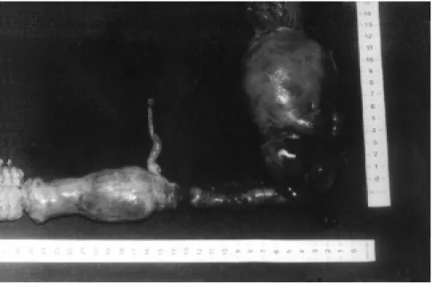
The time of CPB was 84 minutes and circulatory arrest 10 minutes. After closing the thoracic incision, a total hysterectomy and bilateral adnexec-tomy was performed removing the primary site of the tumor and the right ovarian vein. The tumor was a firm; gray-reddish, worm-like mass 25.0 cm in length, with a diameter varying from 1.0 to 4.0 cm and a weight of 120 g. (Fig. 3). Pathologic examination revea-led the uterus to be 400g in weight, 8.0 cm in length, and 3.0 cm in thickness. There were 2 intramural, well-deli-mited nodules of 1.5 and 3.0 cm characterized by interlaced bundles of smooth muscle fibers. Beside the left adnexa, a tumor was found that was 9 cm in length and 3 cm in diameter, was firm, multinodular, gray, worm-like, and had hemorrhagic areas, similar to the tumor in the IVC. The Endome-trium and adnexa were normal.
Microscopic examination showed uterine leiomyoma with elongated smooth muscle fibers, nuclear pleo-morphism and hyperchromatism, slight atypia and mitotic activity, and inva-sion of blood vessels. The diagnosis was neoplasm of undefined
malig-nancy, borderline for leiomyosarcoma and cellular leiomyoma.
The postoperative outcome was uneventful and the patient was dis-charged on the 13th postoperative day.
She remains asymptomatic after 56 months.
DISCUSSION
Of the abdominal tumors that grow in the lumen of the IVC propagating into the heart, the most frequent are renal carcinomas and have been found in 0.7 to 1% of cases.1,2 Intravenous
leiomyomatosis of the uterus (IVL) is rare, and about 10% spread to the heart. 3,4. Leiomyosarcoma (LMS) of
the IVC with cardiac involvement was found in 40 out of 215 cases reported (18.6%).
Primary vascular LMS are rare malignant tumors originating from the media of the vessel wall. They are found five times more often in large veins than in arteries, and the IVC is most frequently involved9. The
in-cidence of LMS of IVC was 2 in 14.00010 in post-mortem examinations.
Leiomyosarcoma of the IVC is found in adults from 15 to 83 years old, mostly in the 6th decade and in females
at a 6:1 ratio. Their length varies from 5 to 38 cm, and they weigh up to 3.6 kg. They present generally as firm, multi-lobulated and gray-reddish masses. Histologically, they have large and hyperchromatic nuclei with great number of mitoses 5,11. Tumors that
arise from the inferior segment of the IVC often grow slowly into adjacent tissue and spread mainly to the kidneys, liver, and aorta in the late stage of the disease. Tumors from the middle and upper segment tend to expand intraluminally, leading to partial, and rarely to total, occlusion of the vascular lumen. The most frequent origin of the tumor is in the middle segment of the IVC (47%), followed by inferior (32%) and superior (21%) segments. Metastases are found in 44% of patients at first observation, most frequently to the liver (42%) and lung (31%) 5,11.
Intravenous leiomyomatosis was first described by Birch-Hirschfeld in 1896 7 and defined by Norris and
Parmley in 1975 12. It is a rare
neo-plasm, characterized by intraluminal growth of benign smooth muscle cell tumor in either venous or lymphatic vessels, that may sometimes spread into inferior vena cava and right cardiac cavities. It is almost exclusively found in women, with the majority of the tumors originating from the uterus. Exceptional cases were found origi-nating from vessels of the skin, IVC, pelvic, or retroperitoneal veins 12,13.
ty, mitotic activity, and nuclear pleo-morphism are observed, the diagnosis of smooth muscle tumor of uncertain malignant potential 13 can be made, as
in the present report.
About 90% of the patients reported have been parous, and 10% have had a history of previous hysterectomy or pelvic surgery 3. The IVL extension to
the heart was first reported by Dürk14
in 1907. Open heart surgery was first reported by Mandelbaum et al.15 in
1974. Up to now, 32 cases have been reported.
These tumors present similar cli-nical findings according to their sites. They can be asymptomatic and found accidentally in abdominal ultrasono-graphy, unrelated operations and necropsy. Abdominal pain is the main complaint found in 68% of the pa-tients, often misdiagnosed as biliary pain. Other findings are nausea, eme-sis, fever, weakness, anorexia, jaun-dice, pallor, dyspnea, and weight loss. The tumor mass in the right atrium and ventricle with intermittent partial obstruction of the tricuspid valve leads to symptoms of dyspnea, palpitation, dizziness, syncope, congestive heart failure with hepatomegaly, ascites, jugular stasis, and peripheral edema, arrhythmias and pulmonary embolism
4,5,11,16. Further growth of the tumor can
worsen the clinical course and increase the risk of sudden death due to total obstruction of the tricuspid valve 17-23.
Tumor and thrombi in the upper seg-ment of IVC lead to Budd-Chiari syndrome and edema of lower extre-mities 4,5,11,16,24.
The bi-dimensional echocardio-gram 25 with emphasis on
transesopha-geal orientation, computed tomo-graphy, and magnetic resonance can be used to confirm the diagnosis. Mag-netic resonance has been found to be superior to computed tomography in
determining the intravascular nature of the tumor and its extension 16,26.
Additional information can be disclo-sed by venacavogram and ultrasono-graphy in a search for metastasis in the abdominal cavity.
The patient status, absence of demonstrable metastases, possibility of resection of the entire tumor, and the risk of sudden death and pulmonary embolism indicate immediate surgical treatment. Early surgical treatment with radical and extensive resection has proven to have the best mid- and long-term results. 5,11,16. Tumors of IVC
can be resected using venous-venous bypass and CPB with normothermia or profound hypothermia and circulatory arrest 16,27,28. The Pringle maneuver may
provide a bloodless field while remo-ving the tumor and thrombus 28.
Re-construction of the IVC can be done through venography or patch angio-plasty 5,16, biological tissue, or with
graft replacement with synthetic tube of polytetrafluorethylene (PTFE) 27,29,
which gives better results. Translo-cation of a segment of the IVC has been proposed and the “en-bloc” resection of the IVC and the adjacent aorta have been used in order to achieve a wider safety margin and decrease the possibility of local recur-rence. Profound hypothermia and total circulatory arrest, reported by Krane et. al.2 and Marshall et al. 30 can be used
to some advantage, such as providing a clear and bloodless operative field, better tumor management, minor risk of embolization, and prevention of uncontrollable hemorrhage. It also obviates the need of ligation of collate-ral vessels, occlusion of porta hepatis, and aortic cross-clamping, and reduces total blood loss. The disadvantage is the coagulopathy induced by deep hypothermia.
Recurrence of LMS of the IVC
after radical resection was found in 38.5% 4,11,16 of the patients, and may
occur for the IVL even after hysterec-tomy and bilateral adnexechysterec-tomy evi-dencing that the tumor growth is independent of the presence of the uterus, and although histologically benign, might be malignant in its mode of behavior31. The long term survival
rates for surgically treated patients was 27.9% and 14.2% after five and ten years, respectively. Tumors of the upper segment are often inoperable and have the worst prognosis 5,11.
Adjuvant therapies, such as radio-therapy and chemoradio-therapy, have been administered without a well established protocol. With the use of different agents, dose, and duration of admi-nistration, it becomes difficult to evaluate the impact of treatment. Given preoperatively, radiotherapy and che-motherapy may control micrometas-tasis, reduce the size of the tumor, prevent progression, and delay recur-rence. However, this therapy has failed to improve the long term survival rate16.
RESUMO RHCFAP/2984
STOLF, NAG et al. - Tumores abdomi-nais raros com crescimento intra-cardíaco. A propósito de dois casos operados com sucesso. Rev. Hosp. Clín. Fac. Med. S. Paulo 54 (5): 159-164;1999.
Tumores abdominais que crescem pela luz dos vasos e atingem o coração são raros. Estes tumores possuem características histológicas distintas porém causam sintomas abdominais e cardíacos semelhantes e apresentam
elevado risco de embolia pulmonar e morte súbita quando atingem o átrio direito e a valva tricúspide. O trata-mento mais eficaz é a ressecção cirúr-gica radical de todo o tumor usando-se circulação extracorpórea com ou sem hipotermia profunda e parada circulatória total. Relatamos os casos de dois pacientes, o primeiro portador de leiomiossarcoma primário da veia cava inferior e o segundo com leiomio-matose intravascular uterina com crescimento pela veia cava inferior
alcançando o átrio e ventrículo direitos, que foram operados com sucesso para ressecção concomitante do tumor primário e de sua extensão intra-cardíaca, com auxílio da circulação extracorpórea. Os aspectos clínicos, laboratoriais e histopatológicos são discutidos e é feita revisão da literatura.
DESCRITORES: Tumor intracar-díaco. Neoplasia da veia cava infe-rior. Procedimento cirúrgico.
REFERENCES
1. NOVICK AC & COSGROVE DM - Surgical approach for removal of renal cell carcinoma extending into the vena cava and the right atrium. J Urol 1980; 123:947.
2. KRANE RJ, DE VERE WHITE R, DAVIS E et al. - Removal of renal cell carcinoma extending into the right atrium using cardiopulmo-nary bypass, profound hypothermia and circulatory arrest. J Urol 1984; 131:945.
3. TO WWK, NGAN HYS & COLLINS RJ - Intravenous leiomyomatosis with intracardiac involvement. Int J Gynaecol Obstet 1993; 42:37. 4. CLEVELAND DC, WESTABY S & KARP RB - Treatment of
intra-atrial cardiac tumors. JAMA 1983; 249:2799.
5. MINGOLI A, FELDHAUS JR, CAVALLARO A et al. - Leiomyosarco-ma of the inferior vena cava: Analysis and search of world literature on 141 patients and report of three new cases. J Vasc Surg 1991;14:688-99.
6. PERL L - Ein Fall von Sarkom de V cava inferior. Virchows Arch Pathol Anat1871;53: 378-83.
7. BIRSH-HIRSHFELD FV - Lehbuch der pathologishen anatomie. 5th ed. Leipzig, FCW Vogel, 1896
8. KNAUER E - Beitrag zur Anatomie der Uterusmyome. Beitr Geburtshilfe Gynaekol 1903; 1:695.
9. KERVOKIAN J & CENTO DP - Leiomyosarcoma of large arteries and veins. Surgery 1973; 73: 390-400.
10. ABELL MR - Leiomyosarcoma of the inferior vena cava. Review of the literature. Report of two cases. Am J Clin Pathol 1957; 28: 272-87. 11. CACOUB P, PIETTE JC, WECHSLER B et al. - Leiomyosarcoma of the Inferior Vena Cava. Experience with 7 patients and Literature Review. Medicine-Baltimore 1991; 70:293-306.
12. NORRIS HJ & PARMLEY T - Mesenchymal tumors of the uterus: Intravenous leiomyomatosis clinical and pathologic study of 14 cases. CANCER 1975; 36:2164.
13. GROVE A & JØRGENSEN A - Intravascular Leiomyomatosis of the Uterus. Path Res Pract 1996; 192:949-56.
14. DÜRK H - Ueber ein kontinvierlich durch die entere Hohlvene in das Herz vorwachsendes. Fibroma des Uterus. MMW 1907; 54:1154.
15. MANDELBAUM I, PAULETTO FJ & NASSER WK - Resection of a leiomyoma of the inferior vena cava that produced tricuspid valvular obstruction . J Thoracic Cardiovasc Surg 1974; 67:561-567. 16. KULAYLAT MN, KARAKOUSIS CP, DOERR RJ et al. -
Leiomyo-sarcoma of the Inferior vena cava: A Clinicopathologic review and report of three cases. J Surg Oncol 1997; 65: 205-217. 17. HOFFBRAND AV & LLOID THG - Leiomyosarcoma of the inferior
vena cava leading to obstruction of the tricuspid valve. Br Heart J 1964; 26: 709-15.
18. GUTIEREZ O, DESAI S & BURGENER F - Leiomyosarcoma of the inferior vena cava with intracardiac extension. Europ J Radiol 1986; 6: 153-55.
19. DEUTSCH V, FRAENKEL O & FREUND U - Leiomyosarcoma of the inferior vena cava propagating into the right atrium. Br Heart J 1968; 30: 571-74.
20. DEMOULIN JC, SAMBON Y, BOUDIOET Y et al. - Leiomyosarcoma of the inferior vena cava. An unusual cause of pulmonary embolism. Chest 1974; 66: 597-99.
21. WILMSHUST P - Leiomyosarcoma in the right atrium and occluding the inferior vena cava. Br Heart J 1995; 73(3): 236.
22. NILI M, LIBAN E & LEVY MJ - Tricuspid stenosis due to intravenous leiomyomatosis – a call for caution: case report e review of the literature. Texas Heart Inst J 1982; 9:231-5.
23. GONZALEZ-LAVIN L, LEE RH, FALK L et al. - Tricuspid valve obstruction due to intravenous leiomyomatosis. Am Heart J 1984;108:1544-6.
24. MONIG SP, GAWENDA M, ERASMI H et al. - Diagnosis, treatment and prognosis of Leiomyosarcoma of the inferior vena cava. Eur J Surg 1996; 4:231-35.
25. MAURER G & NANDA N - Two-dimensional echocardiographic identification of intracardiac leiomyomatosis. Am Heart J 1982; 103:915-17.
27. ANGERMEIER WK, ROSS JH, NOVICK AC et al. - Resection of nonrenal retroperitoneal tumor with large vena caval thrombi using cardiopulmonary bypass and hypothermic circulatory arrest. J Urol 1990; 144:735-9.
28. ROSENTHAL JT, COLONNA JO & DRINKWATER DC - Leiomyo-sarcoma of the inferior vena cava with atrial extension: Long-term survival following resection and caval replacement with circulatory arrest. Urology 1995; 46: 876-78.
29. KASANO Y, TANIMURA H & KUMADA K - Resectable leiomyo-sarcoma of the inferior vena cava extended into the right atrium with the use of cardiopulmonary bypass and graft replacement. Surgery 1995; 117:473-75.
30. MARSHALL FF, REITZ BA & DIAMOND DA - A new technique for management of renal cell carcinoma involving the right atrium: hypothermia and cardiac arrest. J Urol 1984; 131:945.
31. COOPER NM, GUILLEM J, DALTON J et al. - Recurrent intravenous leiomyomatosis with cardiac extension. Ann Thorac Surg 1992; 53:139-41.