1
Arquivos Brasileiros de Cardiologia - Volume 84, Nº 2, Fevereiro 2005
Case Report
Wolff-Parkinson-White Syndrome and Sinus Venosus
Atrial Septal Defect Association
Patrícia Lopes Moraes, Luíz Márcio Gerken, Bayard Gontijo Filho,
Walter Villela de Andrade Vicente, Paulo Roberto Barbosa Evora
Ribeirão Preto, SP / Belo Horizonte, MG - Brazil
CECORP – Ribeirão Preto, BIOCOR – Belo Horizonte and Medical School of Ribeirão Preto - USP
Mailing address: Paulo Roberto B. Evora - Rua Rui Barbosa, 367/15 Cep 14015-120 - Ribeirão Preto, SP, Brazil
E-mail: [email protected] Received for publication: 07/01/2004 Accepted for publication: 07/29/2004
The Wolff-Parkinson-White syndrome (WPW) and sinus veno-sus atrial septal defect (ASD) association is very rare and not yet reported in the literature. It is the main basis for this case report.
One of the larger studies1 looking at the association between WPW and acquired or congenital heart disease was performed in France. WPW was diagnosed in 49 of 10,750 patients with ac-quired cardiac disease (0.45%) and in 24 of 3,761 patients pre-senting with congenital malformations. Of 1,348 cases of ASD, 5 patients had associated pre-excitation syndromes, 3 of them with right posterior pre-excitations associated with an ostium primum defect, but no patient presenting with sinus venosus ASD and WPW was found 1.
We herein report a young lady presenting with sinus venosus ASD and WPW who was staged for percutaneous radiofrequency ablation followed one month later by surgical correction of the ASD.
Case Report
The patient is a 22-year-old female student. Effort tachycardia and cyanosis of the extremities was noticed by 8 years of age. At the age of 12 years, congenital cardiopathy and cardiac arrhythmia were suspected by a pediatric cardiologist based on clinical exa-mination and basal EKG (fig. 1). Heart catheterization and elec-trophysiological study were performed. The former diagnosed an ostium secundum ASD and measured the mean pulmonary artery pressure at 20 mmHg (fig. 2A). The latter was diagnostic for WPW syndrome. An anteroseptal accessory pathway with S1S1 antegrade conduction up to 270 ms was found. Orthodromic pa-roxysmal supraventricular tachycardia (PSVT) with an effective ventricular refractory period of 240 ms (450 bpm) could be induced (fig. 3).
Effort fatigue, dyspnea, and paroxysmal tachycardia ensued over the years. When the patient was 20 years of age, although a surface echocardiogram missed the ASD, a 1.2 x 1.0cm sinus venosus ASD, right chamber dilatation, and pulmonary artery sys-tolic pressure at its superior limit of normality were depicted by transesophageal echocardiography (fig. 2B).
Two years later, symptoms worsened, the resting heart rate increased to 100 bpm, and dyspnea ensued with light effort. Pro-pranolol 40 mg/day was started and the symptoms improved.
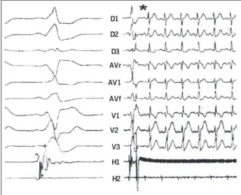
When the patient reached 23 years of age, WPW syndrome with an anteroseptal accessory pathway was evidenced by a per-cutaneous electrophysiological study. Concentric ventricle-atrial (VA) conduction without decrements and with a longer refractory period than the accessory pathway was demonstrated. The ano-malous pathway was ablated 0.5s after starting 6 pulses of radio-frequency (fig. 4).
One month later, the sinus venosus ASD and the associated partial pulmonary anomalous venous drainage to the right atrium were both corrected while the patient was under normothermic extracorporeal circulation and anoxic arrest.
The patient has remained asymptomatic since then.
Discussion
WPW syndrome should be suspected in children with ASD and PSVT, but technical limitations related to the patient’s size as well as the severity of heart failure may prevent a preoperative electrophysiological study.
Attempts to disclose echocardiographic signs of WPW have been disappointing. In one such study, 26 WPW cases were ana-lyzed, and abnormal interventricular septal motion was found to occur with Type I WPW. Other echocardiographic abnormalities were also present ina high percentage of WPW patients but do not seem to be associated withany particular WPW type2.
In Japan, a 7-month-old, 5.3kg girl presenting with ASD and PSVT was surgically treated, the preoperative electrophysiological study not being performed due to severe heart failure. Suspecting a concealed WPW syndrome with retrograde conduction, surgeons performed the operation with the aid of intraoperative epicardial and endocardial mapping. A left-posterior accessory retrograde pathway was found and successfully cryoablated via a transeptal superior approach3.
2
Arquivos Brasileiros de Cardiologia - Volume 84, Nº 2, Fevereiro 2005 Wolff-Parkinson-White Syndrome and Sinus Venosus Atrial Septal Defect Association
included as an adjunct to repair of underlying structural cardiac lesions 4. Based on this perspective, a staged therapeutic strategy was selected for our patient, the electrophysiologic study and accessory pathway ablation being performed before the surgical treatment of the intracardiac defect.
A detail worthy of relative discussion about the WPW syndrome and sinus venosus ASD association is the question of its rarity. This fact has, at least, 2 explanations: a) the difficulty finding reports in the literature could indicate that the association of the 2 entities is really rare and; b) WPW syndrome can happen ran-domly in any cardiopathy or even without cardiopathy, and perhaps interest in clinical practice in such anomalies does not exist. But this sort of statement is broad and a matter of speculation.
The present case is particularly interesting and original because of the unusual association of WPW syndrome with another
con-1. Soria R, Fernandez F, Heller J et al. Wolff-Parkinson-White syndrome and cardio-pathies. Arch Mal Coeur Vaiss 1984 77:1468-80.
2. Chandra MS, Kerber RE, Brown DD, Funk DC. Echocardiography in Wolff-Parkin-son-White syndrome. Circulation 1976;53:943-6.
3. Miyaji K, Furuse A, Kotsuka Y, et al. Successful repair for concealed Wolff-Parkin-son-White syndrome with atrial septal defect in infancy: a case report. Kyobu Geka 1996;49:947-51
References
4. Ashburn DA, Harris L, Downar EH, Siu S, Webb GD, Williams WG. Electrophysio-logic surgery in patients with congenital heart disease. Semin Thorac Cardiovasc Surg Pediatr Card Surg Annu 2003; 6:51-8.
5. Oropeza ES, Martínez-Sánchez A, López CA, Elizondo GV. Wolff-Parkinson-White syndrome associated to Scimitar syndrome. Percutaneous radiofrequency catheter ablation previous to surgical repair. Arch Cardoil Mex 2001; 2:141-5.
DI DII DIII aVR aVL aVF V1 V2 V3 V4 V5 V6
Fig. 1 - EKG tracing depicting a short P-R interval and delta waves typical of Wolff-Parkinson-White syndrome.
Fig. 2 - A) Angiocardiography when the patient was 12 years old. A presumed ostium secundum ASD was diagnosed; B) Echo-Doppler echocardiogram 10 years later. Notice the left to right shunt through a superior sinus venosus ASD (Arrow).
D1
D2
D3
AVr
AV3
AVf
V1
V2
V3
V4
V5
V6
Fig. 3 - EKG recording showing antidromic tachycardia (180 bpm) induced by ventricular stimulation.
D1
D2
D3
AVr
AV1
AVf
V1
V2
V3
H1
H2
Fig. 4 - Endocavitary electrogram at the ablation site. Antidromic tachycardia disappears (*) less than 0.5 s after starting the radiofrequency therapy.