1
Approximately 85% of the patients with tetralogy of Fallot associated with pulmonary atresia have anomalies of the pulmonary vascular tree. The pulmonary arteries are usually of a small caliber, often nonconfluent, and not connected to the right ventricle. In many cases, the proximal branches are stenotic, hypoplastic, or totally absent. The aortopulmonary collateral arteries are frequently present and may supply partially or almost totally the pulmonary blood flow.
The treatment aims at establishing the confluence, normalizing the caliber and distribution of the pulmonary arteries, which may be obtained through the use of shunts and unifocalization of those vessels. Several procedures are usually required before the definitive correction 1-4. Dilation with a balloon catheter and use of stents in the pulmonary arteries may represent an important factor for the success of treatment 5-7. The operative risk ranges from 0 to 20% depending on the number of shunts and thoracotomies before the definitive correction, which is only possible in 60 to 70% of pa-tients3,4. Recently, some researchers have adopted a radical approach with complete pulmonary arterial reconstruction, closure of the interventricular communication, and placement, in the neonatal period 8-10, of a tube in the right ventricular outflow tract. Although the operative risk has been described as small, this approach re-quires more complex surgical techniques and experience of the surgical team, which are available only in certain centers 11,12.
This study aimed at demonstrating the importance of the an-giographic study of the pulmonary blood supply in selecting patients with tetralogy of Fallot and pulmonary atresia for total or partial correction of that malformation.
Methods
From 1980 to 2000, 82 patients with tetralogy of Fallot and pulmonary atresia were studied from the hemodynamic and ci-neangiocardiographic points of view. Twenty-six patients with pul-monary atresia associated with complex cardiac malformation (single atrium, single ventricle, single AV valve, and single outflow tract – pulmonary atresia) were excluded. The remaining 56 pa-tients were between 20 days and 4 years of age, and their weights ranged from 1.7 kg to 10 kg. The pulmonary blood supply was studied by using the following methods: aortography with occlusion of the thoracic aorta with the Berman angiographic catheter 13;
aortography with manual compression of the abdominal aorta 14;
selective injection of contrast medium in the aortopulmonary col-laterals 15; and selective injection of contrast medium in the wedged pulmonary vein 16.
Original Article
Angiographic Study of Pulmonary Circulation
in Tetralogy of Fallot with Pulmonary Atresia
Marco Aurélio Santos e Vitor Manuel Pereira Azevedo
Rio de Janeiro, RJ - Brazil
Instituto Nacional de Cardiologia Laranjeiras
Mailing address: Vitor Manuel Pereira Azevedo - Rua Visconde de Ouro Preto, 39/304 - Cep 22250-180 - Rio de Janeiro, RJ, Brazil E-mail: [email protected]
Received for publication: 01/28/2004 Accepted for publication: 04/01/2004 English version by Stela Maris Costalonga
Objective
To identify the types of pulmonary vascular blood supply in tetralogy of Fallot with pulmonary atresia by the use of hemody-namic studies.
Methods
Fifty-six patients with tetralogy of Fallot and pulmonary atresia, and ages ranging from 20 days to 4 years, underwent cinean-giocardiographic study with contrast medium injections in the following vascular structures: 1) wedged pulmonary vein; 2) aortopulmonary collaterals; 3) thoracic aorta; and 4) ductus arteriosus or systemic-pulmonary shunt.
Results
In the 56 patients studied, pulmonary blood was supplied as follows: in 15, by aortopulmonary collaterals; in 36, only by the ductus arteriosus; and in 5, by the ductus arteriosus and aorto-pulmonary collaterals. The patients were classified into 6 types depending on the type of pulmonary vascular perfusion and the presence or absence of vascular structures that compose the pulmonary circulation in tetralogy of Fallot with pulmonary atresia.
Conclusion
This type of approach enables the obtainment of information necessary for the correct clinicosurgical management of patients, due to the great complexity and extreme variability of the pulmo-nary blood supply in tetralogy of Fallot with pulmopulmo-nary atresia.
Key words
2
The different types of pulmonary blood perfusion were establi-shed based on the presence or absence of the following structures: 1) pulmonary trunk; 2) ductus arteriosus; 3) right and left pulmona-ry arteries and their confluence; and 4) aortopulmonapulmona-ry collaterals. The angiographic study of the pulmonary blood supply aimed at clarifying the following parameters: 1) to identify the way blood reaches the pulmonary circulation from the systemic circulation; 2) to confirm the presence of the pulmonary trunk; 3) to demons-trate the presence of confluence of the right and left pulmonary arteries; 4) to identify the existence of aortopulmonary collaterals and their connection with the pulmonary arteries; 5) to demons-trate the presence of obstruction in the pulmonary arteries and aortopulmonary collaterals; and 6) to infer the diameter of the pulmonary arteries.
Results
Of the 56 patients studied, 15 had pulmonary blood supplied by aortopulmonary collaterals with the following characteristics: in 2 patients, no central pulmonary arteries were identified and, in 13, they were confluent; in 10 patients, the pulmonary arteries had minuscule dimensions; and, in 3, the caliber was “adequate”. All the remaining 41 patients had pulmonary arteries of “adequate” caliber, whose characteristics were as follows: 36 were confluent and pulmonary blood was supplied only through a tortuous, small-caliber ductus arteriosus in the shape of a comma, forming with the descending aorta an acute angle 17. In the 5 remaining patients, the pulmonary arteries were not confluent. Pulmonary blood was supplied through the ductus arteriosus to one lung (right or left) and through an aortopulmonary collateral to the contralateral lung (chart I). No patient had the pulmonary flow bilaterally provided by the ductus arteriosus.
The pulmonary arteries were defined as “adequate” when their caliber was sufficient to accept a systemic-pulmonary shunt. Although arbitrary, that definition is based on the clinical/surgical experience of our unit. Therefore, in our material, a pulmonary artery diameter > 3 mm was considered an adequate caliber.
Because of the complexity and extreme variability of the pul-monary blood supply in this malformation, the investigators, throu-ghout the past decades, have proposed different classifications18-21. In our material, we used a classification based on the presence or
absence of the following vascular structures and the pulmonary vascular perfusion found in the tetralogy of Fallot with pulmonary atresia: pulmonary trunk, ductus arteriosus, pulmonary arteries (con-fluence), and aortopulmonary collaterals (tab. I).
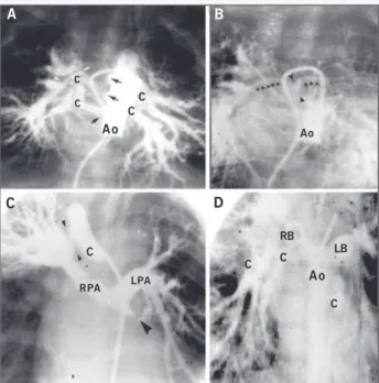
In type I, comprising 25 patients, pulmonary blood flow was supplied exclusively through the ductus arteriosus. All patients had pulmonary trunks, the right and left pulmonary arteries were confluent, the blood supply was unifocal, and no aortopulmonary collaterals were identified (fig. 1). That example depicts vaso-constriction of the pulmonary arteries, but all lobes of both lungs were perfused. The ductus arteriosus is already dilated (infusion of prostaglandin E1),and the pulmonary trunk is not atretic.
Type II was represented by 11 patients. The pulmonary trunk was atretic and pulmonary blood flow was also supplied only by the ductus arteriosus. The pulmonary arteries were confluent, had an adequate caliber, and the blood supply was unifocal. No aortopulmonary collaterals were identified in this group (fig. 2). In figure 2, the right and left pulmonary arteries are confluent and perfused all segments of both lungs. Different degrees of stenosis are observed in several segments of the pulmonary arteries.
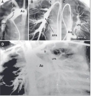
In type III, represented by 13 patients, pulmonary vascular perfusion is exclusively provided by aortopulmonary collaterals, which anastomose with the confluent pulmonary arteries. The pulmonary trunk is atretic and pulmonary perfusion is multifocal
56 patients
15 patients
• 2 absent RPA and LPA • 10 minuscule RPA and LPA • 3 adequate RPA and LPA
15 patients
Multiple AoP-Co
5 patients
nonconfluent RPA and LPA
2 patients
RPA-DA LPA- AoP-Co
3 patients
RPA- AoP-Co LPA - DA
41 patients
adequate RPA and LPA
36 patients
confluent RPA and LPA
36 patients
isolated ductus arteriosus
AoPCo aortopulmonar collateral; RPA right pulmonary artery; LPA left pulmonary artery; DA -ductus arteriosus
Chart 1 - Morphology of the pulmonary arteries and of the pulmonary blood supply in 56 patients with tetralogy of Fallot and pulmonary atresia on angiographic study.
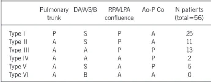
Table I - Types of pulmonary vascular perfusion in tetralogy of Fallot with pulmonary atresia
Pulmonary DA/A/S/B RPA/LPA Ao-P Co N patients
trunk confluence (total=56)
Type I P S P A 25
Type II A S P A 11
Type III A A P P 13
Type IV A A A P 2
Type V A S A P 5
Type VI A B A A 0
Ao-P Co - aortopulmonary collateral; PA - pulmonary artery; DA - ductus arteriosus; RPA and LPA right and left pulmonary artery; P present, A -absent; S - single; B - bilateral.
A
B
RPA A o
LPA
A o
PT
D
LPA RPA
3
(fig. 3). Figure 3 depicts the disparities between the dimensions of the pulmonary arteries in 2 patients. The aortopulmonary col-laterals, 4 in number, originate from the descending aorta and show stenotic areas in their origin, trajectory, or site of anastomosis with the pulmonary arteries.
Type IV was represented by 2 patients. No central pulmonary arteries were identified in this group. Pulmonary blood perfusion is exclusively provided by aortopulmonary collaterals (fig. 4). In
the 2 examples depicted in figure 4, even in the late phase of aortography, the central pulmonary arteries are not visualized, and blood supply is provided from several foci, ie, is multifocal.
In type V, represented by 5 patients, pulmonary vascular perfu-sion is supplied by the ductus arteriosus and aortopulmonary col-laterals. In 2 patients, the right pulmonary artery originates from the ductus arteriosus at the right-hand side of the aortic arch, and the left pulmonary artery originates from one collateral of the descending aorta. In 3 patients, the left pulmonary artery originates from the ductus arteriosus at the left-hand side of the aortic arch, and the right pulmonary artery originates from an aortopul-monary collateral (fig. 5). In figure 5, the pulaortopul-monary arteries are not confluent, and stenosis may be seen in the connection of the ductus arteriosus with the pulmonary artery and also in the con-nection of the collateral with the contralateral pulmonary artery. In this group, pulmonary vascular supply is also multifocal.
Type VI, with perfusion through bilateral ductus arteriosus, was not found in our material, although it has already been reported in the literature in patients with isomeric syndromes.
Of the 56 patients, 20 had their pulmonary blood flow supplied by aortopulmonary collaterals (tab. II). Six patients had one aorto-pulmonary collateral originating from the descending aorta, 7 had 2 aortopulmonary collaterals, 5 had 3 aortopulmonary collaterals, and 2 had 4 aortopulmonary collaterals. An inverse relation was observed between the total number of aortopulmonary collaterals and the size and existence of central pulmonary arteries.
Of the 43 aortopulmonary collaterals, 39 vessels (90.7% – 95%CI = 76.9% to 97.0%) had a defined narrowing at some point of its trajectory or its origin (tab. II, fig. 3, 4, and 5). The most frequent site of stenosis was the connection between the aortopulmonary collateral and the pulmonary artery. In 4 of 20 patients (20% – 95%CI = 6.6% to 44.3%), no aortopulmonary collaterals with stenosis were identified. The other aortopulmonary collaterals without a clearly defined stenosis were frequently
tor-A B C
A o
RPA PT SC D
LPA
RPA
A o
LPA
RPA LPA
Fig. 2 - Type II. A) Aortography in PA view. Through a tortuous ductus arteriosus in the shape of a comma, the right and left pulmonary arteries are seen. The tip of the black arrow indicates the absence of the pulmonary trunk. B) In another patient, critical stenosis is identified at the origin of the right pulmonary artery (white arrow). No pulmonary trunk is visualized. Pulmonary perfusion is performed isolated by the ductus arteriosus, which is not visualized (overlapped by the aorta). C) Selective injection into the Blalock-Taussig shunt. Three sites of obstruction to the pulmonary flow exist as follows: 1) between the Gore-Tex and the upper lobar branch; 2) between the upper lobar branch and the right pulmonary artery (arrow); and 3) between the right and left pulmonary arteries (arrow). This example does not have a pulmonary trunk. Those 3 patients have unifocal pulmonary perfusion with no pulmonary compartmentalization. Ao aorta; PT -pulmonary trunk; D - ductus arteriosus; RPA and LPA - right and left -pulmonary arteries; SC - left subclavian artery.
A
C C A o C C
B
Ao
C
C
RPA LPA
D
C C
RB
A o LB
C
Fig. 3 – Type III. A) Initial phase: multiple collaterals (4) are seen perfusing all segments of both lungs. Those are tortuous well-developed vessels with multiple stenoses at the origin and at the connection with the intrapulmonary arteries of the right lung (black and white arrows). B) Late phase: a minuscule confluent central pulmonary artery is identified (delineated by the black arrows). C) Selective injection of contrast medium in a large collateral that anastomoses with the intrapulmonary artery of the right upper lobe, where an important stenosis at its connection is seen (arrows). From that collateral onwards, the right and left pulmonary arteries, which are confluent and have an “adequate” caliber, are opacified. The left pulmonary artery perfuses the upper lobe and lingula of the left lung. D) Aortography showing the presence of 3 other large collaterals that perfuse the middle and lower lobes of the right lung and the lower lobe of the left lung. In those 2 examples, pulmonary perfusion in both lungs is multifocal with pulmonary compartmentalization. Ao - aorta; RPA and LPA - right and left pulmonary arteries; C - collateral.
A
Ao
B
Ao
C
Ao
D
Ao
4
tuous, elongated, and offered some degree of obstruction to the pulmonary flow.
The most frequent site of pulmonary artery stenosis was in the central pulmonary artery (tab. III). In 5 patients, the stenosis was located at the origin of the right pulmonary artery, and, in 8 patients, at the origin of the left pulmonary artery. Stenosis of the peripheral pulmonary artery was evidenced in 5 patients. Four patients had stenosis of the pulmonary artery at the site of the systemic-pulmonary anastomosis (fig. 2C).
The incidence of incomplete arterial arborization in our material was 29%, and it was particularly found in situations without any confluence of the central pulmonary arteries. When confluence of the right and left branches existed, it did not reach 4%.
Discussion
The capillaries that perfuse the pulmonary acini represent the final common path of the pulmonary vascular blood supply. They connect to an intrapulmonary arterial plexus, which branches inside the bronchopulmonary segments, and the pulmonary blood supply may have one or more systemic sources.
When all intrapulmonary arteries connect to the central pul-monary artery, only one single head of pressure perfuses the entire pulmonary parenchyma of both lungs. This type of blood supply is called unifocal (fig. 1 and 2).
In some situations, the central pulmonary arteries are not confluent, but are perfused by systemic vessels with one single head of pressure. In this case, blood supply is also unifocal. Usually, the unifocal pulmonary blood supply has confluence of the central pulmonary arteries, which are supplied by a ductus arteriosus. However, the unifocal pulmonary blood supply may be performed through aortopulmonary collaterals that anastomose to the central pulmonary arteries.
Finally, in even rarer situations of unifocal supply, the pulmonary arteries are supplied by multiple collateral arteries acquired as intercostal vessels 22.
Multifocal pulmonary blood supply may also occur in the pre-sence of pulmonary arterial confluence (fig. 3C and 3D), in which case the confluent pulmonary arteries are not connected to all intrapulmonary arteries in both lungs. This is the most common form of multifocal pulmonary blood supply. Thus, the confluent pulmonary arteries perfused by one or more aortopulmonary colla-terals are connected to a certain region of the lung, while the rest of the pulmonary parenchyma is perfused by another aorto-pulmonary collateral or others that anastomose with the rest of the pulmonary segments. Another rarer possibility of multifocal supply in the presence of confluent pulmonary arteries is when the confluent pulmonary arteries are perfused by the ductus arte-riosus and the other pulmonary segments are perfused by aorto-pulmonary collaterals.
The concept of unifocal and multifocal pulmonary blood supply, as described by Macartney et al 22, was based on the measurement of total pulmonary resistance relative to the aorta, which is usually elevated. That resistance is consequent to several levels of obstruc-tion of the pulmonary arterial tree (fig. 2B and 2C, and tab. III). From the surgical correction viewpoint, what matters is the relative resistance of the vessel that will be connected to the right ventricle, ie, the central pulmonary artery. Therefore, when the pulmonary blood supply is unifocal, all intrapulmonary arteries are connected to a single head of pressure, as long as no significant stenosis exists in the central and peripheral branches of the pulmonary artery. Fig. 5 - Type V. A) Aortography in PA view. Presence of ductus arteriosus at the
right-hand side of the aortic arch, perfusing the right pulmonary artery. A tubular narrowing is seen at the connection of the ductus arteriosus with the right pulmonary artery. B) Selective injection of contrast medium into the ductus arteriosus. Perfusion of the 3 lobes of the right lung is seen with stenosis in the distal part of the artery of the upper lobe (arrow). C) Semiselective contrast injection in the collateral that originates from the lower portion of the thoracic aorta. Note the important stenosis at the connection of the collateral with the left pulmonary artery (arrows). The right and left pulmonary arteries are noncon-fluent, but perfuse all lobes of both lungs. D) Aortography showing the presence of ductus arteriosus to the left of the aortic arch, which connects with the left pulmonary artery. The right pulmonary artery originates from the collateral that emerges from the middle portion of the thoracic aorta. Stenosis is seen at the connection of both pulmonary arteries. In both examples, the pulmonary blood supply is multifocal, but without compartmentalization of both lungs. Ao -aorta; PA - posteroanterior; PT - pulmonary trunk; D - ductus arteriosus; RPA and LPA - right and left pulmonary arteries; C - collateral.
A D
RPA
A o
B A
RPA
C
A o LPA
C
D
RPA
Ao
D
LPA
Table II - Stenosis of aortopulmonary collaterals in tetralogy of Fallot with pulmonary atresia
N° of N AoP-Co Total AoP-Co AoP-Co Pts with stenotic
patients with stenosis AoP-Co
6 1 6 6 (100%) 6 (100%)
7 2 14 12 (86%) 5 (71%)
5 3 15 13 (87%) 3 (60%)
2 4 8 8 (100%) 2 (100%)
Total=20 - 43 39 (90%) 16 (80%)
AoP-Co - Aortopulmonary collateral; Pts - patients.
Table III - Stenosis of the pulmonary artery in patients with tetralogy of Fallot and pulmonary atresia
Total patients 54
No PAS 32
Central pulmonary artery 13
Origin in the RPA 5
Origin in the LPA 8
Peripheral pulmonary artery 5
Systemic-pulmonary shunt 4
5
When the pulmonary blood supply is multifocal, not all intra-pulmonary arteries are connected to a single head of pressure. Because different foci certainly have different heads of pressure, the pulmonary regional blood flow is extremely variable, resulting in hypo- and hyperperfused pulmonary segments.
It is worth emphasizing that the aortopulmonary collaterals are vascular structures different from the bronchial arteries. The aortopulmonary collaterals originate only from one systemic artery and run towards the origin of one intrapulmonary artery in the region of the pulmonary hilum or its vicinity (fig. 3, 4, and 5). They are conducting arteries with no nutritional function in terms of the pulmonary parenchyma. The aortopulmonary collaterals usually originate from the anterior face of the aorta (opposed to the origin of the intercostal arteries) and, more rarely, from the right brachiocephalic trunk, the subclavian artery, or even a coro-nary artery. They differ from the ductus arteriosus in their histolo-gical structure 23 and also because the ductus arteriosus is cir-cumscribed to a certain region of the aortic arch. Even when the ductus arteriosus originates from the right-hand side of the aortic arch, it is somewhat opposite to the origin of the brachiocephalic trunk or the subclavian artery.
Acquired collateral pulmonary circulation may occur in any cyanotic congenital heart disease. A consensus still exists in the literature regarding the difference existing between that type of pulmonary blood supply and the aortopulmonary collaterals. From the pathophysiological viewpoint, the most important differentia-tion is found in the site of anastomosis with the pulmonary circu-lation. It may be immediately precapillary in the acquired collateral circulation, or in the hilar region in the case of aortopulmonary collateral circulation 24. Both provide effective pulmonary vascular blood supply. Because acquired collateral circulation never produces heart failure, it is clear that high resistance to blood flow is present between the aorta and the pulmonary arteries. Studies in animals have reported that that phenomenon results from intimal proliferation in the small caliber bronchial arteries 25.
The ductus arteriosus is a vascular structure that usually ori-ginates from the contralateral side of the aortic arch, close to the bifurcation of the brachiocephalic trunk (fig. 2A, 5A, and 5D). Rarely, the ductus arteriosus originates from one anomalous sub-clavian artery. In the lack of atrial isomerism, the bilateral ductus arteriosus is exceptionally identified as a form of pulmonary perfusion. Because in our case series the complex forms of pulmonary atresia were excluded, those types of pulmonary perfusion were not iden-tified in our study. According to Macartney et al 22 and, afterwards, to Yoo et al 26, pulmonary perfusion may more rarely be provided by the fifth aortic arch.
Most aortopulmonary collaterals present as tortuous, large ca-liber vessels with multiple stenoses in their origin, their trajectory, or at the site of anastomosis with the intrapulmonary arteries (tab. 2) (fig. 3, 4, and 5). They never connect to the intercostal arteries. In their trajectory, they accompany the bronchi but never form a plexus around them. Their morphology is the same from the neonatal period until a more advanced age.
The acquired collateral circulation is rare in the first year of life, and, therefore, it was not identified in our material. However, over the years, it has become more developed, mainly after tho-racotomy, when adhesion of the visceral and parietal pleurae allows the development of a centripetal collateral circulation, ie, from
the thoracic wall to the lung. In the angiographic study, the acquired circulation appears as innumerous thin vessels originating from any thoracic artery. On the other hand, the bronchial arteries are recognized by their relation with the trachea and main bronchi, and by the way they develop a nutritive plexus with the bronchial walls. Recently, this type of circulation has been valued during total surgical correction, because of the respiratory complications observed in the postoperative period. The latter appear after uni-focalization and are related to an ischemic process in the respiratory airways, due to the interruption of the tracheobronchial blood supply during the mobilization of aortopulmonary collaterals 27.
The central pulmonary arteries are more easily identified when they are confluent, because that is the form in which they usually occur. Their confluence associated with a reduction in or absence of their trunk provides a “seagull” configuration when visualized in the frontal plane 28 (fig. 3B). In our material, they were absent in 2 of the 56 patients (fig. 4). In 22 patients, different degrees of stenoses were observed in their trajectory, and, in 4, they were consequent to systemic-pulmonary shunts (tab. III). When the central pulmonary arteries are not confluent, their identification is often difficult. In most cases, the pulmonary trunk is reduced to a fibrous cord, which is connected to the heart; therefore, during cineangio-cardiography, a concomitance of movement exists between the central pulmonary arteries and the heart, while the remaining arteries in the mediastinum move together with the lungs 29.
In the tetralogy of Fallot with pulmonary atresia, the intrapul-monary arteries have normal configuration and distribution. When no confluence of the central pulmonary arteries exists and the intrapulmonary arteries are connected to the aortopulmonary col-lateral, they may have a distinct configuration, in the shape of a plexus (fig. 5C). This abnormality may result from a hemodynamic disorder consequent to the way they are connected to the aorta30. When a disorder in connection exists, the intrapulmonary ar-teries may remain isolated and not fuse with the hilar arar-teries, as usually occurs 19. As a result, the pulmonary arterial supply to one lobe, or segment, or even part of a segment, remains completely isolated from the rest of the lung. The segmentary or lobar arteries are connected in their proximal portion either to a central pulmonary artery, or to an aortopulmonary collateral, or both (fig. 3C and 3D). The consequence of that disorder is that the pulmonary blood supply becomes compartmentalized. Therefore, each aor-topulmonary collateral perfuses only one segment of the lung. The selective contrast medium injection in the aortopulmonary collateral opacifies only that specific region of the lung.
The combination of pieces of information obtained through selective injection of contrast medium into the aortopulmonary collaterals, into the systemic-pulmonary shunts, and into the central pulmonary arteries will enable the accurate determination of the origin of the pulmonary blood supply of each pulmonary segment, and, more important, of the amount of pulmonary pa-renchyma connected to a certain pulmonary artery. This will reveal whether duplication of the pulmonary blood supply exists, or not, to the same lobe or segment, ie, whether a determined pulmonary region is perfused by more than one source.
6
is connected only to an aortopulmonary collateral artery. In that situation, one shunt to the right central pulmonary artery or even a duct of a corrective surgery will only increase the pulmonary blood supply to the right upper lobe (fig. 3C and 3D). However, if a communication exists between the perfusion of both lobes, then the surgical procedure will supply blood to the entire right lung. The prevalence of incomplete pulmonary arborization was high in our patients, mainly when no confluence existed between the right and left pulmonary arteries. On the contrary, incomplete arborization did not reach 5% when confluence of the right branch with the left one existed 31.
Another point identified in our material was that, when con-fluence of the right and left pulmonary arteries existed, those vessels had a greater caliber. This finding, however, was not con-firmed when association of the aortopulmonary collaterals and the central pulmonary arteries existed (chart 1). In that situation, we identified 10 patients with confluent, but “minuscule”, pulmo-nary arteries and innumerous aortopulmopulmo-nary collaterals (fig. 3A
and 3B). The dimensions of the aortopulmonary collaterals were greater in patients without confluence of the right and left pulmo-nary arteries, and also when the central pulmopulmo-nary arteries did not exist.
Finally, the total number of aortopulmonary collaterals is inversely related to the size and existence of central pulmonary arteries (fig. 4C and 4D), which had already been demonstrated by Shi-mezaki et al 31.
Some of our observations derived from inferences, mainly be-cause of the limitation of the study imposed by our material, which was composed, by more than 50%, of newborn and infant patients. Therefore, the selective contrast medium injection in each collateral in isolation could not be performed, not only due to the excessive amount of contrast medium required, but also due to the excessive prolongation of the procedure in a group of critically ill patients. Therefore, we decided to accept those ob-servations, because a perfect study could hardly be carried out in all patients.
1. Barbero-Marcial M, Atik E, Bancia JA et al. Reconstruction of stenotic or non-confluent pulmonary arteries simultaneously with a Blalock-Taussig shunt. J Thorac Cardiovasc Surg 1988; 95: 82-9.
2. Barbero-Marcial M, Jatene AD. Surgical management of the anomalies of the pulmonary arteries in the tetralogy of Fallot with pulmonary atresia. Semin Thorac Cardiovasc Surg 1990; 1: 93-107.
3. Iyer KS, Mee RBB. Staged repair of pulmonary atresia with ventricular septal defect and major systemic to pulmonary artery collaterals. Ann Thorac Surg 1991; 51: 65-72.
4. Yagihara T, Yamamoto F, Niishigaki JW et al. Unifocalization for pulmonary atresia with ventricular septal defect and major aortopulmonary collateral arteries. J Thorac Cardiovasc Surg 1996; 112: 392-402.
5. Pettersen Md, Ammash NM, Hagler DJ et al. Endovascular stent implantation in a coronary artery to pulmonary fistula in a patient with pulmonary atresia with ventricular septal defect and severe cyanosis. Catheter Cardiovasc Interv 2001; 54: 358-62.
6. El-Said HG, Clapp S, Fagante TE et al. Stenting of stenosed aortopulmonary col-laterals and shunts for palliation of pulmonary atresia/ventricular septal defect. Catheter Cardiovasc Interv 2000; 49: 430-6.
7. Vranicar M, Teitel DF, Moore P. Use of small stents for rehabilitation of hypoplastic pulmonary arteries in pulmonary atresia with ventricular septal defect. Catheter Cardiovasc Interv 2002; 55: 78-82.
8. Reddy VM, Liddicoat JR, Hanley FL. Midline one – stage complete unifocalization and repair of pulmonary artery with ventricular septal defect and major aortopul-monary collaterals. J Thorac Cardiovasc Surg 1995; 109: 832-45.
9. Tchervenkiv CI, Salasidis G, Cecere R et al. One stage midline unifocalization and complete repair in infancy versus multiple-stage unifocalization followed by repair for complex heart disease with major aortopulmonary collaterals. J Thorac Cardio-vasc Surg 1997, 114: 727-37.
10. Lofland GK. The management of pulmonary atresia, ventricular septal defect and multiple aortic pulmonary collateral arteries by definitive single stage repair in early infancy. Eur J Cardiothorac Surg 2000; 18: 480-6.
11. Reddy VM, Mc Elhinney, Amin Zaid et al. Early and intermediate outcomes after repair of pulmonary atresia with ventricular septal defect and major aortopulmonary collateral arteries. Experience with 85 Patients. Circulation 2000; 101: 1826-32. 12. Cho JM, Puga FJ, Danielson GK et al. Early and long-term results of the surgical
treatment of tetralogy of Fallot with pulmonary atresia, with or without major aor-topulmonary collaterals arteries. J Thorac Cardiovasc Surg 2002; 124: 70-81. 13. Herraiz I, Bermudez-Canete R, Cazzaniga M et al. Occlusion aortography.
Evalua-tion of our results with this technique [abstract]. London. World Congress of Pe-diatric Cardiology 1980, N 174.
14. Moll JN, Santos MA, Drumond C et al. Improved visualization of aortopulmonary collateral arteries by abdominal aortic compression during angiography. Circulation 1982; 65: 953-55.
References
15. Garcia DP, Macruz R, Araújo JA et al. Vascularização pulmonar em portadores de cardiopatias congênitas. II – Estudo do componente arterial pulmonar e das anas-tomoses sistêmico-pulmonares. Arq Bras Cardiol 1976; 3: 199-209.
16. Nihill MR, Mullins CE, Namara MC. Visualization of the pulmonary arteries in pseu-dotruncus by pulmonary vein wedge angiography. Circulation 1978, 58: 140-5. 17. Santos MA, Moll JN, Drumond C et al. Development of the ductus arteriosus in
right ventricular outflow tract obstruction. Circulation 1980; 62: 818-22. 18. Somerville J. Management of pulmonary atresia. Br Heart J 1970; 32: 641-51. 19. Haworth SG, Macartney F. Growth and development of pulmonary circulation in
pulmonary atresia with ventricular septal defect and major aortopulmonary col-lateral arteries. Br Heart J 1980; 44: 14-24.
20. Rabinovitch M, Herrera-DeLeon V, Castaneda AR et al. Growth and development of the pulmonary vascular bed in patients with tetralogy of Fallot with or without pulmonary atresia. Circulation 1981; 64: 1234-49.
21. Barbero-Marcial ML. Classification of pulmonary atresia with ventricular septal defect. Ann Thorac Surg 2001; 72: 316.
22. Macartney FJ, Scott O, Deveral PB. Haemodynamic and anatomic characteristics of pulmonary blood supply in pulmonary atresia with ventricular septal defect – in-cluding a case of persistent fifth aortic arch. Br Heart J 1974; 36: 1049-60. 23. Thiene G, Frescura C, Bini RM et al. Histology of pulmonary arterial supply in
pul-monary atresia with ventricular septal defect. Circulation 1979; 60: 1066-74. 24. Macartney FJ. The Haemodynamic of the abnormal outflow tract. In Dyde JA, Smith
RE (eds). Surgery of the Heart. The Coventry Conference, Plenum 1976; 103-17. 25. Haworth SG, de Leval M, Macartney FJ. How the left lung is perfused after ligating
the left pulmonary artery in the pig at birth: clinical implications for the hyperper-fused lung. Cardiovasc Res 1981; 15: 214-26.
26. Yoo SJ, Moes CAF, Burrows PE et al. Pulmonary blood supply by a branch from the distal ascending aorta in pulmonary atresia with ventricular septal defect: dif-ferential diagnosis of fifth aortic arch. Pediatr Cardiol 1993; 14: 230-33. 27. Schulze-Neick I, Ho SY, Bush A et al. Severe airflow limitation after the
unifocali-zation procedure. Clinical and morphological correlates. Circulation 2000; 102 [Suppl III]: III: 142-7
28. Somerville J, Saravalli O, Ross D. Complex pulmonary atresia with congenital systemic collaterals. Classification and canagement. Arch Mal Coeur Vais 1978; 71: 322-28. 29. Fäller K, Haworth SG, Taylor JFN et al. Duplicate sources of pulmonary blood supply in pulmonary atresia with ventricular septal defect. Br Heart J 1981; 46: 263-68.
30. Macartney FJ, Haworth SG. The pulmonary blood supply in pulmonary atresia with ventricular septal defect. In: Goodman MJ, Marquis RM (eds). Paediatric Cardiology. Heart Disease in the Newborn. Churchill Livingstone, Edinburgh 1979; pg 314-38.