1
Acute myocardial infarction results in complex alterations in ventricular architecture, involving both the infarcted and nonin-farcted regions. After coronary occlusion, one may observe acute ventricular dilation characterized by thinning and distension of the impaired region. This alteration is called expansion of the infarction and results from sliding necrotic muscle groups due to disintegration of the interfibrillar collagen 1. In the late phase of acute myocardial infarction, different degrees of cavitary dilation may be observed. This phenomenon results from the process of cardiac hypertrophy of the eccentric type, which seems to mani-fest as a response to an increase in parietal stress. In parallel, an abnormal accumulation was observed in collagen and fibrosis in the viable myocardial areas, both in the infarcted ventricle and the other ventricle. This set of adaptations, in which changes in cardiac composition, mass, volume, and geometry occur, is called myocardial remodeling 2-4.
One of the most striking characteristics of cardiac remodeling is that the process invariably results in a progressive drop in ven-tricular function. Initially, consequently to cell growth, remodeling may contribute to maintain or restore cardiac function. Chronically, however, biochemical, genetic, and structural changes occur, lea-ding to progressive ventricular dysfunction 2-4.
Therefore, several strategies have been used to prevent or attenuate the process of ventricular remodeling after acute myo-cardial infarction 2-4. Experimental and clinical evidence has sug-gested that the administration of growth hormone (GH) in the adult phase may result in morphological and functional alterations capable of modulating cardiac remodeling 5,6. The administration of GH, initiated some weeks after infarction in animals, resulted in morphological and functional improvement 7-10. However, the effects of early administration of growth hormone after infarction have not been studied. Therefore, this study aimed at assessing the effect of the early administration of growth hormone on the process of myocardial remodeling in hearts of rats undergoing experimental infarction caused by coronary ligation.
Methods
The experimental protocol of our study was conducted in ac-cordance with the ethical principles in animal experimentation adopted by the Colégio Brasileiro de Experimentação Animal.
Male Wistar rats weighing between 200 and 250 g were used. Infarction was produced according to the previously described
Original Article
The Early Administration of Growth Hormone
Results in Deleterious Effects on Ventricular
Remodeling After Acute Myocardial Infarction
José G. Mill, Leonardo A. M. Zornoff, Marina P. Okoshi, Katashi Okoshi, Carlos R. Padovani,
Mário Sugisaki, Cláudia M. Leite, Antônio C. Cicogna
Vitória, ES / Botucatu, SP - Brazil
Universidade Federal de Vitória e Faculdade de Medicina de Botucatu Mailing address: Leonardo A. M. Zornoff - Faculdade de Medicina de Botucatu-UNESP - Depto de Clinica Médica - Rubião Jr, S/N Cep 18618-000 - Botucatu, SP, Brazil
E-mail - [email protected] / [email protected] Received for publication: 01/08/2004
Accepted for publication: 07/01/2004 English version by Stela Maris Costalonga
Objective
To assess the effect of growth hormone (GH) on myocardial remodeling in infarcted rats.
Methods
This study comprised 24 Wistar rats divided into 3 groups as follows: 1) AMI-GH group - comprising 8 rats that underwent infarction and were treated with GH; 2) AMI group – comprising 8 rats that underwent infarction and received only the diluent of the GH solution; and 3) control group (C group) – comprising 8 rats that underwent simulated infarction. After 30 days, the animals underwent functional study through echocardiography, and the changes in myocardial contractility of the isolated left ventricular (LV) papillary muscle were studied.
Results
The echocardiography identified an increase in the diastolic (C=7.32±0.49; AMI=8.50±0.73; AMI-GH=9.34±0.73; P<0.05) and systolic (C=3.38±0.47, AMI=5.16±1.24; AMI-GH=5.96±1.54; P<0.05) diameters (mm) in the LV of the infarcted animals. The AMI-GH group animals had a lower ejec-tion fracejec-tion (%) (C=0.9±0.03; AMI=0.76±0.12; AMI-GH= 0.72±0.14; P<0.05 for C vs AMI-GH) compared with those in controls. The study of the isolated left ventricular papillary muscle showed that the AMI-GH group had changes (C= 1.50±0.59; AMI=1.28±0.38; GH=1.98±0.41; P<0.05 for C vs AMI-GH) only in the tension at rest (TR – g/mm2) and in the time
delta for a 50% decrease in the tension developed (TR50, ms) after stimulation with calcium (C=23.75±9.16; AMI=-16.56±14.82; GH=-4.69±8.39; P<0.05 for C vs AMI-GH) and in the delta of tension developed (TD, g/mm2) after
stimulation with isoproterenol (C=0.99±0.17; AMI=0.54±0.62; AMI-GH=0.08±0.75; P<0.05 for C vs AMI-GH) compared with those in control animals.
Conclusion
The early administration of GH in the experimental infarction model in rats may result in adverse effects on the process of ventricular remodeling.
Key words
2
The Early Administration of Growth Hormone Results in Deleterious Effects on Ventricular Remodeling After Acute Myocardial Infarction method 11,12. The rats were anesthetized with ether and underwent
left lateral thoracotomy. After exteriorization of the heart, the left atrium was pushed aside and the left coronary artery was ligated with a 6.0 mononylon thread between the emergence of the pulmonary artery and the left atrium. Then, the heart was returned to the thorax, the lungs inflated with positive pressure, and the thorax closed with 1.0 cotton sutures. The control group underwent a fictitious surgery with no coronary occlusion.
The animals were maintained in cages for recovery, with con-trolled light (12-hour cycles), temperature of approximately 25oC, and fed a standard commercial diet with free access to water.
Immediately after surgery, the surviving animals were divided into 3 groups as follows: 1) control group (C, n = 8), comprising animals that underwent the fictitious surgery and were treated with the diluent of the growth hormone (nonrevealed composition); 2) AMI-GH group (n = 8): comprising infarcted animals treated intramuscularly with growth hormone (0.4 IU/day); and 3) AMI group (n = 8), comprising infarcted animals also treated with the diluent of growth hormone. The treatments with growth hormone or diluent were initiated between 12 and 24 hours after coronary occlusion. Vials of growth hormone and of the diluent were kindly provided by NovoNordisk (São Paulo).
After 30 days of treatment, the animals were anesthetized with intramuscular ketamine hydrochloride (50 mg/kg) and xylidine chlori-de (1 mg/kg) for echocardiographic study. After epilation of the anterior region of the thorax, the animals were placed in the left lateral decubitus position for undergoing echocardiography with the Hewlett-Packard echocardiograph (Sonos 2000 model) equipped with a 7.5-MHz electronic transducer. The flows were assessed with the same transducer operating at 5.0 MHz. For measuring the cardiac structures, M-mode images were used with the ultra-sound beam oriented by the 2-dimensional image with the transducer in the parasternal position of the shorter axis. The image of the left ventricular cavity was obtained by positioning the M-mode cursor right below the mitral valve plane between the papillary muscles. The images of the aorta and left atrium were also obtained in the parasternal position of the short axis with the M-mode cursor posi-tioned at the level of the aortic valve. Recording of the monodi-mensional image (velocity of 100 mm/s) was performed by using a Sony printer (UP-890MD model, Sony Co.). Later, the cardiac struc-tures were manually measured with the aid of a precision pachy-meter, according to the recommendations of the American Society of Echocardiography 13, which had already been validated in the model of infarcted rats 14. The cardiac structures were measured in at least 5 consecutive cardiac cycles. The left ventricular diastolic diameter (LVDD) and the left ventricular posterior wall thickness (LVPWT) were measured at the moment corresponding to the maxi-mum diameter of the cavity. The left ventricular systolic diameter (LVSD) was measured at the moment of the maximum systolic excursion of the posterior wall of the cavity. The left ventricular systolic function was assessed by calculating the percentage of systolic shortening {(LVDD-LVSD)/LVDD x 100} and the ejection fraction {(LVDD3-LVSD3)/LVDD3}. Transmitral diastolic flow (E and A waves) was obtained with the transducer in the 4-chamber apical position. The measurements referring to flows were directly taken in the echocardiographic monitor.
The study of the isolated papillary muscle was performed ac-cording to the previously described procedures 15,16 3 days after echocardiography was performed. The mechanical function was
studied in the left ventricular anterior papillary muscles. After intraperitoneal injection of sodium pentobarbital (50 mg/kg), the rat was beheaded, the chest opened, the heart rapidly removed and placed into the Krebs-Henseleit solution, at the temperature
of 28oC, previously oxygenated (10 min) with 95% oxygen (O
2) and 5% carbon dioxide (CO2). After dissection of the right ventricle and a cut in the interventricular septum, the left ventricle was divided into 2 parts, each one containing its papillary muscle, which was then carefully dissected in a chamber containing the Krebs-Henseleit solution, adequately oxygenated and warmed at 28oC. The papillary muscles, after having their 2 extremities tied to 2 stainless steel rings, were rapidly transferred to a glass chamber containing the Krebs-Henseleit solution, which was
cons-tantly oxygenated with 95% O2 and 5% CO2 and maintained at
the temperature of 28oC, due to the use of a circulating bath. The composition of the Krebs-Henseleit solution, in millimoles
per liter, was as follows: 118.5 NaCl; 4.69 KCl; 1.25 CaCl2;
1.16 MgSO4; 1.18 KH2PO4; 5.50 glucose; and 25.88 NaHCO3. The partial oxygen pressure of the solution was maintained between 550 and 600 mm Hg. The papillary muscles were maintained in the vertical position in glass chambers. The lower ring was atta-ched to a stainless steel thread 0.031 cm in diameter, which was connected to a power transducer (Kyowa 120T-20B). The upper ring was connected to another stainless steel thread, similar to the previously described one, attached to the extremity of the long arm of an isotonic lever. On this extremity, there was a micrometer that, controlling the extension of the movements of the lever, allowed the adjustment of the muscle length in the phase of muscular relaxation. The initial stretching of the muscle fibers was performed with a low-weight load (preload), suspended in the extremity of the short arm of the lever, constructed of aluminum, rigid and light, the ratio between the long and short arms being 4:1.
The muscles were stimulated 12 times per minute by use of platinum needle electrodes, coupled to an electric stimulator pro-grammed to release stimuli in 5-ms square waves. The voltage of the stimulus was adjusted for a value 10% greater than the minimum necessary to cause the maximum mechanical response of the muscle. For functional evaluation, the variables were studied in baseline conditions and after the 3 following different inotropic stimuli: 1) compensatory pause; 2) an increase in the concentration of calcium from 1.25 mM to 5.2 mM; and 3) beta-adrenergic stimu-lation obtained by the addition of isoproterenol to the perfusion bath (final concentration of 10-6 M of isoproterenol in the nutrient solution).
The morphological parameters used for characterizing the papillary muscles were as follows: length (mm) and sectional area (mm2). The in vitro length, in L
3
The Early Administration of Growth Hormone Results in Deleterious Effects on Ventricular Remodeling After Acute Myocardial Infarction The wet weight of the left and right ventricles, normalized for
the final body weight of the rat (BW), was used as an index of ventricular hypertrophy.
The water content of the tissues was assessed through the relation between the wet and dry weights of tissues from the liver, lung, and left ventricle.
The morphometric and echocardiographic data were analyzed through analysis of variance complemented by the Tukey multiple comparisons test. The results of the functional study with the papillary muscle were analyzed through repeated measurements of the mean profiles of the groups complemented by the multiple comparison tests. The significance level was fixed at 5%.
Results
The structural variables of the animals are found in table I. One can see that growth hormone did not interfere with the size of the infarction. The left ventricular weight adjusted for final body weight (mg/g) of the rats was greater in the AMI-GH group compared with that in the AMI group (C = 1.938±0.137; AMI = 1.924±0.135; AMI-GH = 2.076±0.114; P < 0.05 for AMI vs AMI-GH). No statistical difference was observed in the other variables when comparing the 3 groups.
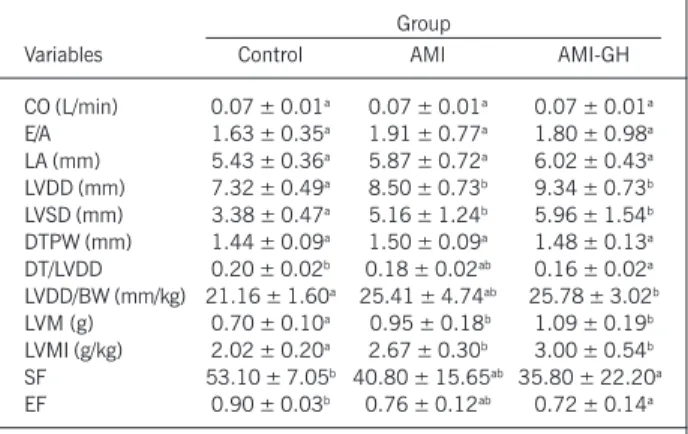
Table II shows the values obtained in the echocardiographic study. The echocardiogram identified an increase in the LVDD (C = 7.32±0.49; AMI = 8.50±0.73; AMI-GH = 9.34±0.73 mm; P < 0.05) and LVSD (C = 3.38±0.47; AMI = 5.16±1.24; AMI-GH = 5.96±1.54 mm; P < 0.05) in infarcted animals, but only the AMI-GH animals had greater LVDD/BW (C = 21.16±1.60; AMI = 25.41±4.74; AMI-GH = 25.78±3.02 mm/kg; P < 0.05 for C vs AMI-GH) compared with those in the control group. The animals in the AMI-GH group had a lower ejection fraction (C = 0.90±0.03; AMI = 0.76±0.12; AMI-GH = 0.72±0.14; P < 0.05 for C vs AMI-GH) and lower shortening fraction (C = 53.10±7.05; AMI = 40.80±15.65; AMI-GH = 35.80±22.20; P < 0.05 for C vs AMI-GH) than the control animals did.
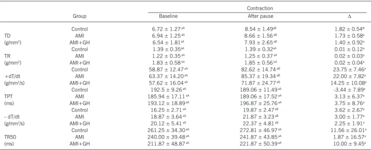
The results of the functional study with isolated papillary muscle and inotropic stimulation with a pause in contraction are shown in table III. The AMI-GH group animals had greater values of tension
at rest than the animals in the other groups did in baseline condi-tions (C = 1.39±0.35; AMI = 1.22±0.35; AMI-GH = 1.83±0.58 g/mm2; P < 0.05). After the pause, one could observe the same behavior as that in the control situation (C = 1.39±0.32; AMI =
1.25±0.37; AMI-GH = 1.85±0.56 g/mm2; P < 0.05). For the
other variables, no differences were found between the groups. The results of the inotropic response after an increase in the calcium concentration in the perfusion medium from 1.25 mM to 5.2 mM are shown in table IV. The AMI-GH group showed an increase in TR in baseline conditions (C = 1.50±0.59; AMI = 1.28±0.38; AMI-GH = 1.98±0.41; P < 0.05 for C vs AMI-GH) and a decrease in the TR50 delta after stimulation with calcium (C = 23.75±9.16; AMI = 16.56±14.82; AMI-GH = 4.69±8.39 ms; P < 0.05 for C vs AMI-GH). For the other variables, no differences were found in the 3 groups.
The results of inotropic stimulation with isoproterenol are shown in table V. The AMI-GH group had changes in the TD delta (C = 0.99±0.17; AMI = 0.54±0.62; AMI-GH = 0.08±0.75; P < 0.05 for C vs AMI-GH) after isoproterenol. The other variables showed no differences in the 3 groups.
Discussion
This study aimed at analyzing the effects of the early adminis-tration of growth hormone in the morphological and functional changes occurring after acute myocardial infarction experimentally produced in rats. For such, the animals were studied by use of echocardiography and preparation of isolated papillary muscle. In the latter, the functional behavior of the muscles was studied in baseline conditions and after 3 types of interventions with positive inotropism: contraction after pause; an increase in calcium con-centration; and addition of isoproterenol to the perfusion bath. These maneuvers allow the identification of functional abnormalities not detected in basal conditions, in addition to the identification of mechanisms involved in the dysfunctions of contractility of the cardiac muscle secondary to several types of injury.
Table I - Morphological variables observed in the different groups
Group
Variable Control AMI AMI-GH
Weight(g) 350.25 ± 34.09ª 357.50 ± 41.97ª 364.25 ± 30.17ª
LV(g) 0.68 ± 0.08ª 0.68 ± 0.09ª 0.75 ± 0.06ª
RV(g) 0.18 ± 0.02ª 0.20 ± 0.06ª 0.23 ± 0.05ª
LV/W (mg/g) 1.94 ± 0.14ªb 1.92 ± 0.13ª 2.08 ± 0.11b
RV/W (mg/g) 0.53 ± 0.04a 0.54 ± 0.10a 0.64 ± 0.18a
lung WW/DW 5.17 ± 0.27a 5.27 ± 0.67a 5.19 ± 0.65a
liver WW/DW 3.10 ± 0.05a 3.02 ± 0.11a 3.06 ± 0.12a
SA (mm2) 0.97 ± 0.13a 1.07 ± 0.16a 1.13 ± 0.19a
LMAX (mm) 7.09 ± 0.59a 7.07 ± 0.71a 7.70 ± 0.83a
%AMI - 22.90 ± 3.10a 24.30 ± 4.90a
GH - growth hormone; AMI - infarcted animals without treatment; AMIGH infarcted animals treated with AMIGH; LV left ventricular weight; RV -right ventricular weight; LV/W - left ventricular weight adjusted for body weight of the rat; RV/W - right ventricular weight adjusted for body weight of the rat; WW/DW relation between wet weight and dry weight; SA -sectional area of the papillary muscle; LMAX - muscle length at maximum performance; %AMI - infarction size. The different letters indicate sta-tistically significant difference (P < 0.05).
Table II - Echocardiographic variables studied in the different groups
Group
Variables Control AMI AMI-GH
CO (L/min) 0.07 ± 0.01a 0.07 ± 0.01a 0.07 ± 0.01a
E/A 1.63 ± 0.35a 1.91 ± 0.77a 1.80 ± 0.98a
LA (mm) 5.43 ± 0.36a 5.87 ± 0.72a 6.02 ± 0.43a
LVDD (mm) 7.32 ± 0.49a 8.50 ± 0.73b 9.34 ± 0.73b
LVSD (mm) 3.38 ± 0.47a 5.16 ± 1.24b 5.96 ± 1.54b
DTPW (mm) 1.44 ± 0.09a 1.50 ± 0.09a 1.48 ± 0.13a
DT/LVDD 0.20 ± 0.02b 0.18 ± 0.02ab 0.16 ± 0.02a
LVDD/BW (mm/kg) 21.16 ± 1.60a 25.41 ± 4.74ab 25.78 ± 3.02b
LVM (g) 0.70 ± 0.10a 0.95 ± 0.18b 1.09 ± 0.19b
LVMI (g/kg) 2.02 ± 0.20a 2.67 ± 0.30b 3.00 ± 0.54b
SF 53.10 ± 7.05b 40.80 ± 15.65ab 35.80 ± 22.20a
EF 0.90 ± 0.03b 0.76 ± 0.12ab 0.72 ± 0.14a
4
The Early Administration of Growth Hormone Results in Deleterious Effects on Ventricular Remodeling After Acute Myocardial Infarction
Our study indicates that the early administration of growth hormone was accompanied by an increase in the mass and di-mensions of the left ventricular cavity in rats with myocardial infarction. These alterations were accompanied by worsening of some cardiac functional variables, such as a drop in the shortening and ejection fractions. Therefore, our results suggest that the early use of growth hormone may result in adverse effects in the process of ventricular remodeling after acute myocardial infarction. It is worth emphasizing that the worsening of remodeling observed in the group receiving growth hormone occurred without an in-fluence from the hormone on the infarction size.
One of the major aspects of our study is the fact that infarction
resulted in an increase in left ventricular mass. Administration of growth hormone was accompanied by an additional increase in left ventricular mass, identified on echocardiography and mor-phological analysis. We also observed that that hypertrophy was accompanied by an increase in the left ventricular cavity, which, similarly to the increase in mass, was greater in the group treated with growth hormone. In addition, in the group receiving growth hormone, a decrease in the relation between thickness of the wall and diameter of the cavity was observed. These data indicate that growth hormone intensified ventricular hypertrophy of the eccentric pattern.
Another relevant aspect of our study was that in the group of
Table III - Effect of contraction after the pause on the functional variables obtained from isolated papillary muscles
Contraction
Group Baseline After pause ∆
Control 6.72 ± 1.27 aA 8.54 ± 1.49aB 1.82 ± 0.54ª
TD AMI 6.94 ± 1.25 aA 8.66 ± 1.56 aB 1.73 ± 0.58a
(g/mm2) AMI+GH 6.54 ± 1.81aA 7.93 ± 2.65 aB 1.40 ± 0.92a
Control 1.39 ± 0.35aA 1.39 ± 0.32aA 0.01 ± 0.12a
TR AMI 1.22 ± 0.35 aA 1.25 ± 0.37 aA 0.02 ± 0.03a
(g/mm2) AMI+GH 1.83 ± 0.58 bA 1.85 ± 0.56 bA 0.02 ± 0.04a
Control 58.87 ± 12.47 aA 82.62 ± 14.74 aB 23.75 ± 7.46a
+dT/dt AMI 63.37 ± 14.20 aA 85.37 ± 19.34 aB 22.00 ± 7.82a
(g/mm2/s) AMI+GH 57.62 ± 16.04 aA 71.87 ± 24.77 aB 14.25 ± 10.08a
Control 192.5 ± 9.26 aA 189.06 ± 11.49 aA -3.44 ± 7.89a
TPT AMI 185.94 ± 17.11 aA 189.06 ± 17.52 aA 3.13 ± 6.37a
(ms) AMI+GH 193.12 ± 18.89 aA 196.87 ± 25.76 aA 3.75 ± 8.76a
Control 16.25 ± 2.71 aA 19.87 ± 2.47 aB 3.62 ± 2.67a
- dT/dt AMI 18.87 ± 3.64 aA 21.87 ± 3.23 aB 3.00 ± 1.77a
(g/mm2/s) AMI+GH 20.12 ± 5.41 aA 22.37 ± 4.81 aB 2.25 ± 1.91a
Control 261.25 ± 34.30 aA 272.81 ± 46.97 aA 11.56 ± 26.01a
TR50 AMI 240.00 ± 39.48 aA 241.87 ± 43.85 aA 1.87 ± 16.57a
(ms) AMI+GH 211.87 ± 48.87 aA 221.87 ± 50.39 aA 10.00 ± 9.45a
Values expressed as mean ± standard deviation; AMI - acute myocardial infarction; AMI+GH - AMI + growth hormone; baseline - contraction before the 30-sec pause; After pause - contraction after the 30-sec pause; TD - maximum tension developed; TR - tension at rest; TPT - time for reaching peak TD; + dT/dt - velocity of variation in TD; -dT/dt - velocity of variation in decreasing TD; TR50 - time for TD to decrease 50% of its maximum value; ∆ - variation between the values of the variables obtained before and after the 30-sec pause; lowercase letters - group effect; capital letters - effect after pause; distinct letters - P < 0.05 (ANOVA, Tukey).
Table IV - Effect of elevation in the extracellular concentration of calcium on the functional variables obtained from isolated papillary muscles
Contraction
Group Baseline Ca+2 5.2 mM
∆
Control 6.77 ± 1.27 aA 7.71 ± 1.31 aB 0.94 ± 0.43ª
TD AMI 7.04 ± 1.24 aA 7.93 ± 1.23 aB 0.88 ± 0.48a
(g/mm2) AMI+GH 6.61 ± 1.82 aA 7.53 ± 2.24 aB 0.92 ±0.59a
Control 1.30 ± 0.41 aA 1.50 ± 0.59 aB 0.20 ± 0.26a
TR AMI 1.16 ± 0.35 aA 1.28 ± 0.38 aA 0.12 ± 0.12a
(g/mm2) AMI+GH 1.88 ± 0.53 bA 1.98 ± 0.41 bA 0.10 ± 0.25ª
Control 59.75 ± 12.51 aA 78.12 ± 12.70 aB 18.37 ± 8.83ª
+dT/dt AMI 62.37 ± 11.37 aA 77.87 ± 14.64 aB 15.50 ± 6.50a
(g/mm2/s) AMI+GH 58.37 ± 16.65 aA 74.00 ± 20.02 aB 15.62 ± 6.43a
Control 186.25 ± 12.46 aB 173.12 ± 14.86 aA -13.12 ± 5.94a
TPT AMI 184.37 ± 13.48 aB 172.5 ± 12.82 aA -11.87 ± 8.42a
(ms) AMI+GH 191.56 ± 23.41 aB 182.5 ± 24.93 aA -9.06 ± 5.97a
Control 16.75 ± 2.43 aA 22.50 ± 3.78 aB 5.75 ± 1.83ª
- dT/dt AMI 19.75 ± 3.57 aA 24.62 ± 3.29 aB 4.87 ± 1.46a
(g/mm2/s) AMI+GH 20.75 ± 4.53 aA 26.12 ± 6.62 aB 5.37 ± 2.82a
Control 245.00 ± 34.64 aB 221.25 ± 35.63 aA -23.75 ± 9.16b
TR50 AMI 231.25 ± 37.01 aB 214.69 ± 26.27 aA -16.56 ± 14.82ab
(ms) AMI+GH 203.44 ± 42.57 aA 198.75 ± 37.41 aA -4.69 ± 8.39a
Values expressed as mean ± standard deviation; AMI - acute myocardial infarction; AMI+GH - AMI + growth hormone; baseline - contraction recorded in extracellular calcium concentration of 1.25 mM; Ca+2 5.2 mM - contraction recorded in extracellular calcium concentration of 5.2 mM; TD - maximum tension
5
The Early Administration of Growth Hormone Results in Deleterious Effects on Ventricular Remodeling After Acute Myocardial Infarction
Table V - Effect of isoproterenol (10-6M) on the functional variables obtained from isolated papillary muscles
Treatment
Baseline Isoproterenol ∆
Control 5.82 ± 0.62 aA 6.81 ± 0.63 aB 0.99 ± 0.17b
TD AMI 6.71 ± 1.03 aA 7.25 ± 1.32 aB 0.54 ± 0.62ab
(g/mm2) AMI+GH 6.74 ± 1.92 aA 6.83 ± 2.06 aA 0.08 ± 0.75a
Control 1.38 ± 0.52 aB 1.15 ± 0.47 aA -0.24 ± 0.10a
TR AMI 1.36 ± 0.41 aA 1.33 ± 0.34 aA -0.02 ± 0.31a
(g/mm2) AMI+GH 1.76 ± 0.50 aA 1.77 ± 0.59 aA 0.01 ± 0.20a
Control 55.57 ± 8.73 aA 75.14 ± 8.19 aB 19.57 ± 6.42a
+dT/dt AMI 63.75 ± 14.67 aA 81.63 ± 18.24 aB 17.88 ± 6.93a
(g/mm2/s) AMI+GH 61.43 ± 17.97 aA 77.43 ± 26.17 aB 16.00 ± 9.61a
Control 173.21 ± 13.59 aB 141.79 ± 10.07 aA -31.43 ± 12.57a
TPT AMI 170.63 ± 21.62 aB 142.50 ± 11.65 aA -28.12 ± 14.12a
(ms) AMI+GH 183.57 ± 28.24 aB 146.43 ± 17.25 aA -37.14 ± 14.96a
Control 19.14 ± 2.34 aA 32.00 ± 3.37 aB 12.86 ± 3.62a
- dT/dt AMI 20.38 ± 2.92 aA 34.13 ± 5.17 aB 13.75 ± 3.06a
(g/mm2/s) AMI+GH 22.14 ± 5.79 aA 35.86 ± 12.21 aB 13.71 ± 7.67a
Control 200.71 ± 31.81 aB 142.50 ± 18.14 aA -58.21 ± 26.76a
TR50 AMI 206.25 ± 23.11 aB 139.06 ± 23.71 aA -67.19 ± 26.13a
(ms) AMI+GH 203.57 ± 32.91 aB 128.93 ± 26.92 aA -74.64 ± 29.94a
Values expressed as mean ± standard deviation; AMI - acute myocardial infarction; AMI+GH - AMI + growth hormone; baseline - contraction recorded before addition of isoproterenol; Isoproterenol - contraction after addition of 10-6M of isoproterenol to the nutrient solution; TD - maximum tension developed; TR - tension
at rest; TPT - time for reaching peak TD; + dT/dt - velocity of variation in TD; -dT/dt - velocity of variation in decreasing TD; TR50 - time for TD to decrease 50% of its maximum value; ∆ - variation between the values of the variables obtained before and after addition of isoproterenol; lowercase letters - group effect; capital letters - effect of isoproterenol; distinct letters - P < 0.05 (ANOVA, Tukey).
infarcted rats treated with growth hormone, cardiac hypertrophy was accompanied by an increase in the ventricular dysfunction produced by infarction. The in vivo functional study with echocar-diography identified lower ejection and shortening fractions in ani-mals treated with growth hormone. On the other hand, the in vitro study of the isolated papillary muscle evidenced changes in some variables. In baseline conditions, alterations were identified in tension at rest in infarcted animals treated with growth hormone. Similarly, the inotropic stimulation also evidenced some changes, because the increase in calcium caused a lower variation (delta) in TR50, while the use of isoproterenol resulted in a lower delta in tension at rest in rats of the growth hormone group.
Other authors have analyzed the effects of the administration of growth hormone in the adult phase in the absence of cardio-vascular disease. In normal rats, the use of growth hormone re-sulted in cardiac hypertrophy without an increase in fibrosis. This response was accompanied by an increase in contractility, altera-tions in the genesis of cardiac action potentials and peripheral vasodilation. In human beings, in the early phases of acromegaly, an increase in cardiac output and heart rate is observed, signs compatible with hyperkinetic syndrome 5.
Considering pathological situations, Yang et al 7, in an experi-mental study, assessed the effects of growth hormone (2 mg/kg/ day) initiated 4 weeks after the induction of large infarcts in rats. After being treated for 13 days, the animals in the treated group showed an increase in cardiac output, in systolic volume, and in the first derivative of the pressure, accompanied by a reduction in the left ventricular end-diastolic pressure compared with the fin-dings in nontreated animals 7. In the same model of large infarcts in rats, the administration of growth hormone resulted in atte-nuation of the remodeling process after 3 weeks of treatment 8. Recently, growth hormone has been observed to cause an increase in the messenger RNA to the calcium pump of the sarcoplasmic reticulum (SERCA-2), a decrease in apoptosis, and an
accumula-tion of collagen in infarcted rats. These alteraaccumula-tions were accompa-nied by functional improvement and prolongation in survival 9,10. It is worth noting, however, that the benefits of GH have not been consistent in all studies. In another study also performed in the model of infarcted rats, the administration of growth hormone (2 mg/kg/day) resulted in a discrete improvement in cardiac function, but with no effects on the variables of ventricular remodeling17.
Clinical studies 18 assessing the role of growth hormone in pathological situations have already reported that patients with acute myocardial infarction have a significant elevation in the levels of growth hormone in the first 24 hours after infarction. Considering that the growth hormone levels related to infarction size and mortality, it has been speculated that growth hormone could participate in the physiopathological events of the remodeling process after infarction in human beings 19, 20.
A relevant aspect is that clinical studies in patients with heart failure also reported conflicting results regarding supplementation with growth hormone. In a study of 7 patients with dilated car-diomyopathy, the administration of growth hormone for 3 months resulted in an increase in cardiac output, with a decrease in the left ventricular end-systolic stress 18. However, these preliminary findings about the beneficial effects of growth hormone in cases of ventricular dysfunction have not been confirmed in randomized studies 21,22. Therefore, the effects of growth hormone in cases of ventricular dysfunction or acute myocardial infarction, or both, have not been completely clarified.
Some theories have been raised to explain the conflicting data reported in the literature. Several factors have been considered important in regard to the effects of the administration of growth hormone in models of cardiac dysfunction. Therefore, the experi-mental use of growth hormone in animals, differences in the dose administered, the duration of treatment, and the etiology of ven-tricular dysfunction could justify the differences observed between clinical and experimental studies.
experi-6
The Early Administration of Growth Hormone Results in Deleterious Effects on Ventricular Remodeling After Acute Myocardial Infarction
1. Matsubara BB, Zornoff LAM. Matriz colágena intersticial e sua relação com a ex-pansão miocárdica no infarto agudo. Arq Bras Cardiol 1995; 64: 559-63. 2. Pfeffer JM, Pfeffer MA, Braunwald E. Influence of chronic captopril therapy on
the infarcted left ventricle of the rat. Circ Res 1985; 57:84-95.
3. Pfeffer MA, Braunwald E. Ventricular remodeling after myocardial infarction: expe-rimental observations and clinical implications. Circulation 1990;81:1161-1172. 4. Cohn JN, Ferrari R, Sharpe N. Cardiac remodeling- concepts and clinical
implica-tions: a consensus paper from an international forum on cardiac remodeling. J Am Coll Cardiol 2000; 35: 569-582.
5. Colao A, Marzullo P, Di Somma C, Lombardi G. Growth hormone and the heart. Clinical Endocrinology 2001; 54: 137-54.
6. Khan AS, Sane DC, Wannenburg T, Sonntag WE. Growth hormone, insulin-like growth factor-1 and the aging cardiovascular system. Cardiovasc Res 2002; 54: 25-35.
7. Yang R, Bunting S, Gillett N, Clark R, Jin H. Growth hormone improves cardiac per-formance in experimental heart failure. Circulation 1995; 92: 262-7.
8. Cittadini A, Grossman JD, Napoli R, et al. Growth hormone attenuates early left ventricular remodeling and improves cardiac function in rats with large myocardial infarction. J Am Coll Cardiol 1997; 29: 1109-16.
9. Tajima M, Weinberg EO, Bartunek J, et al. Treatment with growth hormone enhan-ces contractile reserve and intracellular calcium transients in myocytes from rats with postinfarction heart failure. Circulation 1999; 99: 127-34.
10. Cittadini A, Isgaard J, Monti MG, et al. Growth hormone prolongs survival in ex-perimental postinfarction heart failure. J Am Coll Cardiol 2003; 41: 2154-63. 11. Zornoff LAM, Paiva SAR, Matsubara BB, Matsubara LS, Spadaro J.
Combina-tion therapy with angiotensin converting enzyme inhibiCombina-tion and AT1 receptor inhi-bitor on ventricular remodeling after myocardial infarction in rats. J Cardiovasc Pharmacol Ther 2000; 5: 203-9.
12. Zornoff LAM, Matsubara BB, Matsubara LS, Paiva SAR, Spadaro J. Early rather than delayed administration of lisinopril protects the heart after myocardial infarc-tion in rats. Basic Res Cardiol 2000; 95: 208-14.
13. Sahn DJ, DeMaria A, Kisslo J, Weyman AE. The Committee on M-Mode
Standar-References
dization of the American Society of Echocardiography. Recommendations regar-ding quantitation in M-mode echocardiography: results of a survey of echocardio-graphic measurements. Circulation 1978; 58: 1072-83.
14. Litwin SE, Katz SE, Morgan JP, Douglas PS. Serial echocardiographic assessment of left ventricular geometry and function after large myocardial infarction in the rat. Circulation 1994; 89: 345-54.
15. Cicogna AC, Padovani CR, Okoshi K, Aragon FF, Okoshi MP. Myocardial function during chronic food restriction in isolated hypertrophied cardiac muscle. Am J Med Sci 2000; 320: 244-8.
16. Cicogna AC, Padovani CR, Okoshi K, Matsubara LS, Aragon FF, Okoshi MP. The in-fluence of temporal food restriction on the performance of isolated cardiac muscle. Nutr Res 2001; 21: 639-48.
17. Bollano E, Bergh CH, Kjellstrom C, et al. Growth hormone alone or combined with metoprolol preserves cardiac function after myocardial infarction in rats. Eur J Heart Fail 2001; 3: 651-60.
18. Fazio S, Sabatini D, Capaldo B, et al. A preliminary study of growth hormone in the treatment of dilated cardiomyopathy. N Engl J Med 1996; 334: 809-14. 19. Friberg L, Werner S, Eggertsen G, Ahnve S. Growth hormone and insulin-like growth
factor-1 in acute myocardial infarction. Eur Heart J 2000; 21: 1547-54. 20. Wollert KC, Drexler H. Growth hormone and insulin-like growth factor-friend of the
infarct heart? Eur Heart J 2000; 21: 1499-501.
21. Isgaard J, Bergh C-H, Caidhal K, et al. A placebo-controlled study of growth hor-mone in patients with congestive heart failure. Eur Heart J 1998; 19: 1704-11. 22. Osterziel KJ, Strohm O, Schuler J, et al. Randomised, double-blind,
placebo-con-trolled trial of human recombinant growth hormone in patients with chronic heart failure due to dilated cardiomyopathy. Lancet 1998; 1233-7.
23. Mill JG, Vasalo DV, Leite CM. Influence of sarcoplasmic reticulum on the inotropic responses of the rat myocardium resulting from changes in rate and rhythm. Braz J Med Biol Res 1994; 1455-65.
24. Zornoff LAM, Cicogna AC, Paiva SAR, Spadaro J. Remodelamento e seu impacto na progressão da disfunção ventricular. Rev Soc Cardiol Estado de São Paulo 2002; 12: 371-8.
mental studies about the effects of growth hormone after acute myocardial infarction. First of all, the studies cited used animals with large infarctions, greater than 35% of the total left ventricular circumference. Consequently, the animals had signs of significant cardiac dysfunction. Our study used animals with small infarctions (range: from 16.7 to 30.6%), and, therefore, with mild alterations in ventricular function. Therefore, our data together with those reported in the literature suggest that the degree of ventricular dysfunction may be a determining factor in the effects of growth hormone after acute myocardial infarction. Another peculiarity of our study is that, while other studies introduced growth hormone weeks after infarction induction, our animals began to receive growth hormone immediately after infarction. This fact suggests that growth hormone may interfere in a deleterious manner with the physiopathological phenomena that occur in the early phase after acute myocardial infarction, particularly the healing process and infarction expansion.
In regard to the mechanisms involved in the genesis of ventri-cular dysfunction induced by growth hormone, our study showed no change in functional behavior after the maneuver to pause the
contraction. Considering that that maneuver analyzes the integrity
of the sarcoplasmic reticulum 23, one can conclude that that
system is not affected by the growth hormone. On the other hand, the administration of growth hormone resulted in a smaller delta for some functional variables with the increase in calcium concentration, as well as after isoproterenol administration. Thus, our data indicate that a decrease in responsiveness to calcium and in the beta-adrenergic pathway may have occurred. It is worth noting, however, that these alterations in the papillary muscle occurred only in some isolated variables, which could indicate preserved myocardial function. This way, although the muscular function was not severely impaired, the growth hormone caused depression in left ventricular function. Our results are in accordance with the concept that geometry plays a relevant role in the pa-thophysiology of ventricular dysfunction in the remodeled heart, probably due to an increase in parietal stress 24.