TRICKS OF THE TRADE
The expanding spectrum of
clinically-distinctive, immunotherapy-responsive
autoimmune encephalopathies
O espectro em expansão das encefalopatias autoimunes clinicamente distintas e que
respondem à imunoterapia
Sarosh R Irani1, Angela Vincent2
For around 30 years, it has been recognised that limbic encephalitis (LE) can be associated with a distant cancer. Such paraneoplastic LE is often refractory to even aggressive immunotherapies and tumour removal. Using brain-section immunohistochemistry and western blotting techniques, a number of encephalitis-associated autoantibody targets was subsequently discovered. Some showed relatively speciic as-sociations with the type of underlying tumour, for instance, Ma2 antibodies and testicular tumours1. As these techniques
employed denatured neural antigens, and revealed intrac-ellular targets, it was diicult to conceive that the detected antibodies were directly pathogenic2.hey are still
consid-ered likely bystanders in a predominantly T-cell mediated syndrome.
More recently, a number of antibodies have been discovered targeting the extracellular domain of neural proteins in their native conformations3,4. Most frequently, these antibodies are
directed against the voltage-gated potassium channel (VGKC)
1 DPhil MRCP; Nuffield Department of Clinical Neurosciences, West Wing, John Radcliffe Hospital, Oxford, UK;
2 FRS FRCPath; Nuffield Department of Clinical Neurosciences, West Wing, John Radcliffe Hospital, Oxford, UK.
Correspondence: Sarosh R Irani; Nuffield Department of Clinical Neurosciences, West Wing, John Radcliffe Hospital, Oxford, UK; E-mail: [email protected] Support: S.R.I was supported by the National Institute for Health Research (NIHR), Department of Health, UK. This work has been supported by the NIHR-funded Oxford Biomedical Research Centre.
Conflict of interest: A.V. and the Department of Clinical Neurology in Oxford receive royalties and payments for Ab assays. A.V. is the inventor on patent application WO/2010/046716 entitled “Neurological Autoimmune Disorders”. S.R.I is a co-inventor. The patent has been licensed to Euroimmun AG for the development of assays for Lgi1 and other VGKC-complex Abs. A.V and S.R.I. may receive royalties for testing of VGKC complex Abs.
Received 11 January 2012; Accepted 18 January 2012
ABSTRACT
The autoimmune encephalopathies are a group of conditions that are associated with autoantibodies against surface neuronal proteins, which are likely to mediate the disease. They are established as a frequent cause of encephalitis. Characteristic clinical features in individual patients often allow the specificity of the underlying antibody to be confidently predicted. Antibodies against the VGKC-complex, mainly LGI1(leucine-rich glioma-inactivated 1), CASPR2 (contactin-associated protein 2), and contactin-2, and NMDA (N-methyl, D-aspartate) -re-ceptor are the most frequently established serological associations. In the minority of cases, an underlying tumour can be responsible. Early administration of immunotherapies, and tumour removal, where it is relevant, offer the greatest chance of improvement. Prolonged courses of immunotherapies may be required, and clinical improvements often correlate well with the antibody levels. In the present article, we have summarised recent developments in the clinical and laboratory findings within this rapidly expanding field.
Key words: autoantibody, encephalitis, leucine-rich glioma-inactivated 1, contactin-associated protein 2, N-methyl, D-aspartate.
RESUMO
As encefalopatias autoimunes constituem um grupo de condições associadas à presença, no soro, de anticorpos contra proteínas de super-fície neuronais. Acredita-se que esses anticorpos sejam mediadores da ocorrência da doença, sendo reconhecidos atualmente como causas frequentes de encefalite. Apresentações clínicas características permitem, muitas vezes, predizer o grupo específico de anticorpos subja-centes. Anticorpos contra o complexo VGKF, especialmente LGI1 (leucine-rich glioma-inactivated1), CASPR2 (contactin-associated protein 2) e contactina-2, e contra o receptor NMDA(N-methyl, D-aspartate) são as associações sorológicas mais frequentemente estabelecidas. Na minoria dos casos, pode ser detectado um tumor subjacente. As maiores chances de melhora estão relacionadas à administração precoce de imunoterapia e à remoção do tumor, quando presente. A duração da imunoterapia pode se prolongada e a melhora se correlaciona, muitas vezes, com os níveis séricos de anticorpos. Neste artigo, estão resumidos os avanços recentes nos achados clínicos e laboratoriais neste campo que está em tão rápida expansão.
complex and the N-methyl, D-aspartate (NMDA) receptor3-6.
hese antibodies are likely to be pathogenic, the diseases are typically immunotherapy-responsive, and they are only associ-ated with cancer in a minority of patients. he antibodies and related clinical features will be the focus of this article.
THE VGKC-COMPLEX: LGI1, CASPR2, AND CONTACTIN-2
LE is a term often used to describe the triad of amnesia, disorientation, and seizures. A minority of patients may de-velop additional features such as delusions, hallucinations, and sleep-cycle disturbances. he onset is typically over a few days or weeks and the main diferential diagnoses include viral encephalitis and metabolic derangements3. he most
common serological association is with VGKC-complex an-tibodies. It often predicts a <10% risk of an underlying tu-mour and a good response to early immunotherapies, typi-cally with a parallel reduction in VGKC-complex antibody levels7. A useful clue to VGKC-complex antibody positivity is
serum hyponatraemia and around half of the patients show T2/FLAIR high-signal in the medial temporal lobes7,8.
Morvan’s syndrome (MOS) represents another en-cephalopathy associated with VGKC-complex antibodies9.
Neuromyotonia (NMT) is a peripheral nerve hyperexcitability disorder, which was the irst syndrome to be associated with VGKC-complex antibodies10. NMT is a critical part of MOS.
When compared with LE, patients with MOS have more fre-quent tumours and psychiatric features, with fewer seizures and magnetic resonance image (MRI) changes (SRI and AV, unpublished observations). Despite the higher frequency of tumours, many cases present a clear response to immuno-therapies accompanied by a paralleled reduction in antibody levels in the few cases examined prospectively.
A highly-distinctive seizure semiology has been observed in association with VGKC-complex antibodies. Firstly de-scribed in three cases, in 200811, and subsequently in 26
addi-tional cases, in 201112, faciobrachial dystonic seizures (FBDS)
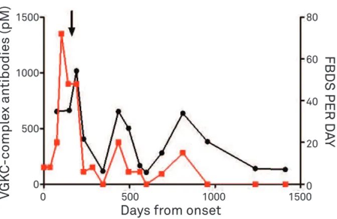
manifest as frequent, brief, dystonic epileptic events that typ-ically afect the arm and the ipsilateral face12. Importantly,
patients are commonly (anti epileptic drugs) AED-refractory and AEDs are associated with signiicant side efects. By con-trast, the FBDS are immunotherapy-responsive, often exqui-sitely (Fig 1)12. Importantly, in at least 75% of cases, FBDS
seem to precede the onset of amnesia and confusion that typify VGKC-complex antibody LE12. herefore, we have
pro-posed that clinical recognition of FBDS may allow initiation of immunotherapies which prevent LE. Prospective studies are awaited to examine such hypothesis.
he four clinical syndromes (NMT, MOS, LE and FBDS) show consistencies in their subacute onset and immunotherapy-re-sponses. However, the individual phenotypes appear distinct
from one another. herefore, a key question has arisen: how can one antibody mediate the four syndromes? To address the ques-tion, the concept of the ‘VGKC-complex’ was irst established in 200913. Experiments have shown that VGKCs found within
the mammalian brain membranes used to deine the antibodies are tightly-complexed with a number of other proteins. Some of these proteins – leucine-rich glioma-inactivated 1 (LGI1), con-tactin-associated protein 2 (CASPR2) and contactin-2 – were identiied as targets of the patient antibodies (Fig 2)8,13,14.
LGI1-antibodies are predominantly observed in patients with LE and FBDS8,14,15, and CASPR2-antibodies in MOS and NMT8,13,14. here
is some overlap with LGI1-antibodies in some patients with MOS and, less frequently, NMT. Also, CASPR2-antibodies are found in few patients with LE8.
VGK
C
-compl
ex antibodies (pM)
Days from onset
FBDS PER D
AY
1500
1000
500
0
80
60
40
20
0
0 500 1000 1500
Fig 1. Close correlation between faciobrachial dystonic seizure (FBDS) frequency and VGKC-complex antibodies in a single patient. Clinical relapses are associated with discontinuation of immunotherapies, which were commenced as depicted by the downward arrow. Adapted from Irani et al.12.
C
A
S
P
R
2
CONT
AC
TIN2
LGI1
125I-DTXTable. Differences between VGKC-complex and NMDAR-antibody associated encephalitis (modified from Irani and Vincent, Discovery Medicine 2011; 11(60):449-458. With permission from Discovery Medicine).
VGKC-complex antibody (usually LGI1 antibody)
NMDAR-antibody
Gender ratio (M:F) 2:1 1:3
Age Usually >50 years-old Usually <50 years-old
Target antigen LGI1>>>CASPR2 NR1 subunit of NMDAR
Is there tumour associated? Rarely (<10%). If so, SCLC and thymoma. Ovarian teratoma (20 to 50%), others rarely.
Clinical features Amnesia, disorientation, and medial temporal
lobe seizures.
Psychiatric features, amnesia, disorientation, and seizures progress to movement disorder, dysautonomia, and central hypoventilation.
Distinctive clinical features Faciobrachial dystonic seizures, typically precede
amnesia and confusion.
Choreoathetoid movement disorder, usually starting days to weeks after the psychiatric features.
Blood tests (other than antibody) Hyponatraemia (consistent with SIADH, in around 60%).
Nil
MRI Bilateral hipocampal high signal (in around 60%).
In remaining 40%, changes can be unilateral or absent.
Often normal. Occasionally, non-specific high signal or medial temporal lobe high signal.
CSF Most commonly no cells or oligoclonal bands.
Positive antibodies in many patients.
Early lymphocytic pleocytosis and later
oligoclonal bands. Antibodies detected in majority of patient CSFs.
Immunotherapy regime Usually good response to one to two
immunotherapies (steroids +/-IvIg/plasma exchange).
Slow response, often over months; typically requires >2 immunotherapies.
MRI: magnetic resonance imaging; CSF: cerebrospinal fluid; SCLC: small cell lung cancer; SIADH: syndrome of inappropriate antidiuretic hormone.
Contactin-2 antibodies were least commonly detect-ed8. As LGI1 is almost exclusively expressed in central
ner-vous system neurons, and CASPR2 in both the central and peripheral nervous systems, the antibodies are considered likely mediators of the diseases. While careful correlations between antibody levels and clinical outcomes in individual patients, in combination with passive transfer experiments in animals, are required to formally assess VGKC-complex anti-body pathogenicity, it has been proven that LGI1-antibodies can generate seizure activity in hippocampal slice cultures16.
Interestingly, further evidence for the disease relevance of these proteins come from known genetic variations. Human LGI1-mutations produce a lateral temporal lobe epilepsy syndrome17 and mice lacking LGI1 show a variety of motor
semiologies18. Humans with CASPR2 mutations present
au-tism, seizures, and peripheral neuropathy19.
THE NMDA-RECEPTOR
A different characteristic set of clinical features has been linked to another autoantibody (Table). On this oc-casion, the antibodies are directed against an ion-channel: the NMDA-receptor. The initial article described antibod-ies in young females with ovarian teratomata20. Patients
showed clinical improvements with a combination of tu-mour removal and immunotherapies. Since this impor-tant finding, it has become clear that the disease affects both males and females and it is more commonly non-paraneoplastic21-23. Tumours are most frequently detected
in women between 20 and 40 years-old, and they are al-most always ovarian teratomata.
he typical disease appears to progress though two major, well-characterized stages21. he irst one is associated with
prom-inent psychiatric features, such as delusions, behavioural change, wandering, and hallucinations. In addition, patients develop language diiculties, amnesia, disorientation, and seizures21,24.
After a lag of 10 to 20 days, patients develop movement disorder, decreased level of consciousness, and lorid dysautonomia. he latter two features frequently take patients to the Intensive Care Unit (ICU), where this disease is not an uncommon diagnosis25.
he movement disorder characteristically involves orofacial dyskinesias and choreoathetoid limb movements. However, aki-netic-rigid-mute presentations can predominate. Many of these patients are reminiscent of von Economo’s encephalitis lethar-gica (EL) cases and, indeed, around half of the children originally given a diagnosis of EL have NMDAR-antibodies26. In children,
the movement disorder may be the presenting feature27, there is
a very low frequency of tumours and often a preceding infection, which is associated with a variety of organisms21.
herapy for NMDAR-antibody encephalitis should in-volve early removal of a tumour, if present, and early insti-tution of irst-line immunotherapies21,24. hese two measures
1. Gultekin SH, Rosenfeld MR, Voltz R, Eichen J, Posner JB, Dalmau J. Paraneoplastic limbic encephalitis: neurological symptoms, immunological findings and tumour association in 50 patients. Brain 2000;123:1481-1494.
2. Vincent A, Lily O, Palace J. Pathogenic autoantibodies to neuronal proteins in neurological disorders. J Neuroimmunol 1999;100:169-180.
3. Vincent A, Bien CG, Irani SR, Waters P. Autoantibodies associated with diseases of the CNS: new developments and future challenges. Lancet Neurol 2011;10:759-772.
4. Irani SR, Bien CG, Lang B. Autoimmune epilepsies. Curr Opin Neurol 2011;24:146-153.
5. Granerod J, Ambrose HE, Davies NW, et al. Causes of encephalitis and differences in their clinical presentations in England: a multicentre, population-based prospective study. Lancet Infect Dis 2010;10:835-844.
6. Wandinger KP, Klingbeil C, Gneiss C, et al. New serological markers for the differential diagnosis of autoimmune limbic encephalitis. J Lab Med 2011;35:329-342.
7. Vincent A, Buckley C, Schott JM, et al. Potassium channel antibody-associated encephalopathy: a potentially immunotherapy-responsive form of limbic encephalitis. Brain 2004:127;701-712.
8. Irani SR, Alexander S, Waters P, et al. Antibodies to Kv1 potassium channel-complex proteins leucine-rich, glioma inactivated 1 protein and contactin-associated protein-2 in limbic encephalitis, Morvan’s syndrome and acquired neuromyotonia. Brain 2010;133:2734-2748.
9. Liguori R, Vincent A, Clover L, et al. Morvan’s syndrome: peripheral and central nervous system and cardiac involvement with antibodies to voltage-gated potassium channels. Brain 2001;124:2417-2426. References
Taken together, this disease appears to involve predomi-nantly cortical regions early in the disease and later subcorti-cal areas, such as the brainstem pattern generators, basal gan-glia, ascending activating reticular system, and hypothalamus. Indeed, when combined with data from sequential EEGs and MRIs, the disease progression could be explained by a corti-cal-to-subcortical transition in the burden of the pathology21,28.
his appears to involve a lymphocytic pleocytosis, however the lack of evidence of inlammation on imaging suggests that NMDA-receptor downregulation, without complement ixa-tion, is the predominant pathophysiological mechanism24.
OTHER ANTIGEN TARGETS
Additionally, a variety of other autoantibody targets has been discovered in rarer encephalopathic syndromes. hese are directed against the glycine, GABAB and AMPA
recep-tors. Progressive encephalomyelitis with rigidity and myoclo-nus (PERM) has long been associated with GAD-antibodies, however as GAD is an intracellular enzyme, these antibodies are unlikely to be causative. herefore, it is more disease-rele-vant that PERM has recently been associated with antibodies that target the extracellular domain of the glycine receptor29. A
few patients with LE, without VGKC-complex antibodies, may have antibodies directed against the glycine, GABAB or AMPA
receptors. Although in a recent unselected screen, the latter two are very rarely found in cases with autoimmune encephali-tis6. Furthermore, there remain a minority of LE cases without
an associated antibody identiied to date: further antibodies will surely be discovered in many of these cases.
ANTIBODY DETERMINATION AND SERUM-TO-CSF RATIOS
For all the antibodies discussed, the most able method to mimic the native antigen is the cell-based assay (CBA). his
achieves surface expression of the antigen and in non-perme-abilized mammalian cells, it only permits the antibody access to the extracellular domain of the antigen3,4,20,30. All antibodies
can be measured in this manner. However, there are still anti-bodies that target the VGKC-complexes, which do not have a known antigenic target8. herefore, for sensitivity, we still
rec-ommend using the traditional VGKC-complex radioimmu-noassay. Conversely, the CBAs may detect CASPR2 and LGI1-antibodies when the radioimmunoassay is negative (Irani and Vincent, unpublished observations), and it may be that a combination of both assays ofers optimal coverage.
It is a consistent inding (>95% of cases), for all the con-ditions described, that antibody concentrations in the se-rum are higher than in the CSF8,21,28. his implies that using
the relatively concentrated serum ofers the most sensitive diagnostic assay. However, as intrathecal synthesis of anti-body is established in some of these conditions21,24, it may
be that the measurement of CSF antibodies plays a role in the follow-up of patients, and this possibility needs to be investigated.
CONCLUSIONS
In summary, there is a growing number of CNS-disorders which are associated with potentially pathogenic autoanti-bodies. Many of these antibodies are directed against CNS receptors (e.g. NMDA, glycine, AMPA, and GABAB), but also
10. Shillito P, Molenaar PC, Vincent A, et al. Acquired neuromyotonia: evidence for autoantibodies directed against K+ channels of peripheral nerves. Ann Neurol 1995;38:714-722.
11. Irani SR, Buckley C, Vincent A, et al. Immunotherapy-responsive seizure-like episodes with potassium channel antibodies. Neurology 2008;71:1647-1648.
12. Irani SR, Michell AW, Lang B, et al. Faciobrachial dystonic seizures precede Lgi1 antibody limbic encephalitis. Ann Neurol 2011;69:892-900.
13. Vincent A. Antibodies to Contactin-Associated Protein 2 (Caspr2) in thymoma and Morvans syndrome. Ann Neurol 2009;66 (Suppl 13):S3.
14. Irani SR, Waters P, Kleopa KA, et al. Antibodies to components of the voltage-gated potassium channel-associated complex: LGI1 and Caspr2 as antigenic targets in limbic encephalitis, Morvan’s and neuromyotonia. Neurology 2010;75:379-381.
15. Lai M, Huijbers MG, Lancaster E, et al. Investigation of LGI1 as the antigen in limbic encephalitis previously attributed to potassium channels: a case series. Lancet Neurol 2010;9:776-785.
16. Lalic T, Pettingill P, Vincent A, Capogna M. Human limbic encephalitis serum enhances hippocampal mossy fiber-CA3 pyramidal cell synaptic transmission. Epilepsia 2010;52:121-131.
17. Morante-Redolat JM, Gorostidi-Pagola A, Piquer-Sirerol S, et al Mutations in the LGI1/Epitempin gene on 10q24 cause autosomal dominant lateral temporal epilepsy. Hum Mol Genet 2002;11:1119-128.
18. Chabrol E, Navarro V, Provenzano G, et al. Electroclinical characterization of epileptic seizures in leucine-rich, glioma-inactivated 1-deficient mice. Brain 2010;133:2749-2762.
19. Strauss KA, Puffenberger EG, Huentelman MJ, et al. Recessive symptomatic focal epilepsy and mutant contactin-associated protein-like 2. N Engl J Med 2006;354:1370-1377.
20. Dalmau J, Tüzün E, Wu HY, et al. Paraneoplastic anti-N-methyl-D-aspartate receptor encephalitis associated with ovarian teratoma. Ann Neurol 2007;61:25-36.
21. Irani SR, Bera K, Waters P, et al. N-methyl-D-aspartate antibody encephalitis: temporal progression of clinical and paraclinical observations in a predominantly non-paraneoplastic disorder of both sexes. Brain 2010;133:1655-1667.
22. Gabilondo I, Saiz A, Galán L, et al. Analysis of relapses in anti-NMDAR encephalitis. Neurology 2011;77:996-999.
23. Abdullah S, Lim SY, Goh KJ, Lum LCS, Tan CT. Anti-N-methyl-D-aspartate receptor (NMDAR) encephalitis: a series of ten cases from a university hospital in Malaysia. Neurology Asia 2011;16:241-246.
24. Dalmau J, Gleichman AJ, Hughes EG, et al. Anti-NMDA-receptor encephalitis: case series and analysis of the effects of antibodies. Lancet Neurol 2008;7:1091-1098.
25. Davies G, Irani SR, Coltart C, et al. Anti-N-methyl-D-aspartate receptor antibodies: a potentially treatable cause of encephalitis in the intensive care unit. Crit Care Med 2010;38:679-682.
26. Dale RC, Irani SR, Brilot F, et al. N-methyl-D-aspartate receptor antibodies in pediatric dyskinetic encephalitis lethargica. Ann Neurol 2009;66:704-709.
27. Florance NR, Davis RL, Lam C, et al. Anti-N-methyl-D-aspartate receptor (NMDAR) encephalitis in children and adolescents. Ann Neurol 2009;66:11-18.
28. Irani SR, Vincent A. NMDA Receptor Antibody Encephalitis. Curr Neurol Neurosci Rep 2011;11:298-304.
29. Hutchinson M, Waters P, McHugh J, et al. Progressive encephalomyelitis, rigidity, and myoclonus: a novel glycine receptor antibody. Neurology 2008;71:1291-1292.