ARTICLE
HAM/TSP: association between white matter
lesions on magnetic resonance imaging,
clinical and cerebrospinal fluid findings
HAM/TSP: associação entre lesões de substância branca à ressonância magnética,
achados clínicos e do líquido cefalorraquidiano
Marzia Puccioni-Sohler1, Emerson Gasparetto2, Mauro Jorge Cabral-Castro3, Carla Slatter4, Cecilia M. Vidal5,
Romeu Domingues Cortes6, Bruce R. Rosen7, Caterina Mainero8
Human T-cell lymphotropic virus type 1 (HTLV-1) was the irst retrovirus to be isolated in humans1. It is endemic in
southeastern Japan, the Caribbean, South America and parts of Africa, and also in Melanesia and the Middle East2. here
are approximately 20 million people infected worldwide3,4.
Most infected patients remain asymptomatic. Approximately 1–2% develop a chronic spastic paraparesis called as HTLV-1 associated myelopathy/tropical spastic paraparesis (HAM/ TSP). It predominantly involves the spinal cord at the thorac-ic level. Laboratory diagnosis is based on the demonstration
1 Professor and Chief, Clinical Pathology Service/Cerebrospinal Fluid Laboratory, University Hospital Clementino Fraga Filho of the Federal University of Rio de
Janeiro (HUCFF/UFRJ), Rio de Janeiro RJ, Brazil, and Cerebrospinal Fluid Laboratory Neurolife and Neuroinfection Clinic, University Hospital Graffrée e Guinle of the Federal University of the State of Rio de Janeiro (HUGG/ UNIRIO), Rio de Janeiro RJ, Brazil;
2 Professor and Radiologist, Center for Imaging Diagnosis (CDPI), Rio de Janeiro RJ, Brazil;
3 Microbiologist, Clinical Pathology Service/Cerebrospinal Fluid Laboratory, HUCFF/UFRJ, and Institute of Microbiology Paulo de Góes, UFRJ, Rio de Janeiro RJ, Brazil; 4 Neurologist, Neuroinfection Clinic, HUGG/ UNIRIO, Rio de Janeiro RJ, Brazil;
5 Medical Student, Neuroinfection Clinic, HUGG/ UNIRIO, Rio de Janeiro RJ, Brazil; 6 Director and Radiologist, CDPI, Rio de Janeiro RJ, Brazil;
7 Professor and Director of the Athinoula A. Martinos Center Harvard Medical School, Boston, USA; 8 Assistant in Neuroscience, Harvard Medical School, Boston, USA.
Correspondence: Marzia Puccioni-Sohler; Praia do Flamengo 66 / Bloco B / conj. 219-220; 22210-903 Rio de Janeiro RJ - Brasil; Email: [email protected] Support: Fundação de Amparo à Pesquisa do Rio de Janeiro (FAPERJ), Brazil, and National Institutes of Health, USA.
Conflict of interest: There is no conflict of interest to declare.
Received 07 September 2011; Received in final form 25 November 201; Accepted 02 December 2011
ABSTRACT
Objective: To investigate the association between clinical data, white matter lesions and inflammatory cerebrospinal fluid (CSF) findings in HTLV-1
associated myelopathy/tropical spastic paraparesis (HAM/TSP). Method: We studied brain and cervical spinal cord on magnetic resonance im-aging (MRI) and CSF examinations of 28 Brazilian HAM/TSP patients. Results: The majority of patients had severe neurological incapacity with EDSS median of 6.5 (3-8). The brain MRI showed white matter lesions (75%) and atrophy (14%). The preferential brain location was periventricular. Cervical demyelination lesions occurred in 11% of the cases, and cervical atrophy in 3.5%. One patient had enhancement lesions on T1 cervical spinal cord MRI. Cases with spinal cord lesions had signs of acute CSF inflammation. The brain white matter lesions predominated in the patients with higher age. Conclusion: Our data suggest that an active inflammatory process is associated with the cervical spinal cord lesions in HAM/TSP. The brain abnormalities are not related to the clinical picture of HAM/TSP.
Key words: human T-lymphotropic virus 1, HTLV-1 associated myelopathy, cerebrospinal fluid, magnetic resonance imaging.
RESUMO
Objetivo: Analisar a associação entre aspectos clínicos, lesões de substância branca e reação inflamatória aguda no líquido
cefalorraquidia-no (LCR) na mielopatia associa ao HTLV-1 (HAM/TSP). Método: Foram estudadas ressonâncias magnéticas (RM) do encéfalo/medula espin-hal cervical e exame do LCR de 28 pacientes com HAM/TSP. Resultados: A maioria dos pacientes apresentava grave incapacidade neurológi-ca, com EDSS 6,5 (3-8). A RM revelou lesões da substância branca (75%) com predominância periventricular e atrofia cortical (14%). Lesões desmielinizantes cervicais ocorreram em 11% dos casos e atrofia em 3,5%. Um paciente apresentou lesão cervical na T1 com captação de contraste. Sinais de inflamação aguda no LCR ocorreram em situações de lesão da medula espinhal cervical. As alterações de substância branca do encéfalo predominaram nos indivíduos com maior faixa etária. Conclusão: Nossos achados sugerem que processo inflamatório com atividade clínica na HAM/TSP está associado a lesões da medula espinhal cervical. As anormalidades da substância branca encefálicas não são relacionadas ao quadro clínico de HAM/TSP.
Palavras-Chave: vírus linfotrópico de células T humanas tipo 1, mielopatia associa ao HTLV-1, líquido cefalorraquidiano, imagem por
of anti-HTLV-1 antibodies in serum and cerebrospinal luid (CSF)5-7.
Inlammatory CSF occurs in 95% of cases of HAM/TSP. Pleocytosis, high protein concentration and dysfunction of blood-CSF barrier are cardinal signs of an acute inlamma-tory process8. Among the chronic inlammatory parameters,
intrathecal synthesis of immunoglobulins (total and anti-HTLV-1IgG) remains stable throughout the disease9.
Studies of magnetic resonance image (MRI) demonstrate atrophy, focal lesions and difuse white matter abnormalities in the spinal cord and more discrete in brain of HAM/TSP pa-tients10. he pathogenic and clinical signiicance of the brain
lesions is not completely understood. he abnormalities could be due to the inlammatory process induced by virus-es, such as occurs in spinal cord, or represent lacunar infarc-tions associated with degenerative microangiopathy, charac-teristic of the predominant age group in patients11.
he investigation of the association between clinical ind-ings, the CSF and imaging was performed to clarify the sig-niicance of white matter lesions found on MRI in patients with HAM/TSP.
METHODS
his descriptive and prospective study included 28 cases of HAM/TSP7, seen in the Neuroinfection Outpatient Clinic
of the University Hospital Grafrée e Guinle of the Federal University of the State of Rio de Janeiro (HUGG/UNIRIO) for the period between August 2006 and August 2011. All patients underwent MRI of the brain, cervical spine and CSF analysis. he Kurtzke Expanded Disability Status Scale (EDSS) was used to grade the severity of the neurological incapacity12.
Normal neurological examination in the EDSS corresponded to zero, while the progression to death was represented by 10. All patients had positive serum and CSF for HTLV-1 de-termined by enzyme-linked immunosorbent assay (ELISA) (ADALTIS, FDA, Montreal, Quebec), conirmed by Western blot in serum (Diagnostic Biotechnology, Singapore City, Singapore). he presence of cerebrovascular disease was ex-cluded. he study was approved by the Ethics in Research of HUGG/Ministry of Health. All patients signed a consent form.
CSF analysis
It includes cytology, assessment of the total protein con-centration and CSF glucose, albumin and IgG in paired CSF and serum by nephelometric method (Dade Behring), and oligoclonal IgG bands in the CSF and serum by isoelectric focusing method (Pharmacia). he albumin quotient (CSF/ serum) assessed the presence of dysfunction of the blood-CSF barrier (Q alb≥8x10⁻³), whereas the IgG index (Q IgG/Q
albumin) ≥0.7 and/or detection oligoclonal IgG bands in CSF showed intrathecal synthesis of total IgG13. We used the
speciic antibody index (AI) to determine the intrathecal syn-thesis of anti-HTLV-1 (AI≥1.5)14,15.
MRI findings
he patients underwent brain and cervical spinal cord on a 3.0 Tesla scanner (TIM Trio, Siemens, Germany) with the following protocol: 1) brain – sagital T1-weighted images, coronal T2-weighted images, axial FLAIR sequence, axial dif-fusion-weighted images and axial T1-weighted images after intravenous injection of gadolinium; 2) spinal cord – sagit-tal T1- and T2-weighted images, axial T2*-weighted images and axial T1-weighted images post-gadolinium administra-tion. Two board certiied radiologists evaluated the images and deined the following indings by consensus: presence of white matter T2 hyperintense lesions and its distribution, and presence of indirect signal of brain (proeminence of cor-tical sulci and issures, and ventricular enlargement) and spi-nal cord (diameter reduction) atrophy.
Statistical analysis
It included the Mann-Whitney test to compare numer-ic data between two subgroups and the Fisher exact test to compare categorical data. he level of signiicance was 5%.
RESULTS
Characteristics of patients with HAM/TSP -he group of 28 patients with HAM/TSP was predominantly composed by females (2:1); median age: 54 years (range, 35-74 years); medi-an disease duration: 9.5 years (rmedi-ange, 1-28 years). he major-ity of patients (79%) reported ive or more years of symptoms. All patients required support to walk and 46% of them were conined to a wheelchair. he median EDSS was 6.5 (constant bilateral assistance required to walk about 20 meters without resting), ranging from 3 ( fully ambulatory) to 8 (restricted to wheelchair). he onset and progression of disease were bi-modal: six patients had an abrupt onset with rapid progres-sion (<5 years) to severe neurological disability ( four paraple-gics conined to a wheelchair and two paraparetic, requiring support). In the remaining 22 patients, the beginning was in-sidious with chronic evolution.
CSF analysis - Abnormal test occurred in 96.5% of the pa-tients with HAM/TSP: 7% had protein isolated high level of protein in CSF, and 89% had inlammatory reaction in CSF. he association between the variables showed that patients with cardinal signs of an acute inlammatory process, such as pleocytosis, had similar duration of symptoms compared to the group with intrathecal synthesis of total IgG and an-ti-HTLV-1 (Table 1). here was no association between CSF indings, patient age and disability.
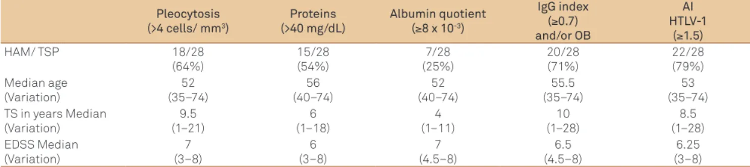
Signs of brain atrophy (prominence of cortical sulci and issures, and ventricular enlargement) occurred in four pa-tients, three of them with concomitant white matter lesions. One of them had pronounced atrophy in the convexity, par-ticularly in the frontal regions, as well in the cisternas and sylvian issures. Another patient had cerebellar atrophy asso-ciated with slight accentuation of cisternas. Regarding MRI of the cervical spinal cord, hyperintense lesions were found in three patients (11%). One of them had hyperintense signal with contrast enhancement. Spinal cord atrophy was found in only one case, characterized by thinning of the cervical spine and the proximal portion of the dorsal medulla (Fig 2).
A signiicant association was found between the pres-ence of cerebral white matter lesions and periventricular lo-cation with older patients, as well as between periventricular
location and absence of acute signs of inlammation in CSF (pleocytosis, protein concentration and dysfunction of blood-CSF barrier) (Tables 2 and 3). here was no associa-tion between other brain locaassocia-tions, duraassocia-tion of symptoms and disability (Table 2). No patients had brain gadolinium enhancement lesions. Spinal cord hyperintense lesions in the white substance of the cervical spine occurred in three paraplegic patients, all conined to a wheelchair: 1. MJRS, 66 years old, 18 years of disease, had acute inlammatory CSF (pleocytosis, high protein concentration and intrathecal syn-thesis of total IgG), and MRI revealed multiple foci of hyper-intensity on T2 difusely involving the cervical spine; 2. MRI, 40 years old, one year of illness, presented CSF pleocytosis with (17 cells/mm3) high protein concentration (84 mg/dl),
dysfunction of blood-CSF barrier (14.7x10⁻³) and intrathecal
synthesis of HTLV-1 antibodies (73.3), MRI image showed an oval hyperintense signal on STIR with contrast enhancement, measuring about 5 mm in diameter in the earlier portions of the cervical cord at C1 (Fig 2); 3. MDT, 54 years old, 24 years of disease, normal CSF examination, MRI showed hyperintense signal on T2 images in the C4-C5-C6-C7 associated with at-rophy of the cervical spinal cord. Only the patient 2 had gado-linium enhancement in the cervical spinal cord in MRI.
DISCUSSION
Changes in MRI of the brain and spinal cord have been found in patients with HAM/TSP10. In sense to clarify the
ori-gin of and the signiicance of these lesions, we studied the brain and spinal cord MRI of 28 HAM/TSP patients7. We
compared the MRI indings with clinical and CSF inding. In the whole patients group studied, 75% of subjects had hy-perintense lesions in brain white matter (50% periventricu-lar) and 11%, spinal cord. Indirect signals of atrophy were observed in 14% of MRI of the brain and only 3% of the spi-nal cord. he frequency of these changes has been variable in the literature: hyperintense lesions in brain (52.4% -84%) and in spinal cord (7.9%)16-20. Atrophy of the spinal cord
oc-curred in 20-74% of the cases. hese diferences may be due to individual characteristics of the population, such as age, Table 1. Association between CSF findings and clinical aspects of 28 HAM/TSP patients.
Pleocytosis
(>4 cells/ mm3)
Proteins (>40 mg/dL)
Albumin quotient (≥8 x 10⁻³)
IgG index (≥0.7) and/or OB
AI HTLV-1
(≥1.5)
HAM/ TSP 18/28
(64%)
15/28 (54%)
7/28 (25%)
20/28 (71%)
22/28 (79%) Median age
(Variation)
52 (35–74)
56 (40–74)
52 (40–74)
55.5 (35–74)
53 (35–74) TS in years Median
(Variation)
9.5 (1–21)
6 (1–18)
4 (1–11)
10 (1–28)
8.5 (1–28) EDSS Median
(Variation)
7 (3–8)
6 (3–8)
7 (4.5–8)
6.5 (4.5–8)
6.25 (3–8) TS:Time of symptoms; EDSS: Expanded Disability Status Scale; OB: Oligoclonal band; AI: Specific antibody index; HTLV-1: Human T-cell lymphotropic virus type 1; HAM/TSP: HTLV-1 associated myelopathy/tropical spastic paraparesis; CSF: cerebrospinal fluid.
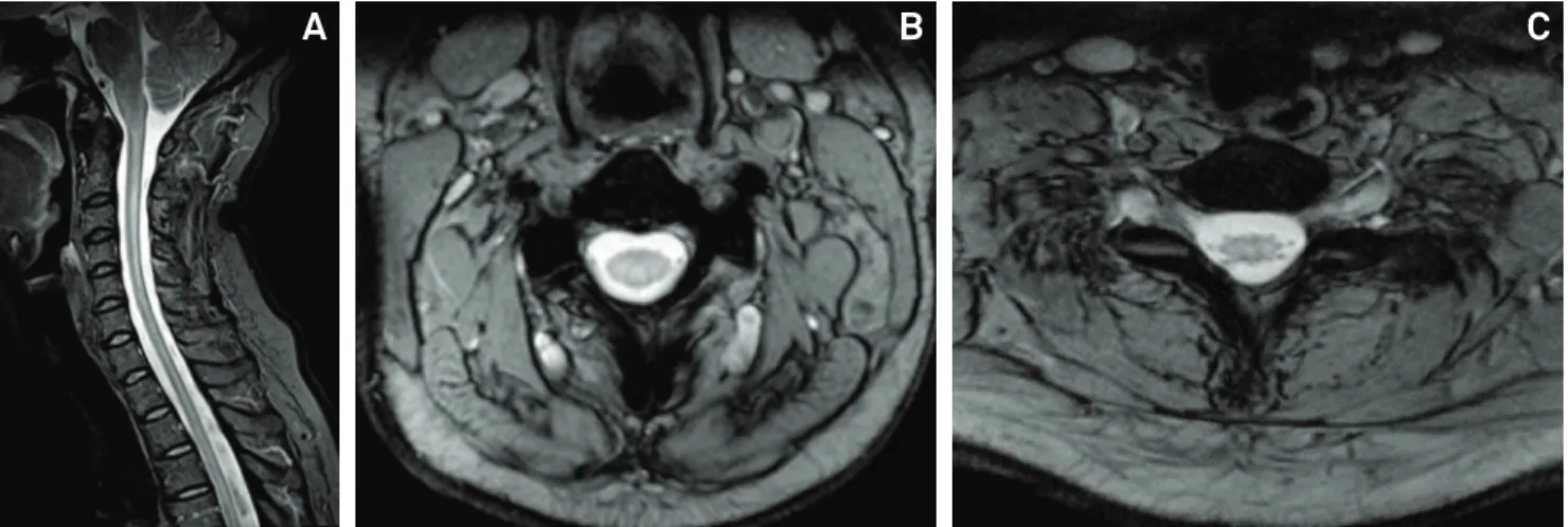
Fig 1. Brain MRI findings in HTLV-1 infection. (A) Axial T2-weighted image shows multiple foci of high signal intensity on the white matter, mainly in the periventricular region. (B) Axial FLAIR demonstrates a similar pattern of high signal intensity foci, predominating in the subcortical region. (C) Sagittal T1-weighted image shows no significant abnormalities. (D) Sagittal T1-weighted image demonstrates multiple foci of low signal intensity in the white matter.
D
A
C
sex, degree of neurological disability, degree of disease pro-gression and duration of symptoms. We observed association between the age and the frequency of T2 hyperintense brain MRI lesions, but not with disease duration, disability and CSF indings. Based on our data, we speculate whether the brain MRI T2 hyperintense lesions could be related with a micro-degenerative process associated to the age. Additionally, the presence of acute inlammatory CSF (pleocytosis, high con-centration of protein and blood-CSF barrier dysfunction) was found in patients with MRI T2 hyperintense cervical lesions. Interesting, this location is closest to the CSF space and may relect the cervical cord inlammatory lesion
Neurologic disability was severe in our population. All of them depended on support to walk or were conined to wheelchairs, with a median duration of symptoms of 9.5 years (1–28 years). Gessain and Gout reported that, about 10 years after onset of symptoms, 30% of patients were conined to bed and 45% needed assistance to walk21. here was
vari-ability of the clinical forms: rapidly progressive evolution to severe disability in six patients with <5 years of symptoms and chronic forms. he progression of acute symptoms could relect a marked inlammatory process that occurs in the spi-nal cord22. Despite the bimodal progression observed in our
population of HAM /TSP, there was no signiicant associa-tion between the degree of disability assessed by EDSS and duration of symptoms and indings of brain MRI. One out of six patients with rapidly evolving had enhancement lesion in MRI of the cervical spine and signs of acute inlammation in CSF. his is considered by some authors a malignant form, variant HAM/TSP. Anatomopathological indings appear to be associated with the duration of the myelopathy, in which shorter length of illness is associated with marked inlamma-tion in the spinal cord, whereas predominance of degenera-tion occurs in patients with more than nine years of illness. he coexistence of chronic and active lesions is described in the intermediate period of symptoms22.
he CSF examination revealed the inlammatory origin of this disease in 89% of patients, consistent with the litera-ture (95%)13,15. here was a higher frequency of acute signs of
inlammation characterized by pleocytosis (64%) compared to previous studies (26%)15. his is in agreement with the
clinical indings of severe outcome. In general, patients with signs of acute inlammation in the CSF had a shorter dura-tion of symptoms [median, 6.5 years (1-21 years)]. By deini-tion, it is speculated that, despite the chronic disease, could have occurred reactivation of inlammatory signs of disease activity. Similar to this situation, Iwasaki et al. reported ana-tomical and pathological changes of active inlammatory le-sions in the spinal cord in a case of HAM/TSP with 29 years of disease23.
Morgan et al. observed no diferences between the num-ber, size and location of lesions in patients with HAM/TSP and seropositive controls19. he authors found no correlation
between volume and number of lesions and clinical charac-teristics of patients, EDSS, viral load in peripheral blood and production of gamma-globulin. hese suggested that the high frequency of abnormalities in white matter in patients infect-ed with HTLV-1 is relatinfect-ed to the early presence of inlamma-tion of the central nervous system. However, as patients were matched by the same age, the high frequency of white matter lesions on MRI in both groups could be related to demyelina-tion secondary to degenerative microangiopathy. Kira et al. and Ogata et al. demonstrated, through MRI and autopsy, correlation between the periventricular white matter lesions and cerebral spinal posterior column24,25. he study revealed
similar pathologic changes to the brain and spinal cord, with demyelination, loss of axons and thickening of small vessels. he authors explained the absence of inlammatory iniltra-tion by the use of corticosteroids. However, autopsy studies show that inlammatory changes are perivascular iniltration, rarely occurred activated T lymphocytes, as well as damage in the parenchyma25. In this study, no signiicant associations
Fig 2. Cervical spinal cord MRI findings in HTLV-1 infection. (A) Sagittal STIR and (B) axial T2* GRE (at the level of C3) demonstrate extensive high signal intensity from C2 to the level of the dorsal spinal cord, predominating on the posterior region of the spinal cord, as well as mild spinal cord atrophy. (C) Axial T2* GRE (at the level of D3) shows the spinal cord atrophy and diffuse high signal intensity.
Variable White matter lesions
(T2) Median (range) p-value
AGE Pos (21) 56 (40–74) <0.05
Neg (7) 49 (35–55)
TS (yrs.) Pos (21) 8 (1–28) >0.05
Neg (7) 16 (4–21)
EDSS Pos (21) 7 (3–8) >0.05
Neg (7) 6.5 (3–7)
Variable Periventricular lesions
AGE Pos (14) 59 (40–74) <0.05
Neg (14) 49 (35–66)
TS (yrs.) Pos (14) 9.5 (1–28) >0.05
Neg (14) 10 (3–21)
EDSS Pos (14) 6.25 (3–8) >0.05
Neg (14) 6.5 (3–7)
Variable Subcortical lesions
AGE Pos (10) 50.5 (42–70) >0.05
Neg (18) 54.5 (35–74)
TS (yrs.) Pos (10) 6 (3–14) >0.05
Neg (18) 14 (1–28)
EDSS Pos (10) 6.5 (3–8) >0.05
Neg (18) 6.5 (6–8)
Variable Semioval centre
AGE Pos (9) 59 (40–74) >0.05
Neg (19) 52 (35–70)
TS (yrs.) Pos (9) 8 (1–18) >0.05
Neg (19) 13 (3–28)
EDSS Pos (9) 6.5 (3–8) >0.05
Neg (19) 6.5 (3–8)
Variable Corona radiata
AGE Pos (9) 57 (40–74) >0.05
Neg (19) 52 (35–70)
TS Pos (9) 8 (1–24) >0.05
Neg (19) 10 (3–28)
EDSS Pos (9) 7 (4.5–8) >0.05
Neg (19) 6 (3–8)
Table 2. Association between white matter lesions of the brain with clinical findings in 28 HAM/TSP patients.
TS:Time of symptoms, EDSS: Expanded Disability Status Scale; HAM/TSP: HTLV-1 associated myelopathy/tropical spastic paraparesis.
between the presence and location of white matter lesions on brain MRI and duration of illness or disability were found. Nevertheless, we identiied an association between older age and the indings of brain MRI, and the periventricular loca-tion and no signs of acute inlammaloca-tion in CSF.
Cerebral atrophy was observed in 14% of our patients with HAM/TSP. In the study by Griith et al., only 5% (1/19) patients with HAM/TSP had cerebral atrophy, showing that this is not frequent, although the authors suggest that
Findings on Brain MRI Presence of pleocytosis/
Hyperproteinorrachia/ Disf. Blood-CSF barrier
Absence of pleocytosis/
Hyperproteinorrachia/ Disf. Blood-CSF barrier p-value
White matter lesions 10 (67%) 6 (100%) >0.05
Periventricular lesions 4 (27%) 6 (100%) <0.05
Subcortical lesions 6(40%) 0 >0.05
Semioval centre 5(33%) 1(17%) >0.05
Corona radiata 5 (33%) 1 (17%) >0.05
Table 3. Association between presence and location of brain white matter lesions of brain and active inflammatory CSF findings in 28 HAM/TSP patients.
HAM/TSP: HTLV-1 associated myelopathy/tropical spastic paraparesis; CSF: cerebrospinal fluid.
the mechanisms of brain atrophy are the same responsible for spinal cord atrophy secondary to degeneration of the parenchyma26.
Cervilla et al. did not show hyperintense lesions in the spinal cord of patients with HAM/TSP, but 87% had atrophy of the dorsal segment18. he median range of age of the
pa-tients consisted of 56 years (26-73), and the evolution time was 11.6 years (3-33). In the study by Bagnato et al., 14% of pa-tients had hyperintensity on spinal cord images16. Moreover,
Umehara et al. studied 11 patients with HAM/TSP with le-sions in the spinal cord10. he variation in the duration of the
disease consisted of three to seven months, 72% had pleocy-tosis and high protein concentration. MRI showed changes in the cervical, thoracic and cervico-thoracic, characterized by swelling and T2 hyperintense lesions in the posterior or lat-eral columns. herefore, the authors suggest the existence of a variant of HAM/TSP and also that the changes observed on MRI would be the relection of an early and active inlamma-tion in the spinal cord27,28. In our series, three patients (15%)
had demyelinating lesions in cervical cord, all with the same degree of disability, paraplegic, conined to a wheelchair, but with diferences in duration of illness, the CSF proile and the characteristics of image. In a patient with 18 years of symp-toms, CSF examination showed signs of inlammatory activ-ity associated with local immune reaction and multiple foci of demyelination difusely involving the cervical spine. his is consistent with a pathologic inding of HAM/TSP, in which signs of activity appear throughout disease progression22.
he other patient had only one year of illness, CSF examina-tion showed signs of inlammatory activity, as well as MRI, showing a single image with hyperintense signal on STIR and contrast enhancement in the cervical spinal cord, consis-tent with the indings of early active lesions of HAM/TSP10.
Finally, the third patient, with 24 years of disease, normal CSF, showed atrophy of the cervical and thoracic cord associated with hyperintense signal on T2 image at C4-C5-C6-C7 com-patible with the chronic form of HAM/TSP22. An interesting
aspect is that, although the population studied was found with advanced disease, there was only one case of spinal cord atrophy.
reported lesions are similar to those found in degenerative microangiopathy. In this study, consistent with the literature, there was no association between the presence of cerebral white matter lesions with sex, disease duration and degree of neurological disability of patients with HAM/TSP. he pro-ile of the CSF, similar to other published studies, showed an inlammatory pattern in the majority of the patients. Spinal cord lesions were associated with active inlammation in the
CSF. Moreover, signs of active and acute inlammation (lin-fomonuclear pleocytosis, high protein concentration and dysfunction of the blood-CSF) were associated with more severe form of HAM/TSP and shorter duration of disease. However, the presence of acute inlammatory CSF indings in patients with longer time of symptoms suggests the per-sistence of active disease.
1. Poiesz BJ, Ruscetti FW, Gazdar AF, Bunn PA, Minna JD, Gallo RC. Detection and isolation of type-C retroviris particles from fresh an cultured lymhocytes of a patient with cutaneous T-cell lymphoma. Proc Natl Acad Sci 1980,77:7415-7419.
2. Proietti FA, Carneiro-Proietti AB, Catalan-Soares BC, Murphy EL. Global epidemiology of HTLV-I infection and associated diseases. Oncogene 2005,24:6058-6068.
3. Edlich RF, Arnette JA, Williams FM. Global epidemic of human T-cell lymphotropic virus type I (HTLV-I). J Emerg Med 2000;18:109-119.
4. Mueller N, Okayama A, Stuver S, Tachibana N. Findings from the Miyazaki Cohort Study. J Acquir Immune Defic Syndr Hum Retrovirol 1996;13(Suppl 1):S2-S7.
5. Kitagawa T, Fujishita M, Taguchi H, Miyoshi I, Tadokoro H. Antibodies to HTLV-I in Japanese immigrants in Brazil. JAMA 1986;256:2342.
6. Gessain A, Vernant JC, Maurs L, et al. Antibodies to human T-lymphotropic virus type–I in patients with tropical spastic paraparesis. The Lancet 1985;24:407-410.
7. Osame M. Review of WHO Kagoshima meeting and diagnostic guidelines for HAM/TSP. In: Blattner WA (Ed). Human retrovirology: HTLV. New York: Raven Press 1990;191-197.
8. Reiber H, Felgenhauer K. The diagnostic significance of antibody specificity indices in multiple sclerosis and herpes virus induced diseases of the nervous system. Clin Investig 1992;70:28-37.
9. Puccioni-Sohler M, Yamano Y, Rios M, et al. Differentiation of HAM/TSP from patients with multiple sclerosis infected with HTLV-1. Neurology 2007;68:206-213.
10. Umehara F, Nose H, Saito M, et al. Abnormalities of spinal magnetic resonance images implicate clinical variability in human T-cell lymphotropic vírus type I – associated myelopathy. J NeuroVirol 2007;13:260-267.
11. Godoy AJ, Kira J, Hasuo K, Goto I. Characterization of cerebral white matter lesions of HTLV-I-associated myelopathy/tropical spastic paraparesis in comparison with multiple sclerosis and collagen-vasculitis: a semiquantitative MRI study. J Neurol Sci 1995;133: 102-111.
12. Kurtzke JF, Beebe GW, Nagler B, Kurland LT, Auth TL. Studies on the natural history of multiple sclerosis--8. Early prognostic features of the later course of the illness. J Chron Dis 1977;30:819-830.
13. Link H, Cruz M, Gessain A, Gout O, Thé G, Kam-Hansen S. Chronic progressive myelopathy associated with HTLV-I: oligoclonal IgG and anti-HTLV-1 IgG antibodies in cerebrospinal fluid and serum. Neurololgy 1989;39:1566-1572.
14. Reiber H, Felgenhauer K. Protein transfer at the blood cerebrospinal fluid barrier and the quantification of the humoral immune response within the central nervous system. Clin Chim Acta 1987;163:319-328.
15. Puccioni-Sohler M, Rios M, Carvalho S, et al. Diagnosis of HAM/ TSP based on CSF proviral HTLV-I DNA and HTLV-I antibody index. Neurology 2001;57:725-727.
16. Bagnato F, Butman JA, Mora CA, et al. Conventional magnetic resonance imaging features in patients with tropical spastic paraparesis. J Neurovirol 2005;11:525-534.
17. Kuroda Y, Matsui M, Yukitake M, et al. Assessment of MRI criteria for MS in Japanese MS and HAM/TSP. Neurology 1995;45:30-33.
18. Cervilla J, Cartier L, García L. [Brain and spinal cord magnetic resonance imaging in spastic paraparesis associated to human T-lymphotropic virus]. Rev Med Chil 2006;134:1010-1018.
19. Morgan DJ, Caskey MF, Abbehusen C, et al. Brain magnetic resonance imaging white matter lesions are frequent in HTLV-I carriers and do not discriminate from HAM/ TSP. AIDS Res Human Retroviruses 2007;23:1499-1503.
20. Yukitake M, Takase Y, Nanri Y, et al. Incidence and clinical significances of human T-cell lymphotropic virus type I-associated myelopathy with T2 hyperintensity on spinal magnetic resonance images. Intern Med 2008;47:1881-1886.
21. Gessain A, Gout O. Chronic myelopathy associated with human T-lymphotropic virus type I (HTLV-I). Ann Intern Med 1992;117:933-946.
22. Iwasaki Y. Pathology of chronic myelopathy associated with HTLV-I infection (HAM/TSP). J Neurol Sci 1990;96:103-123.
23. Iwasaki Y, Sawada K, Aiba I, et al. Widespread active inflammatory lesions in a case of HTLV-I-associated myelopathy lasting 29 years. Acta Neuropathol 2004;108:546-551.
24. Kira J, Fujihara K, Itoyama Y, Goto I, Hasuo K. Leukoencephalopathy in HTLV-I-associated myelopathy/tropical spastic paraparesis: MRI analysis and a two year follow-up study after corticosteroid therapy. J Neurol Sci 1991;106:41-49.
25. Ogata A, Nagashima K, Tashiro K, Miyakawa A, Mikuni C. MRI-pathological correlate of brain lesions in a necropsy case of HTLV-I associated myelopathy. J Neurol Neurosurg Psychiatry 1993;56:194-196.
26. Aye MM, Matsuoka E, Moritoyo T, et al. Histopathological analysis of four autopsy cases of HTLV-I-associated myelopathy/tropical spastic paraparesis: inflammatory changes occur simultaneously in the entire central nervous system. Acta Neuropathol 2000;100:245-252.
27. Griffith C, Bagnato F, Gupta S, et al. Brain volume measurements in patients with human T- cell lymphotropic virus-1-associated tropical spastic paraparesis. J Neurovirol 2006;12:349-355.
28. Yamamoto F, Yamashita S, Yamamura A, et al. Abnormal spinal MRI findings in human T-cell lymphotropic virus type I-associated myelopathy. Clin Neurol Neurosurg 2009;111:624-628.
29. Izumo S. Neuropathology of HTLV-1-associated myelopathy (HAM/ TSP). Neuropathology 2010;30:480-485.