DOI: 10.1590/0004-282X20150097
ARTICLE
Effects of aging on nitrergic neurons in human
striatum and subthalamic nucleus
Efeitos do envelhecimento sobre os neurônios nitrérgicos no estriado e núcleo
subtalâmico humano
Bruno Lopes dos Santos-Lobato1,4, Elaine Aparecida Del-Bel1,2,4, José Eymard Homem Pittella3, Vitor Tumas1,4
Basal ganglia (BG) are complex structures responsible for motor control, motor learning, executive functions, be-haviors and emotion1. his system is regulated by many neu
-rotransmitters, including nitric oxide (NO), a gaseous
mol-ecule recently reported to be responsible for many essential physiologic processes, including signaling in central nervous
system2. NO is generated by neuronal NO synthase (nNOS) in
neurons. After its synthesis in neuronal cells, NO difuses to
neighboring cells and binds to soluble guanylate cyclase, trig
-gering signaling pathways which modulate synaptic activity
and other function3,4. Nitrergic neurons are present in many
regions of central nervous system, particularly in BG.
Striatum and subthalamic nucleus (STN) are nuclei which contain high densities of nitrergic cells. Striatum is the main input nucleus of BG and receives topographical excitatory
projections from almost the entire cerebral cortex. A
specif-ic class of striatal GABAergspecif-ic aspiny interneurons synthesiz
-es NO5. STN is another crucial nucleus for indirect pathway
of BG, and also receives cortical inputs through hyperdirect pathway. Messenger RNA of nNOS was detected in more than
95% of subthalamic neurons6.
Presence of nNOS-expressing neurons in BG suggests ni
-trergic neurotransmitter system has a role on regulation of functions processed by these nuclei.
1Universidade de São Paulo, Faculdade de Medicina de Ribeirão Preto, Departamento de Neurociências e Ciências do Comportamento, Ribeirao Preto SP, Brazil; 2Universidade de São Paulo, Faculdade de Odontologia de Ribeirão Preto, Departamento de Fisiologia, Ribeirao Preto SP, Brazil;
3Universidade de São Paulo, Faculdade de Medicina de Ribeirão Preto, Departamento de Patologia, Ribeirao Preto SP, Brazil; 4Universidade de São Paulo, Núcleo de Apoio à Pesquisa em Neurociência Aplicada, Ribeirão Preto SP, Brazil.
Correspondence: Vitor Tumas; FMRP/USP, Departamento de Neurociências e Ciências do Comportamento; Av. Bandeirantes, 3900; 14049-900 Ribeirão Preto SP, Brasil; E-mail: [email protected]
Conflict of interest: There is no conlict of interest to declare.
Support: Conselho Nacional de Desenvolvimento Cientíico e Tecnológico.
Received 08 January 2015; Received in inal form 04 April 2015; Accepted 24 April 2015. ABSTRACT
Nitric oxide (NO) is a major neurotransmitter associated with motor control in basal ganglia. Movement disorders, as essential tremor and Parkinson’s disease, are more prevalent on aged individuals. We investigated the effects of aging on neuronal density and diameter/area of nitrergic neurons in samples of striatum (caudate and putamen) and subthalamic nucleus of 20 human brains from normal subjects, stained by histochemistry for NADPH-diaphorase and immunohistochemistry for neuronal NO synthase. Our data showed aging does not modify the neuronal density and size of nitrergic neurons in striatum and subthalamic nucleus. These indings suggest a lack of association between aging and morphologic changes on nitrergic neurons.
Keywords: nitric oxide, basal ganglia, striatum, subthalamic nucleus, aging.
RESUMO
O óxido nítrico (NO) é um importante neurotransmissor associado ao controle motor nos núcleos da base. Os distúrbios de movimento, como tremor essencial e a doença de Parkinson, são mais prevalentes em indivíduos idosos. Nós investigamos os efeitos do envelhecimento sobre a densidade neuronal e diâmetro/área dos neurônios nitrérgicos em amostras de estriado (caudado e putâmen) e núcleo subtalâmico de 20 encéfalos humanos de indivíduos normais, corados pela técnica histoquímica da NADPH-diaforase e imunohistoquímica para a sintase do NO neuronal. Nossos resultados mostraram que o envelhecimento não modiica a densidade neuronal e as dimensões dos neurônios nitrérgicos no estriado e núcleo subtalâmico. Estes achados sugerem uma falta de associação entre envelhecimento e mudanças morfológicas nos neurônios nitrérgicos.
Aging brain is related to morphological and functional
changes, however, efects of aging on nitrergic neurons of the BG are not extensively studied. In this study, our objective
was to analyze inluence of aging on density and size of ni
-trergic neurons in human striatum and STN.
METHOD
Subjects and sample collection
Post-mortem brain samples were obtained from twenty adult individuals (i.e., ten individuals under 60 years of age and ten individuals over 60 years of age) who exhibited no
clinical or pathological evidence of neurological or psychi
-atric disorders, brain death or HIV infection. Demographic
and clinical data from subjects, such as age, gender, pre-vious diseases, medications, agonic state before death,
death-sample ixation interval (i.e., post-mortem interval),
sample ixation-sample cryoprotection interval, and sam
-ple cryoprotection-sam-ple freezing interval, were record
-ed. Tissue was collected at autopsy and was free of signii
-cant macroscopic pathological alterations, as indicated by hematoxylin-eosin and Nissl staining. Brains were cut into slices with a thickness of 0.5 cm and ixed in a solution of 4% paraformaldehyde in 0.1 M phosphate bufer (PB) (pH 7.4) for 48 hours at 4°C. Slices were immersed in 30% sucrose in PB at 4°C until sedimentation, frozen with isopentane and dry ice, and stored at -80°C. Samples from striatum and STN were cut with a cryostat to generate sections with a thickness of 30
μm. Sections were serially collected in a cryoprotective solu
-tion until staining.
he study was approved by the Ribeirão Preto Medical School Ethic Committee, and tissue was kindly provided by Department of Pathology of Ribeirão Preto Medical School.
NADPH-diaphorase histochemistry
Sections of striatum were processed to visualize NADPH-diaphorase (NADPHd) enzymatic activity, which
correlates with presence of nNOS in neurons, using a pro
-tocol adapted from Vincent and Kimura7,8. Free-loating
sections were rinsed for 30 minutes in PB and
subsequent-ly incubated in a staining solution containing 0.1 M PB (pH
8.0) with 0.3% Triton X-100, 1 mg/ml of β-NADPH (Sigma,
Saint Louis, Missouri, U.S.A.) and 0.1 mg/ml of Nitroblue Tetrazolium (NBT) (Sigma). Incubation was performed in the dark at 37°C for 180 minutes. After incubation, sections were rinsed thoroughly in PB, mounted onto gelatin-coated slides,
and air-dried overnight. Sections were dehydrated with dif
-ferent grades of alcohol, and covered with Entellan® mount
-ing medium (MERK, Darmstadt, Germany).
Immunohistochemistry
NADPHd technique for STN sections showed poor staining, and use of immunohistochemistry was chosen to
label nNOS, with a better signal-noise ratio. Following an
-tigenic recovery and blocking with bovine serum albumin 2% (BSA – NGS; Jackson Immuno Research, U.S.A.), free-loating sections were incubated overnight with a 1:15,000 dilution of a sheep anti-nNOS antibody (kindly donated by Prof. Dr. Piers C. Emson, Babraham Institute, Cambridge,
U.K.). To detect primary antibody, it was used rabbit an
-ti-sheep biotinylated secondary antibody, at a dilution of 1:400 (Vector Laboratories; Burlingame, California, U.S.A.), incubated for 90 minutes. Secondary antibody was used in
combination with an avidin-biotin-peroxidase complex for
120 minutes (Vector Laboratories) and 3,3’-diaminoben
-zidine for 10 minutes (Sigma). NADPHd staining was not performed on the same sections labeled by nNOS or NeuN immunohistochemistry.
Analysis of striatum and subthalamic nucleus
Striatum (i.e., caudate and putamen) was subdivided into
10 levels along the anteroposterior axis, from the most ante-rior portion of caudate to the most posteante-rior portion of puta-men, using the following coordinates, with anterior
commis-sure as point zero11: section 1, -20.0 mm; section 2, -15.0 mm;
section 3, -10.0 mm; section 4, -5.8 mm; section 5, -2.0 mm; section 6, +2.0 mm; section 7, +9.3 mm; section 8, +14.6 mm; section 9, +19.9 mm; and section 10, +25.2 mm. Caudate and
putamen were also subdivided, using anterior commissure as a landmark, into three (i.e., precommisural head,
postcom-missural head and body of caudate) and two territories (i.e.,
precommissural and postcommissural putamen)
respective-ly, as described previously5.
STN was subdivided into 3 anteroposterior levels at
specific coordinates9: anterior: at the level of mammilla
-ry bodies, +13.3 mm; middle: next to SN pars compacta, +16.0 mm; and posterior: at the level of red nucleus, +18.6
mm) and into three functional territories (i.e.,
sensorimo-tor, associative and limbic), based on method proposed by
Parent and Hazrati10.
We examined sections with an Axiophot II microscope
(Carl Zeiss, Jena, Germany) equipped with a 3.0 mega
-pixel Axiocam MRc digital camera and a microcomput
-er with AxioVison LE software v-ersion 4.8.1.0 (Carl Zeiss
MicroImaging GmbH, Oberkochen, Germany). Whole sec
-tion image was scanned at high resolu-tion and 100x
magni-ication using “MosaiX” module of AxioVison LE software.
Final rendering of each drawing was generated with Adobe
Photoshop CS5 64-bit for Windows (Adobe Systems, San Jose, California, U.S.A.). Images were saved on a computer in TIFF format.
Measurement of neuronal density and morphometric parameters
Neuronal counts were performed manually, using a vir
-tual grid (i.e., squares with an area of 1 mm2), which was
of Health, U.S.A.), on image ile. Grid squares were analyzed
alternately, by a scanning method11. Counting was performed
by a single investigator blinded to clinical data. Only neurons
with clear boundaries and recognizable neuronal morphol
-ogy were included. Neuronal density was deined as mean
number of labeled neurons per square of a region or nucleus,
and this value was represented as cell/mm2. Some nitrergic
neurons from striatum and STN were selected for measure
-ment of area of soma (μm2) and maximum diameter of soma
(μm). Approximately 40 neurons per brain in striatum and 24
neurons per brain in STN were analyzed for these morpho
-metric parameters.
Statistics
All values were described as mean ± SEM. T-test or Mann-Whitney test was used to compare two independent
samples. Pearson’s correlation coeicients were used to veri
-fy association between age and neuronal density. Diferences were considered signiicant when p < 0.05.
RESULTS
NADPHd histochemical staining and immunohistochem
-ical staining resulted in reliably stained nitrergic neurons in
all studied brains. Pre- and post-mortem factors (i.e., agonic
state before death and post-mortem intervals) did not inlu
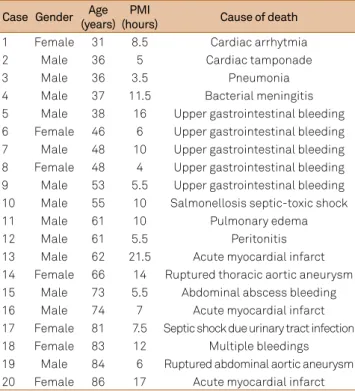
-ence nitrergic neuronal counts (backwise method; general linear model with all factors: p = 0.309). Mean post-mortem interval was 9.3 hours (Table).
In striatum, there was no correlation between age and ni
-trergic neuronal density in caudate or putamen, and no dif
-ference on area/diameter of soma between > 60 years-old and < 60 years-old subjects (Figure 1).
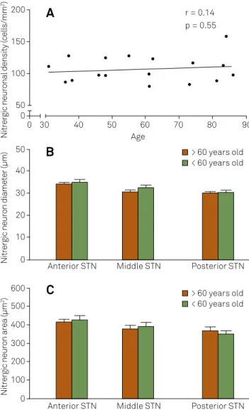
In STN, there was no correlation between age and nitrergic neuronal density and no diference on area/diameter of soma between > 60 years-old and < 60 years-old subjects (Figure 2).
Table. Clinical data of patients. The mean PMI was 9.3 hours.
Case Gender Age (years)
PMI
(hours) Cause of death
1 Female 31 8.5 Cardiac arrhytmia
2 Male 36 5 Cardiac tamponade
3 Male 36 3.5 Pneumonia
4 Male 37 11.5 Bacterial meningitis
5 Male 38 16 Upper gastrointestinal bleeding 6 Female 46 6 Upper gastrointestinal bleeding 7 Male 48 10 Upper gastrointestinal bleeding 8 Female 48 4 Upper gastrointestinal bleeding 9 Male 53 5.5 Upper gastrointestinal bleeding 10 Male 55 10 Salmonellosis septic-toxic shock
11 Male 61 10 Pulmonary edema
12 Male 61 5.5 Peritonitis
13 Male 62 21.5 Acute myocardial infarct 14 Female 66 14 Ruptured thoracic aortic aneurysm 15 Male 73 5.5 Abdominal abscess bleeding 16 Male 74 7 Acute myocardial infarct 17 Female 81 7.5 Septic shock due urinary tract infection
18 Female 83 12 Multiple bleedings
19 Male 84 6 Ruptured abdominal aortic aneurysm 20 Female 86 17 Acute myocardial infarct PMI: post-mortem interval.
Body CN: body of caudate; Prec Head CN: precommissural head of caudate; Prec Put: precommissural putamen; Postc Head CN: postcommissural head of caudate; Postc Put: postcommissural putamen; PV Put: posteroventral putamen.
Figure 1. Effects of aging on neuronal density and morphology in striatum. Regression analysis between nitrergic neuronal density and age (in years) in caudate nucleus (A) and putamen (B). Histograms of nitrergic neuron area (C) and diameter (D) found within territories of striatum in young adults and aged individuals. t = p between 0.05 and 0.1.
Nitrergic neuronal density
(cells/mm
2)
Nitrergic neuronal density
(cells/mm
2)
Age
t
r = -0.07 p = 0.74
> 60 years old < 60 years old 20
15
10
5
0 0
Nitrergic neuron area (µm
2) 400 350
50 100 150 200 300 250
0
40 30
Prec Head CN Postc Head CN Body CN Prec Put Postc Put
t
Prec Head CN Postc Head CN Body CN Prec Put Postc Put
60
50 70 80 90
Age
r = 0.05 p = 0.82 20
15
10
5
0
0 30 40 50 60 70 80 90
Nitrergic neuron diameter (µm)
300
250
50 100 150 200
0
> 60 years old < 60 years old
A
C
B
DISCUSSION
Our study did not show any efect of aging on density and morphology of nitrergic neurons in human striatum and
STN. Although a previous study had shown changes in neuro
-nal density of the striatum and STN of aged rats, a work with
human brains did not show any signiicant change on mor
-phometric parameters with aging in striatum11,12.
Aging is associated with classic morphological changes
on BG. Striatum and substantia nigra show reduction on its
volumes, but a noteworthy loss of neurons is only seen on sub
-stantia nigra13. Age-related progressive atrophy on striatum is
attributed to a combination of neuronal loss and reduction of
neuronal area. here are few data about role of aging on STN,
globus pallidus and pedunculopontine tegmental nucleus.
here are some evidences suggesting nitrergic neurons in human striatum may be less vulnerable to neurodegenerative process of Huntington’s disease. Neuropathology of this disease
shows a massive loss of medium spiny neurons and striatal at
-rophy which spares nNOS-expressing neurons14. Our results
in-dicate nitrergic neurons may be resistant also to aging process.
Our study had some limitations. Regarding to counting is
-sues, a two-dimensional method was chosen for cell
count-ing, instead of a three-dimensional stereological and “unbi
-ased” method, which could bring some doubt to validity of results. his option was done due the large volume of human striatum, high number of neurons counted, low variability in
nitrergic neuron size and homogeneous pattern for neuro
-nal distribution in striatum and STN. In these settings, both
two-dimensional and three-dimensional cell-counting are
ca-pable of providing estimates of neuronal density of cells in brain
and each approach has its own strengths, weaknesses and bi-ases14,15. herefore, we must interpret these results carefully be
-fore assume a lack of causality between the aging process and
morphologic changes on nitrergic neurons. Considering
pos-sible biases of a two-dimensional cell analysis, these data must be conirmed also by a three-dimensional-based study.
Furthermore, this study did not investigate subtle neuronal
morphology (as the length and complexity of dendritic branch
-ing), recently associated with the process of brain aging16.
In conclusion, our results suggest aging does not change
density and diameter/area of nitrergic neurons in human stri
-atum and STN. Further works are needed to conirm absence of association between aging and nNOS-expressing neuronal
density and size. hese data are essential for a wider knowl
-edge of BG circuitry, as well the modulation mediated by NO in striatum and STN and possible therapeutic interventions
on neurological and psychiatric diseases involving the nitrer
-gic neurotransmitter system.
ACKNOWLEDGMENTS
We would like to thank the Applied and Experimental Neurology Laboratory of Ribeirão Preto Medical School for
image processing, to Mrs. Célia Aparecida da Silva and au
-topsy technicians for technical support.
References
1. Lanciego JL, Luquin N, Obeso JA, Functional neuroanatomy of the basal ganglia. Cold Spring Harb Perspect Med. 2012;2(12):a009621. doi:10.1101/cshperspect.a009621
2. Garthwaite J, Charles SL, Chess-Williams R. Endothelium-derived relaxing factor release on activation of NMDA receptors suggests role as intercellular messenger in the brain. Nature. 1988;336(6197):385-8. doi:10.1038/336385a0
STN: subthalamic nucleus.
Figure 2.Effects of aging on neuronal density and morphology in subthalamic nucleus. Regression analysis between nitrergic neuronal density and age (in years) in subthalamic nucleus (A). Histograms of nitrergic neuron diameter (B) and area (C) found within territories of subthalamic nucleus in young adults and aged individuals.
Nitrergic neuronal density (cells/mm
2)
Age
r = 0.14 p = 0.55
> 60 years old < 60 years old 200
150
100
50 0 0
Nitrergic neuron area (µm
2)
600
200 100 300 400 500
0
40 30
Anterior STN Middle STN Posterior STN 60
50 70 80 90
> 60 years old < 60 years old
Nitrergic neuron diameter (µm)
50
20 10 30 40
0
Anterior STN Middle STN Posterior STN
A
B
3. Benarroch EE. Nitric oxide: a pleiotropic signal in the nervous system. Neurology. 2011;77(16):1568-76. doi:10.1212/WNL.0b013e318233b3e4
4. Del-Bel E, Padovan-Neto FE, Raisman-Vozari R, Lazzarini M. Role of nitric oxide in motor control: implications for Parkinson’s disease pathophysiology and treatment. Curr Pharm Des. 2011;17(5):471-88. doi:10.2174/138161211795164176
5. Bernácer J, Prensa L, Giménez-Amaya JM. Morphological features, distribution and compartmental organization of the nicotinamide adenine dinucleotide phosphate reduced-diaphorase interneurons in the human striatum. J Comp Neurol. 2005;489(3):311-27. doi:10.1002/cne.20616
6. Nisbet AP, Foster OJ, Kingsbury A, Lees AJ, Marsden CD. Nitric oxide synthase mRNA expression in human subthalamic nucleus, striatum and globus pallidus: implications for basal ganglia function. Brain Res Mol Brain Res. 1994;22(1-4):329-32. doi:10.1016/0169-328X(94)90062-0
7. Matsumoto T, Nakane M, Pollock JS, Kuk JE, Förstermann U. A correlation between soluble brain nitric oxide synthase and NADPH-diaphorase activity is only seen after exposure of the tissue to ixative. Neurosci Lett. 1993;155(1):61-4. doi:10.1016/0304-3940(93)90673-9
8. Vincent SR, Kimura H. Histochemical mapping of nitric oxide synthase in the rat brain. Neuroscience. 1992;46(4):755-84. doi:10.1016/0306-4522(92)90184-4
9. Mai K, Assheuer J, Paxinos G. Atlas of the human brain. London: Academic Press; 2004.
10. Parent A, Hazrati LN. Functional anatomy of the basal ganglia. I. The cortico-basal ganglia-thalamo-cortical loop. Brain Res Brain Res Rev. 1995;20(1):91-127. doi:10.1016/0165-0173(94)00007-C
11. Selden N, Geula C, Hersh L, Mesulam MM. Human striatum: chemoarchitecture of the caudate nucleus, putamen and ventral striatum in health and Alzheimer’s disease. Neuroscience. 1994;60(3):621-36. doi:10.1016/0306-4522(94)90491-X
12. Cha CI, Sohn SG, Chung YH, Shin C, Baik SH. Region-speciic changes of NOS-IR cells in the basal ganglia of the aged rat. Brain Res. 2000;854(1-2):239-44. doi:10.1016/S0006-8993(99)02279-9
13. Fearnley JM, Lees AJ. Ageing and Parkinson’s disease: substantia nigra regional selectivity. Brain. 1991;114(5):2283-301.
doi:10.1093/brain/114.5.2283
14. Ferrante RJ, Kowall NW, Beal MF, Richardson EP Jr, Bird ED, Martin JB. Selective sparing of a class of striatal neurons in Huntington’s disease. Science. 1985;230(4725):561-3. doi:10.1126/science.2931802
15. Matricon J, Bellon A, Frieling H, Kebir O, Le Pen G, Beuvon F et al. Neuropathological and Reelin deiciencies in the hippocampal formation of rats exposed to MAM: differences and similarities with schizophrenia. PloS One. 2010;5(4):e10291. doi:10.1371/journal.pone.0010291