Increased Intima-Media Thickness is Independently Associated with
Ischemic Stroke
Dário Freitas
1, Ana Alves
1, Alexandre Pereira
1, Telmo Pereira
1, 2Escola Superior de Saúde Dr. Lopes Dias do Instituto Politécnico de Castelo Branco - Curso de Licenciatura em Cardiopneumologia1, Escola Superior de Tecnologia da Saúde de Coimbra do Instituto Politécnico de Coimbra - Curso de Licenciatura em Cardiopneumologia2 - Portugal.
Abstract
Background: Stroke is one of the major causes of death worldwide. The importance of increased intima-media thickness in cardiovascular risk stratification has been recurrently studied. The relationship between them, however, is still controversial.
Objectives: To determine whether increased common carotid artery (CCA) intima-media thickness can be used as an independent high-risk marker for the occurrence of stroke.
Methods: This study sample comprised 948 patients consecutively assessed by use of cervical triplex scan from January 2004 to June 2009. Those patients were divided into groups according to the presence or absence of recent stroke as follows: a group of patients with ischemic stroke (n = 452, 48%); a group of patients with hemorrhagic stroke (n = 22, 2%); and a group of patients with no events (n = 474, 50%).
Results: On logistic regression analysis adjusted for the classic cardiovascular risk factors, increased CCA intima-media thickness associated significantly and on an approximately linear way with ischemic stroke (Odds Ratio = 1.808, confidence interval: 1.291–2.534, p = 0.01), but not with hemorrhagic stroke (p = ns). A significant interaction with age was also found, showing a greater discriminative capacity for the risk of ischemic stroke in individuals aged less than 50 years.
Conclusions: The increased CCA intima-media thickness was identified as an independent predictor of the risk for ischemic stroke, but not for hemorrhagic stroke, emphasizing the usefulness of its assessment on clinical practice. (Arq Bras Cardiol 2012;98(6):497-504)
Keywords: stroke; increased intima-media thickness; common carotid artery; ultrasound; risk factors.
Mailing Address: Dário Luís Leal de Freitas • Rua Dr. Mariano Roque Laia, nº 2, Rés-do-Chão A. Postal Code 2650-051, Amadora - Portugal. E-mail: [email protected]
Manuscript received September 10, 2011; manuscript revised September 28, 2011; accepted January 01, 2012.
concluded that the use of other intima-media measurements, namely in the common carotid artery (CCA), bears a stronger correlation with the occurrence of stroke11-14.
Ultrasonography, especially the cervical triplex scan, is the most effective and used non-invasive method to assess the presence of intima-media thickening. That method provides information about arterial wall changes, and progression and regression of extracranial carotid lesions15-21. On the ultrasound
assessment of the intima-media thickness, according to Stein et als., the stratification of values should be always adjusted for the patient’s age, gender and race17. According to the
authors of the EDUCATE Study, regarding the occurrence of vascular events, an excellent clinical prognosis is associated with a negative carotid ultrasound study22.
This investigation study was aimed at assessing the discriminative capacity of the increased CCA intima-media thickness for the risk of stroke.
Methods
Data included in the sample refer to all patients undergoing cervical triplex scan between January 1st, 2004, and June 30,
2009. The methodology of data collection was structured
Introduction
Currently, one of the major consequences of the atherosclerotic disease is stroke, which is one of the major causes of death in the adult population in Portugal and Brazil, and one of the major causes of disability in industrialized countries worldwide1-3. Stroke is an important
public health problem, requiring urgently the early clinical diagnosis of its manifestations4-7. The relationship between
stroke and increased intima-media thickness has been widely discussed in the scientific community; however, the value of that parameter and, consequently, its clinical usefulness are still controversial.
and objective, and a table was created for the records. Data collection was retrospective, by use of direct consultation of the database, and the target population comprised patients with no events, patients diagnosed with hemorrhagic stroke and patients diagnosed with ischemic stroke. In the “no event” group, ultrasonography was performed preventively, while, in the groups with events, ultrasonography was performed close to the event time. The “no event” group comprised individuals with the following characteristics: clinical information regarding the assessment of extracranial circulation, syncope, dizziness, ataxia, headaches, dementia, vertebrobasilar insufficiency, and tinnitus. The ischemic stroke group comprised those with clinical information regarding ischemic stroke and cerebellar stroke. The hemorrhagic stroke group comprised those with clinical information regarding hemorrhagic stroke and subarachnoid hemorrhage.
Patients with the following characteristics were excluded: insufficient clinical data; multiple strokes; reassessment after stroke; and incomplete reports due to lack of patient collaboration.
All patients underwent cervical triplex scan on an Aloka 5500 ultrasound equipment, with a 7.5 MHz linear probe®
(Siemens Healthcare®, Germany), according to adequate
procedures defined by the manufacturer. The measurements were always taken by the same examiner on the distal CCA at approximately 20 mm from the carotid bulb, the intima-media thickness being defined as the maximum distance between the lumen-intima interface and the media-adventitia interface. The intima-media thickness was the mean value obtained from the six measurement points.
The quantitative variables studied were age and the right and left CCA intima-media thickness. The qualitative variables studied were gender, clinical data (“no event”, ischemic stroke, hemorrhagic stroke), and the presence of risk factors for atherosclerotic disease (cardiac disease, hypercholesterolemia, arterial hypertension, type 2 diabetes mellitus, smoking , and increased CCA intima-media thickness). It is worth noting that of the mentioned risk factors for atherosclerotic disease, the existence of cardiac disease, hypercholesterolemia, arterial hypertension, type 2 diabetes mellitus, and smoking was considered based on
the clinical data obtained at the time of the examination and registered on the patients’ medical records.
The patients’ data were processed with the Statistical Package for the Social Sciences (SPSS) software for Windows, version 17.0. The distribution of the variables was tested regarding normality by use of the Kolmogorov-Smirnov test, and regarding homogeneity of the variances by use of the Levene’s test.
For the general characterization of the sample and distribution of the qualitative and quantitative variables, simple descriptive statistics was used. The values of the quantitative variables are shown as mean ± standard deviation and minimum – maximum. The values of the qualitative variables are shown as absolute values and percentages. Parametric variables were compared between groups by use of ANOVA (post-hoc Tukey test), while categorical variables were compared by use of the chi-square test.
Logistic regression was used for data statistical analysis, and ischemic stroke and hemorrhagic stroke were defined as dichotomous dependent variables. The variables defined as independent were gender, age, cardiac disease, hypercholesterolemia, arterial hypertension, type 2 diabetes mellitus, smoking, and right and left CCA intima-media thickness. The criterion for statistical significance adopted was p ≤ 0.05, with a 95% confidence interval.
Results
The sample comprised 948 patients as follows: 452 (48%) with ischemic stroke; 22 (2%) with hemorrhagic stroke; and 474 (50%) with no events (Figure 1).
Table 1 shows the sample distribution of age (years) and right and left CCA intima-media thickness values (mm) according to gender.
Table 2 shows the sample distribution of the risk factors for atherosclerotic disease according to the events considered.
The ischemic stroke group had the highest mean age (69.83 years), and statistically significant differences were observed between that group and the “no event” group (p < 0.001). The morphological study of the left CCA showed the highest intima-media thickness values in the ischemic stroke
Figure 1 – Distribution of the sample according to the type of events
Ischemic stroke
No events
group (1.14 ± 0.2 mm), and statistically significant differences were also observed between that group and the “no event” group (1.06 ± 0.2 mm; p < 0.001). The morphological study of the right CCA showed the highest intima-media thickness values in the hemorrhagic stroke group (1.15 ± 0.2 mm), and statistically significant differences were observed between the ischemic stroke group and the “no event” group (p < 0.001). Regarding risk factors (Table 2), increased CCA intima-media thickness was the most prevalent, present in 355 (78.5%) and 288 (60.8%) patients of the ischemic stroke and “no event” groups, respectively. In the hemorrhagic stroke group, arterial hypertension was the most prevalent risk factor, present in 19 patients (86.4%). In all groups, the smoking habit was the least prevalent risk factor. Compared with the other two groups, the ischemic stroke group showed the greatest proportion of the following: male gender; presence of increased CCA
intima-media thickness; and presence of other risk factors, such as arterial hypertension and hypercholesterolemia.
Table 3 shows the results of univariate logistic regression for the hemorrhagic stroke event.
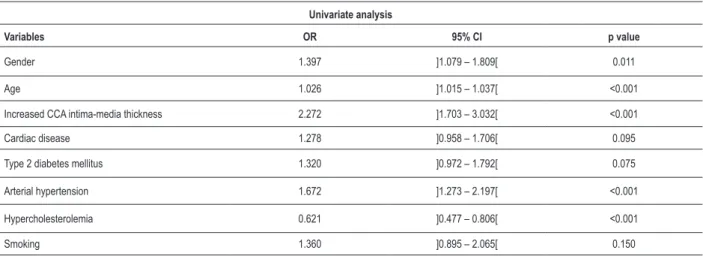
The univariate logistic regression models adopted showed that only arterial hypertension had a marginally significant effect on the likelihood of developing hemorrhagic stroke (OR = 3.344; CI: 0.982–1.386, p = 0.053). Table 4 shows the results of univariate logistic regression for the ischemic stroke event.
The univariate logistic regression models adopted showed that the following variables have a statistically significant predictive effect on the likelihood of developing ischemic stroke: hypercholesterolemia (OR = 0.621; CI: 0.477–0.806, p < 0.001); age (OR = 1.026; CI: 1.015– 1.037, p < 0.001); gender (OR = 1.397; CI: 1.079–1.809,
Table 2 - Distribution of the risk factors for atherosclerotic disease according to the events considered
Variables
Clinical data
Ischemic stroke Hemorrhagic stroke No events Total
n % n % n % n %
Gender
Male 274 60.6 15 68.2 245 51.7 534 56.3 Female 178 39.4 7 31.8 229 48.3 414 43.7 Cardiac disease No 320 70.8 19 86.4 356 75.1 695 73.3 Yes 132 29.2 3 13.6 118 24.9 253 26.7 Type 2 diabetes mellitus No 339 75.0 18 81.8 378 79.7 735 77.5 Yes 113 25.0 4 18.2 96 20.3 213 22.5 Arterial hypertension No 127 28.1 3 13.6 193 40.7 323 34.1 Yes 325 71.9 19 86.4 281 59.3 625 65.9 Hypercholesterolemia No 295 65.3 12 54.5 255 53.8 562 59.3 Yes 157 34.7 10 45.5 219 46.2 386 40.7 Smoking No 398 88.1 22 100.0 429 90.5 849 89.6 Yes 54 11.9 0 0.0 45 9.5 99 10.4 CCA intima-media thickening No 97 21.5 4 18.2 186 39.2 287 30.3 Yes 355 78.5 18 81.8 288 60.8 661 69.7
CCA = common carotid artery
Table 1 - Distribution of age and right and left CCA intima-media thickness values according to gender
Gender
Male - 534 (56.3%) Female - 414 (43.7%)
Mean ± SD Minimum - Maximum Mean ± SD Minimum - Maximum
Age 67.93 ± 12.49 27 – 96 67.44 ± 13.18 25 - 99 Left CCA intima-media thickness 1.1 ± 0.2 0.7 – 1.4 1.1 ± 0.2 0.5 – 1.4 Right CCA intima-media thickness 1.1 ± 0.2 0.7 – 1.4 1.1 ± 0.2 0.6 – 1.4
p = 0.011); arterial hypertension (OR = 1.672; CI: 1.273–2.197, p < 0.011); and increased CCA intima-media thickness (OR = 2.272; CI: 1.703–3.032, p < 0.001).
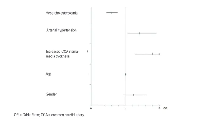
On the multivariate analysis with all the previously mentioned variables, only increased CCA intima-media thickness, arterial hypertension and hypercholesterolemia showed a statistically significant and independent effect on the likelihood of developing ischemic stroke. Thus, increased CCA intima-media thickness was independently associated with ischemic stroke (OR = 1.808; CI: 1.291– 2.534, p = 0.01) (Figure 2).
Considering that increased left and right CCA intima-media thickness might have a statistically significant effect on the likelihood of developing ischemic stroke, once more the univariate logistic regression models were used. They showed an increase in the likelihood of developing ischemic stroke as follows: of 28.1% for each 0.1-mm increase in the left CCA intima-media thickness; and of 27.7% for each 0.1-mm increase in the right CCA intima-media thickness.
Still regarding the univariate logistic regression study for the ischemic stroke event, left and right CCA intima-media thickness values were considered according to previously defined age groups (under 50 years of age; from 50 to 65 years; and over 65 years). The results showed that the left and right CCA intima-media thickness values had a statistically significant predictive effect on the likelihood of developing ischemic stroke at all age groups, being more significant at ages under 50 years (OR = 1.697; CI: 1.192–2.417, p = 0.003 on left CCA, and OR = 2.048; CI: 1.371–3.059, p < 0.001 on right CCA). Figures 3 and 4 show the type of relationship between the occurrence of ischemic stroke and the increased right and left CCA intima-media thickness, by use of the univariate logistic regression model.
The analysis of the graphs in Figures 3 and 4 shows that, for the right and left CCA, the relationship between the risk of ischemic stroke and the intima-media thickness is approximately linear.
Table 3 – Univariate logistic regression for the hemorrhagic stroke event
Univariate analysis
Variables OR 95% CI p value
Gender 0.595 ]0.240 – 1.473[ 0.262
Age 1.006 ]0.972 – 1.041[ 0.733
Increased CCA intima-media thickness 1.981 ]0.664 – 5.905[ 0.220 Cardiac disease 0.427 ]0.125 – 1.455[ 0.174 Type 2 diabetes mellitus 0.762 ]0.255 – 2.277[ 0.627 Arterial hypertension 3.344 ]0.982 – 11.386[ 0.053 Hypercholesterolemia 1.219 ]0.521 – 2.850[ 0.648
OR = Odds Ratio; CI = conidence interval; CCA = common carotid artery.
Table 4 – Univariate logistic regression for the ischemic stroke event
Univariate analysis
Variables OR 95% CI p value
Gender 1.397 ]1.079 – 1.809[ 0.011
Age 1.026 ]1.015 – 1.037[ <0.001
Increased CCA intima-media thickness 2.272 ]1.703 – 3.032[ <0.001 Cardiac disease 1.278 ]0.958 – 1.706[ 0.095 Type 2 diabetes mellitus 1.320 ]0.972 – 1.792[ 0.075 Arterial hypertension 1.672 ]1.273 – 2.197[ <0.001 Hypercholesterolemia 0.621 ]0.477 – 0.806[ <0.001
Smoking 1.360 ]0.895 – 2.065[ 0.150
Figure 2 – Multivariate logistic regression for the ischemic stroke event. OR = Odds Ratio; CCA = common carotid artery.
Hypercholesterolemia
Arterial hypertension
Increased CCA intima-media thickness
Age
Gender
OR = Odds Ratio; CCA = common carotid artery.
R
is
k of i
sc
hem
ic
s
trok
e
Figure 3 – Relationship between the risk of ischemic stroke and the left CCA intima-media thickness. 1st tertile; 1
2nd tertile; 1.732
3rd tertile; 2.209
OR = Odds Ratio; CCA = common carotid artery.
R
is
k of i
sc
hem
ic
s
trok
Discussion and conclusions
Several studies have reported that increased CCA intima-media thickness can be used as a marker of high risk for the occurrence of stroke.
Our study showed that, in fact, there is an association between the occurrence of stroke and increased CCA intima-media thickness.
Regarding hemorrhagic stroke, the increased CCA intima-media thickness showed no statistically significant results, unlike arterial hypertension, which increases 3.34 fold the risk for that event. Ariesen et als.23 have shown that arterial hypertension
increases 3.68 fold the risk for intracerebral hemorrhage. Regarding ischemic stroke, the results differed completely; the increased CCA intima-media thickness, along with the other risk factors for atherosclerotic disease, has a statistically significant and independent effect on the likelihood of developing ischemic stroke. There is an 80.8% increase in the likelihood of the occurrence of that event. These results are in accordance with those of other studies, such as the ARIC Study24, CH Study10, CAP Study13, Rotterdam Study25,26, and
PARC Study27, whose results were similar. However, the analysis
of those results require some considerations. According to Touboul et als.11,28, the reason why some individuals develop
a greater intima-media thickness increase might be related to other factors not approached here, such as anatomic/ hemodynamic factors and genetic susceptibility. In addition, the fact of defining the variables as binary, for the purpose of statistical analysis, might interfere with data interpretation, because, in some cases, the duration and severity of the risk factors are worth assessing.
Another finding was the increase in the likelihood of developing ischemic stroke of 28.1% for each 0.1-mm increase in the left CCA intima-media thickness and of 27.7% for each 0.1-mm increase in the right CCA
intima-media thickness. Those results are in accordance with those of other similar studies. In the ARIC Study24, a 0.18-mm
increase in the CCA intima-media thickness leads to a 60% and 31% increase in the risk of stroke in women and men, respectively. The CH Study10 has reported that a 0.2-mm
increase in the CCA intima-media thickness results in a 37% increase in that risk. On the other hand, the Rotterdam Study25 has reported that a 0.163-mm increase in the CCA
intima-media thickness is associated with a 41% increase in the stroke occurrence.
The present study also showed that the left and right CCA intima-media thickness value has a statistically significant predictive effect on the higher likelihood of developing ischemic stroke for ages under 50 years as compared with ages over 50 years. Regarding left CCA, there is a 69.7% increase, while for right CCA, the increase is of 104.8%. In addition, the relationship between the risk of stroke occurrence and the CCA intima-media thickness value is approximately linear, being higher between the 1st and 2nd tertiles, for both the right and left CCAs. Other studies, such as the CAPS Study13
and the PARC Study27, have reported similar results. That result
is worth noting and should be carefully assessed, because vascular events in young individuals are rare, and because a statistically significant difference does not always translate clinical significance. Lorenz et als.29 have also suggested that
further studies should be performed on young populations. The ARIC Study24 hasconcluded that the relationship between
the CCA intima-media thickness values and the likelihood of developing stroke is greater for values under 1.0 mm; thus, the risk for stroke occurrence will be greater in the presence of lower intima-media thickness reference values. Because young individuals usually have absolute intima-media thickness values under 1.0 mm, their risk for the stroke occurrence might be greater. In the elderly, the relationship found can be explained by the medication they usually take (beta-blockers,
Figure 4 – Relationship between the risk of ischemic stroke and the right CCA intima-media thickness. OR = Odds Ratio; CCA = common carotid artery.
R
is
k of i
sc
hem
ic
s
trok
e
1st tertile; 1
2nd tertile; 1.982
statins, angiotensin-converting-enzyme inhibitors), which can influence the intima-media thickness values 13,27.
This study has also some limitations. Although all individuals included in the database are part of the Portuguese National Healthcare System and were cared for and underwent tests at the same hospital, they do not represent the general population. Thus, all conclusions should be interpreted with due care. The representative number of individuals of the hemorrhagic stroke population is reduced, which might have influenced somehow the results obtained for that event.
Thus, we leave the following recommendations for future studies: to include a higher number of individuals with hemorrhagic stroke; to include other risk factors for atherosclerotic disease, such as body mass index, which will enrich the results; and to carry out a similar study, but of prospective nature, including individuals of the general population and youngsters. In addition to the presence of increased CCA intima-media thickness, it would be worth assessing the behavior of several risk factors for atherosclerotic disease over time to evaluate the behavior of the relationship here described. In addition to stroke, it would also be worth considering acute myocardial infarction, to verify in which of those events the relationship
with the increased CCA intima-media thickness would be most significant.
In conclusion, the increased CCA intima-media thickness showed to be independently associated with ischemic stroke but not with hemorrhagic stroke. Although several authors have claimed that increased CCA intima-media thickness bears a moderate correlation with stroke occurrence, they have emphasized the need for further studies so that it can be considered a risk marker for theoccurrence of vascular events, such as stroke27. This study reinforces that association.
Potential Conflict of Interest
No potential conflict of interest relevant to this article was reported.
Sources of Funding
There were no external funding sources for this study.
Study Association
This study is not associated with any post-graduation program.
References
1. Hallström B, Jönsson AC, Nerbrand C, Norrving B, Lindgren A. Stroke incidence and survival in the beginning of the 21st century in southern Sweden: comparisons with the late 20th century and projections into the future. Stroke. 2008;39(1):10-5.
2. Araújo D, Teich V, Passos RB, Martins SC. Análise de custo-efetividade da trombólise com alteplase no acidente vascular cerebral. Arq Bras Cardiol. 2010;95(1):12-20.
3. Monteiro I, Vaz Almeida MD. [Dietary fat and ischemic stroke risk in Northern Portugal]. Acta Med Port. 2007;20(4):307-18.
4. Engelhorn CA, Engelhorn AL, Cassou MF, Zanoni CC, Gosalan CJ, Ribas E, et al. Intima-media thickness at the origin of the right subclavian artery as an early marker of cardiovascular risk. Arq Bras Cardiol. 2006;87(5):609-14. 5. Ederle J, Brown MM. Stroke prevention. Herz. 2008;33(7):518-23. 6. Prado SS, Ribeiro ML, Cardoso GP, Bousquet-Santos K, Velarde LG, Nóbrega AC.
Carotid artery structural and functional evaluation in relatives of type 2 diabetic patients. Arq Bras Cardiol. 2009;92(3):186-192, 190-6.
7. Goldstein LB, Adams R, Becker K, Furberg CD, Gorelick PB, Hademenos G, et al. Primary prevention of ischemic stroke: a statement for the health care professionals from the stroke council of the American Heart Association. Circulation. 2001;103(1):163-82.
8. Rosa EM, Kramer C, Castro I. Association between coronary artery atherosclerosis and the intima-media thickness of the common carotid artery measured on ultrasonography. Arq Bras Cardiol. 2003;80(6):589-92, 285-8.
9. Romero JR, Beiser A, Seshadri S, Benjamin EJ, Polak JF, Vasan RS, et al. Carotid artery atherosclerosis, MRI indices of brain ischemia, aging, and cognitive impairment: the Framingham Study. Stroke. 2009;40(5):1590-6.
10. O’Leary DH, Polak JF, Kronmal RA, Manolio TA, Burke GL, Wolfson SK Jr. Carotid-artery intima and media thickness as a risk factor for myocardial infarction and stroke in older adults. Cardiovascular Health Study Collaborative Research Group. N Engl J Med. 1999;340(1):14-22.
11. Touboul PJ, Labreuche J, Vicaut E, Amarenco P; GENIC Investigators. Carotid intima-media thickness, plaques, and Framingham risk score as independent determinants of stroke risk. Stroke. 2005;36(8):1741-5.
12. Mackinnon AD, Jerrard-Dunne P, Sitzer M, Buehler A, von Kegler S, Markus HS. Rates and determinants of site-specific progression of carotid artery intima-media thickness: the carotid atherosclerosis progression study. Stroke. 2004;35(9):2150-4.
13. Lorenz MW, von Kegler S, Steinmetz H, Markus HS, Sitzer M. Carotid intima-media thickening indicates a higher vascular risk across a wide age range: prospective data from the Carotid Atherosclerosis Progression Study (CAPS). Stroke. 2006;37(1):87-92.
14. Dijk JM, van der Graaf Y, Bots ML, Grobbee DE, Algra A. Carotid intima-media thickness and the risk of new vascular events in patients with manifest atherosclerotic disease: the SMART Study. Eur Heart J. 2006;27(16):1971-8.
15. de Groot E, van Leuven SI, Duivenvoorden R, Meuwese MC, Akdim F, Bots ML, et al. Measurement of carotid intima-media thickness to assess progression and regression of atherosclerosis. Nat Clin Pract Cardiovasc Med. 2008;5(5):280-8.
16. Touboul PJ, Hennerici MG, Meairs S, Adams H, Amarenco P, Bornstein N, et al. Mannheim carotid intima-media thickness consensus (2004-2066). An update on behalf of the Advisory Board of the 3rd and 4th Watching the Risk Symposium, 13th and 15th European Stroke Conferences, Mannheim, Germany, 2004, and Brussels, Belgium, 2006. Cerebrovasc Dis. 2007;23(1):75-80.
18. Coll B, Feinstein SB. Carotid intima-media thickness measurements: t e c h n i q u e s a n d c l i n i c a l r e l e v a n c e . C u r r A t h e r o s c l e r R e p . 2008;10(5):444-50.
19. Baldassarre D, Amato M, Bondioli A, Sirtori CR, Tremoli E. Carotid artery intima-media thickness measured by ultrasonography in normal clinical practice correlates well with atherosclerosis risk factors. Stroke. 2000;31(10):2426-30.
20. Stein JH. Carotid intima-media thickness and vascular age: you are only as old as tour arteries look. J Am Soc Echocardiogr. 2004;17(6):686-9.
21. Hurst RT, Ng DW, Kendall C, Khandheria B. Clinical use of carotid intima-media thickness: review of the literature. J Am Soc Echocardiogr. 2007;20(7):907-14.
22. Akosah KO, McHugh VL, Barnhart SI, Mathiason MA, Schaper AM, Perlock PA. Pilot results of the Early Detection by Ultrasound of Carotid Artery Íntima-Media Thickness Evaluation (EDUCATE) study. Am J Hypertens. 2007;20(11):1183-8.
23. Ariesen MJ, Claus SP, Rinkel GJ, Algra A. Risk factors for intracerebral hemorrhage in the general population: a systematic review. Stroke. 2003;34(8):2060-5.
24. Chambless LE, Folsom AR, Clegg LX, Sharrett AR, Shahar E, Nieto FJ, et al. Carotid wall thickness is predictive of incident clinical stroke: the Atherosclerosis Risk in Communities (ARIC) study. Am J Epidemiol. 2000;151(5):478-87.
25. Bots ML, Hofman A, Grobbee DE. Increased common carotid intima-media thickness: adaptive response or a reflection of atherosclerosis? Findings from the Rotterdam Study. Stroke. 1997;28(12):2442-7.
26. Hollander M, Hak AE, Koudstaal PJ, Bots ML, Grobbee DE, Hofman A, et al. Comparison between measures of atherosclerosis and risk of stroke: the Rotterdam study. Stroke. 2003;34(110):2367-72.
27. Touboul PJ, Vicaut E, Labreuche J, Belliard JP, Cohen S, Kownator S, et al; PARC study participating physicians. Correlation between the Framingham risk score and intima media thickness: the Paroi Artérielle et Risque Cardio-vasculaire (PARC) study. Atherosclerosis. 2007;192(2):363-9.
28. Touboul PJ, Elbaz A, Koller C, Lucas C, Adraï V, Chédru F, Amarenco P. Common carotid artery intima-media thickness and brain infarction: the Etude du Profil Génétique de l’Infarctus Cerebral (GÉNIC) case control study. The GENIC Investigators. Circulation. 2000;102(3):313-8.