Keywords
Calcium / analysis; risk assessment; coronary angiography; coronary disease; tomography; x ray computed.
Mailing Address: Carlos Eduardo Rochitte •
Av. Dr. Enéas de Carvalho Aguiar 44 - Andar AB - Ressonância & Tomografia Cardiovascular – Cerqueira Cesar - 05403-000 – São Paulo, SP - Brazil E-mail: [email protected], [email protected]
Manuscript received February 28, 2012; manuscript revised February 28, 2012; accepted March 15, 2012.
Coronary Artery Calcium Score and Coronary Computed
Tomographic Angiography for Cardiovascular Risk Stratification
Clerio F. Azevedo
1,2, Carlos E. Rochitte
3, João A.C. Lima
4Instituto D’Or de Pesquisa e Ensino (IDOR)1; Universidade do Estado do Rio de Janeiro2, Rio de Janeiro, RJ; Instituto do Coração (InCor) da
Faculdade de Medicina da Universidade de São Paulo3, São Paulo, SP, Brazil; Johns Hopkins University 4, Estados Unidos, USA
Abstract
Cardiovascular disease is the leading mortality cause worldwide. The capacity to identify among the asymptomatic individuals the subgroup at greater risk for developing cardiovascular events is fundamental in any strategy aimed at reducing the rate of cardiovascular events. The first step in cardiovascular risk stratification is the use of global risk scores, the Framingham risk score being the most frequently used. However, previous studies have shown that, although very useful, clinical scores, when used alone, have a limited capacity for stratifying cardiovascular risk in a significant part of the population.
In that context, coronary artery calcium score (CACS) and coronary computed tomographic angiography might play an important role as complementary tools for risk stratification of asymptomatic patients. The CACS provides important prognostic information that is incremental to clinical scores based on traditional risk factors and other diagnostic modalities, such as C-reactive protein measurement. In addition, CACS has the potential to change and help the patients’ clinical management. On the other hand, coronary computed tomographic angiography provides a detailed assessment of the anatomy of the coronary arteries, allowing visualizing not only the lumen, but also the coronary arterial walls. Compared with conventional invasive coronary angiography, coronary computed tomographic angiography has excellent accuracy to identify and mainly exclude the presence of significant obstructive lesions. In addition, it proved to be able to provide incremental prognostic information to traditional risk factors and CACS.
Introduction
Cardiovascular disease is the leading mortality cause worldwide. In only one year, 17.3 million individuals died due to cardiovascular diseases, 7.3 million secondary to
coronary artery disease (CAD) and 6.2 million secondary to cerebrovascular diseases1. Only in Brazil, over 900,000 cases
of acute myocardial infarction (AMI) occurred per year, of which over 300,000 were fatal2. Reducing those numbers and,
consequently, the CAD-related morbidity and mortality has been one of the major objectives of public health policies in Brazil and worldwide. However, more than half of the acute coronary syndromes and sudden deaths are known to occur in previously asymptomatic individuals3. Thus, the capacity
to identify among asymptomatic individuals the subgroup at greater risk for developing cardiovascular events represents a fundamental step in any strategy aimed at reducing the rate of cardiovascular events4.
The first step in the cardiovascular risk stratification of asymptomatic individuals is the use of global risk scores, such as the Framingham, PROCAM (Münster Heart Study), SCORE, and Reynolds risk scores, of which, the Framingham risk score is the most frequently used. That type of assessment is relatively simple to be performed, bears no risk for patients, provides valuable prognostic information and bears a great potential to change clinical management. Thus, global risk assessment scores should be always the first step in cardiovascular risk stratification of asymptomatic individuals5.
A fundamental question is whether using only clinical scores suffices. Previous studies have shown that those scores have limitations. According to Greenland et al3, 50% of the patients
with acute coronary syndromes would have been classified as at intermediate risk by use of the Framingham score if they had been assessed before the acute event3. In that same
study, the authors have shown that approximately 40% of the general population would also be classified as at intermediate risk. In addition, they have shown that 75% of the previously asymptomatic patients with acute coronary syndromes had not met the criteria for treatment with statins before the acute event. Those data illustrate the concept that, although very useful, the clinical scores, when used alone, have a limited capacity for stratifying the cardiovascular risk in a significant part of the population. In fact, such limitations are even more significant in younger individuals and those of the female sex. It is exactly in that context that some laboratory and imaging tests, such as coronary artery calcium score (CACS) and coronary computed tomographic angiography (CCTA), might play an important role as complementary tools to clinical scores for risk stratification of asymptomatic patients.
the coronary artery anatomy and atherosclerotic burden, previously only possible by use of invasive exams, such as invasive coronary angiography and intracoronary ultrasound, can now be obtained in a non-invasive way and with excellent accuracy by use of cardiac CT. In clinical practice, CT can be used to assess CAD in determining CACS and in CCTA.
Although CCTA has excellent accuracy to identify or exclude the presence of significant obstructive lesions, being able to provide valuable prognostic information6, the most robust and
validated diagnostic modality to stratify global cardiovascular risk is CACS5,12,13.
This review approaches the basic technical notions, major clinical applications and scientific evidence available on both methods, CACS and CCTA, focusing mainly on their roles as tools for global cardiovascular risk stratification.
Coronary artery calcium score
The test
Initially, prior to the advent of multidetector CT devices, electron-beam CT was used to assess CACS. In fact, a good part of the scientific literature on CACS has been based on that technique14. However, currently, electron-beam CT
represents an outdated and virtually unavailable diagnostic modality. With the introduction of multidetector CT devices at the end of the 1990s, that became the method to assess CACS in clinical practice, proving to be at least comparable, and, in many aspects, superior to electron-beam CT for that purpose. Thus, in this article, all technical considerations about CACS image acquisition refer to multidetector CT.
Determining CACS is based on a non-contrasted acquisition of a series of 3-mm axial slices covering the entire heart extension. Images are acquired synchronously with the signal of the electrocardiogram (ECG). The protocol of synchronization with the ECG can be prospective or retrospective. The radiation dose used in the retrospective acquisition protocols is significantly higher; thus, prospective protocols are more commonly used. In practice, the effective radiation dose in a prospective acquisition is low, being usually under 1.5 mSv, and, on average, around 0.9 mSv to 1.1 mSv.
Calcification is defined as a hyperattenuating lesion with
signal intensity over 130 Hounsfield units (HU) and area ≥
3 adjacent pixels (at least 1 mm2). It can be calculated based
on the weighted sum of densities over 130 HU (Agatston score) or by use of methods determining calcium volume or mass. Although calcium volume or mass scores have better reproducibility, the large population data banks that describe the distribution of coronary calcification according to the patients’ age, ethnicity and sex are based on the Agatston score, which is, thus, the most often used score in clinical practice (Figure 1).
One question still remains: How are the results of CACS classified and interpreted?
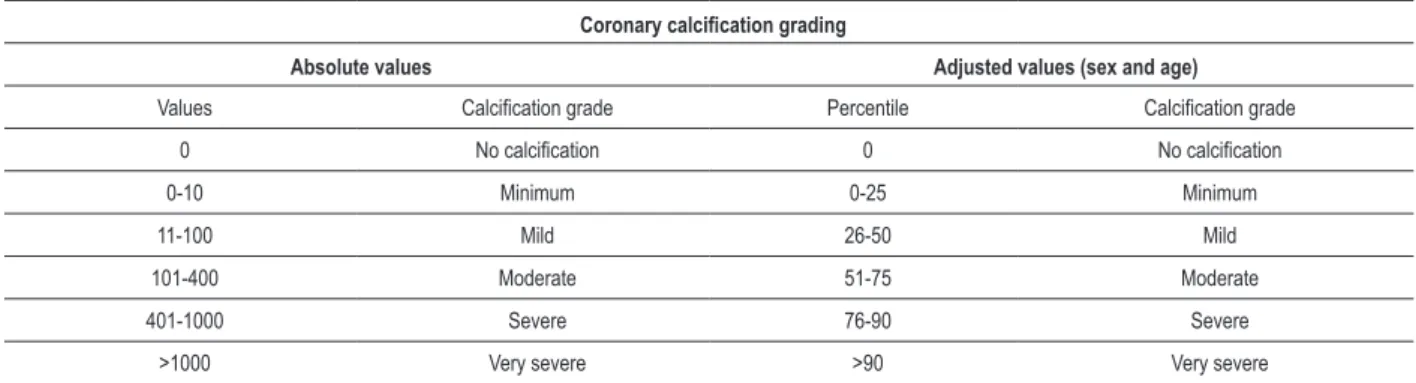
The CACS values obtained can be classified by using either fixed cut-off points or cut-off points adjusted for the patients’ age, sex, and, more recently, ethnicity. The more accepted limit values in the two classification types are shown in table 1.
In the classification using fixed cut-off points, the patients are categorized according to pre-established limits. In the classification using adjusted cut-off points, the patients are categorized according to the percentiles of distribution of the CACS values of the general population (Figure 2). It is worth noting, however, that both classification types provide valuable prognostic information and are present in the reports provided by diagnostic centers trained in performing cardiac imaging. Initially, CACS was used as a diagnostic tool aimed at identifying or excluding the presence of obstructive CAD in symptomatic patients. The initial studies have shown that CACS has an excellent negative predictive value (NPV) to exclude the presence of significant CAD (96% to 100%), but the positive predictive value (PPV) was, at most, moderate14.
In contrast, more recent studies have shown that, particularly in younger patients or in a population with a high prevalence of significant CAD, in addition to the inadequate PPV, CACS shows a NPV insufficient to exclude the presence of significant obstructive disease.
In fact, Gottlieb et al.15 have shown that, in a population
with a 56% prevalence of significant CAD, even 19% of the
patients with CACS = 0 had at least one obstructive lesion ≥
50% on invasive coronary angiography15. Considering the only
moderate PPV and the conflicting data regarding the NPV, the
Figure 1 - Images illustrating the coronary artery calcium score of three patients with increasing calciication grades in the territory of the anterior descending artery: A.
current concept is that, in general, CACS should not be used as a tool to diagnose significant obstructive CAD in symptomatic patients. The actual value of CACS in clinical practice consists in its capacity to provide valuable prognostic information and to serve as an important tool for risk stratification of asymptomatic patients.
Prognostic value
An important concept to understand why CACS bears the potential to play a relevant role in cardiovascular risk stratification is that coronary artery calcification represents a marker of the presence of atherosclerotic plaques in coronary arteries. In the coronary arterial bed, calcification occurs almost exclusively in the context of atherosclerotic disease. In a certain individual, the amount of calcification in the coronary arteries has a good correlation with the total coronary atherosclerotic burden. However, not the entire atherosclerotic plaque is calcified. In fact, previous reports have shown that the calcified portion corresponds to only 20% of the total plaque volume, that is, the coronary artery calcification would represent only the “iceberg tip” of CAD, and the non-calcified portion would account for 80% of the total coronary atherosclerotic burden. It is worth noting that the presence or absence of calcium has not been significantly associated with the propensity to rupture of a certain plaque, and that the presence of calcification is a signal of neither “stability” nor “instability” of any plaque.
Several population longitudinal studies involving a large number of patients have shown that CACS is significantly associated with the occurrence of major cardiovascular events (all-cause death, cardiac death, and non-fatal AMI) in the medium- and long-term follow-up. The higher the amount of calcium in the coronary arteries, the greater the chance of the patient having a cardiovascular event in the future.
Even more important, a series of previous studies have shown that the capacity of CACS to predict the occurrence of cardiovascular events is additional to the risk stratification by use of the Framingham score and other methods, such as C-reactive protein measurement. In fact, a classical study16 has shown that
patients at intermediate risk according to the Framingham score and with CACS > 300 have a rate of cardiovascular events of 2.8%/year (approximately equivalent to a 28% rate over ten years), which would place them in the high-risk group (> 20% over ten years).
A recent consensus published by the ACCF/AHA on the CACS use to assess global cardiovascular risk12 has analyzed
the combined data of six large studies that included 27,622 asymptomatic patients and has assessed the major predictors of a total of 395 cardiovascular events. Of the 11,815 patients with CACS = 0, the rate of events was very low, only 0.4% over the three to five subsequent years. When compared with patients with a CACS = 0, those with a CACS between 100 and 400 had a relative risk (RR) of 4.3 (95% confidence interval [95% CI]: 3.5 – 5.2; p < 0.0001), those with a CACS between 400 and 1000 had a RR of 7.2 (95% CI: 5.2 – 9.9; p < 0.0001), and those with a CACS > 1000 had a RR of 10.8 (95% CI: 4.2 – 27.7; p < 0.0001).
Since the publication of that consensus, other prospective studies have confirmed those findings and have demonstrated that the association between CACS and prognosis is similar regardless of the patients’ sex or ethnicity14,17. Even more
important, all such studies have also confirmed that CACS can provide additional prognostic information as compared with the assessment by use of the traditional risk factors alone. On the ROC analysis, the area under the curve to predict the occurrence of cardiovascular events was significantly greater with CACS than with the Framingham or PROCAM risk scores. In the MESA study, considering only the traditional risk factors, the C statistics was 0.79 to predict major cardiovascular events, and 0.83 when the CACS information was associated with that of risk factors (p = 0.006)17.
Previous studies have also shown that CACS provides additional prognostic information to the C-reactive protein assessment. In four studies based on multivariate models, CACS remained as an independent predictor of major cardiovascular events, while the C-reactive protein no longer was a significant predictor on multivariate analyses18-21. When compared with
the carotid intima/media thickness (IMT) measurement, CACS proved to be a better predictor of subsequent events; the hazard ratio (HR) of the IMT was 1.7 (95% CI: 1.1 – 2.7; p = 0.07), while the HR of the CACS was 8.2 (95% CI: 4.5 – 15.1; p = 0.001)22.
Potential to change clinical management
In addition to its role as a risk stratification tool, another important aspect of CACS relates to its potential to change and help with the clinical management of patients suspected
Table 1 – Classiication of the coronary artery calcium score values according to the severity of atherosclerotic involvement
Coronary calciication grading
Absolute values Adjusted values (sex and age)
Values Calciication grade Percentile Calciication grade
0 No calciication 0 No calciication
0-10 Minimum 0-25 Minimum
11-100 Mild 26-50 Mild
101-400 Moderate 51-75 Moderate
401-1000 Severe 76-90 Severe
of having CAD. In an observational study, Kalia et al.23 have
shown that the use rate of lipid-lowering drugs in dyslipidemic patients increased from 44% to over 90% in those with severe calcification on CACS assessment23. Other preventive
behavioral measures, such as the regular practice of physical exercises and the adoption of healthier diets, have also
shown an improvement in the subgroup with higher CACS values. Moreover, in a prospective study following 1,640 patients up for a mean period of six years, the use rates of statins and aspirin were 3.5 and 3 times greater, respectively, in the subgroup with calcification plaques on the CACS24.
That significant increase did not depend on other risk factors
Figure 2 - Graphs illustrating the distribution of coronary artery calciication measured by the Agatston score according to patients’ age and sex. It is worth noting that
the scale of the graphs referring to the female (A) and male (B) sexes are different, i.e., the mean magnitude of the calciication in men is much more accentuated, independently of the age bracket.
Female sex
Male sex
Ca
lci
um
sc
or
e
Ca
lci
um
sc
or
e
Age (years)
Age (years)
Percentile 25
Percentile 50
Percentile 75
Percentile 90
and demonstrated the CACS potential to change clinical management in a sample of the general population.
According to the results of the JUPITER study25, a
large number of asymptomatic patients with LDL levels considered normal have begun to have indication for statin treatment. In that study, the major criterion to indicate statin treatment was ultra-sensitive C-reactive protein test. However, recent data of the MESA study have shown that the subgroup of patients with indication for statin treatment according to the criteria of the JUPITER study, but who had no coronary calcification on CACS assessment, were at low risk for cardiovascular events. Considering that in the JUPITER study the reduction in the absolute risk of the entire population was low, the authors of the MESA study have concluded that CACS might play a relevant role in selecting those with the greater potential to benefit from the treatment, among all patients who met the criteria for receiving statins. In other words, not C-reactive protein alone, but its combination with CACS would be a better strategy to select patients with the greatest potential to benefit from the treatment with statins.
Current recommendations
In an expert consensus on the use of CACS for assessing the global cardiovascular risk published by the ACCF/AHA in 200712, asymptomatic patients classified as at intermediate
risk by the Framingham risk score were considered good candidates to the CACS assessment aiming at refining the stratification, and potentially changing clinical management. In that document, the authors have not classified the recommendation grade.
More recently, in the 2010 ACCF/AHA guideline for assessment of cardiovascular risk in asymptomatic patients5, the CACS quantification for cardiovascular risk
stratification in individuals classified as at intermediate risk by the Framingham risk score was considered a class IIa indication, with level of evidence B. It is worth noting that in that document, the only intervention considered as class I was the stratification using global risk scores based on the traditional risk factors, such as the Framingham risk score, and the clinical assessment regarding the presence of early CAD family history. Neither imaging nor laboratory diagnostic exams were classified as class I recommendation grade.
For the purpose of comparison, in that document, echocardiography, exercise test, and myocardial perfusion scintigraphy were considered class IIb or III recommendation grade indications.
Finally, in the ACCF/SCCT/ACR/AHA/ASE/ASNC/NASCI/ SCAI/SCMR 2010 appropriate use criteria for cardiac CT report8, the use of CACS for stratifying the global
cardiovascular risk of patients classified as at intermediate risk by the Framingham risk score was rated as a level 7 (A7) appropriate indication. That report used a scale ranging from 1 to 9 to designate appropriateness as follows: 1-3, inappropriate use; 4-6, uncertain; and 7-9, appropriate. In that document, the CACS use was also considered a level 7 (A7) appropriate indication in patients classified as at low
risk by the Framingham risk score, but who had an early CAD family history.
Regarding the Brazilian recommendations, the 2006 Brazilian Society of Cardiology guidelines on the use of cardiac CT7 defined the use of CACS for stratifying the global
cardiovascular risk of patients classified as at intermediate risk by the Framingham risk score as a class I indication. That same document defined as a class IIa indication the use of CACS in patients classified as at low risk by the Framingham risk score, but who had an early CAD family history.
Coronary computed tomographic angiography
The test
Coronary computed tomographic angiography (CCTA) became possible with the advent of multidetector CT devices at the end of the 1990s. It began to be clinically used from the beginning of the following decade, after the introduction of 16-row detector CT devices. Since then, the technological development in that area has been vertiginous, and currently devices with 256 rows and even 320 rows of detectors are available, allowing the acquisition of all images necessary for coronary assessment at one single heartbeat.
The CCTA is based on the acquisition of a series of axial slices with submillimetric thickness covering the entire cardiac extension. Similarly to CACS, the images are acquired synchronously with the ECG signal. The ECG synchronization protocol can be prospective sequential or retrospective helical. Considering the 64-row devices currently available, while the amount of radiation of one retrospective acquisition with dose modulation is around 9.0 mSv, the effective dose of a prospective acquisition is around 3.0 mSv26.
For the purpose of comparison, the radiation dose of a conventional technetium or sestamibi myocardial perfusion scintigraphy is also around 9.0 mSv. The radiation dose of a thallium myocardial perfusion scintigraphy is around 18.0 mSv. It is worth noting that, in general, the medical community has made a great effort to progressively reduce the amount of radiation used in cardiologic tests,
and that the current devices with ≥ 256 rows already
allow performing coronary tomographic angiography with effective radiation doses lower than 1.0 mSv.
Diagnostic value
There are some systematic reviews assessing the diagnostic value of CCTA since the advent of 64-row devices in 200427-30. In general, those studies have shown
due to its greater longitudinal coverage and better spatial and temporal resolution (Figure 3).
Currently there are more than 50 single-center studies29 and three multicenter studies31-33 assessing the
accuracy of CCTA with 64-row devices for the assessment of stable symptomatic patients suspected of having significant CAD. In all cases, CCTA has been compared with the current reference method, invasive conventional coronary angiography. In general, those studies have very consistently demonstrated that CCTA has an excellent accuracy to detect or exclude the presence of significant CAD. In particular, they have shown a particularly high NPV, making the test especially useful to exclude the presence of significant obstructive lesions in that population.
In the most recent systematic review assessing the single-center studies carried out until the end of 2007 using 64-row devices29, the mean sensitivity to identify
the presence of significant CAD was 98% and the mean specificity was 88%. The mean prevalence of significant CAD in those studies was 61% and the PPV and NPV were 93% and 96%, respectively.
In the multicenter CORE 64 study32, assessing 291
patients with clinical indication for invasive coronary angiography, the prevalence of significant CAD was 56%. In that study, the sensitivity and specificity of CCTA, in an analysis per patient, were 85% and 90%, respectively. The area under the ROC curve was 0.93. The PPV was 91% and the NPV was 83%.
In the multicenter ACCURACY trial31, assessing 245
symptomatic patients, the prevalence of significant CAD
was 25% for stenoses ≥ 50% and only 14% for stenoses ≥
70%. In that study, sensitivity was 94% or 95%, depending on the threshold chosen to define the presence of significant CAD. Specificity was 82%. The NPV was 99% for both the 50% and 70% thresholds. The PPV was only 48% for the 70% threshold, and 64% for the 50% threshold.
Finally, a multicenter study performed at three Dutch university-affiliated hospitals has assessed 360 patients with indication for invasive coronary angiography33. The
prevalence of significant CAD was 68%. The sensitivity and specificity of CCTA, in an analysis per patient, were 100% and 64%, respectively. The PPV was 86% and the NPV was 97%.
Prognostic value
In recent years, a large number of studies have assessed the role played by magnetic resonance and CT not only for the diagnostic evaluation13,31-39, but also for the prognostic
evaluation40-47 of patients with cardiovascular diseases.
Regarding cardiac CT, a recent series of studies have shown that, not only the CACS assessment, but also the assessment of coronary anatomy by use of CCTA provide important prognostic information in patients suspected of having significant CAD42,44,45,47-49. Using a 16-row detector CT device,
Min et al.44 have studied 1,127 patients and assessed the
relationship between all-cause mortality and the results of CCTA. After a mean 15-month follow-up, the authors have
shown that the CAD severity on CCTA was an important predictive factor of total mortality, providing additional prognostic information to the traditional risk factors.
In another important study, Ostrom et al.45 have assessed
2,538 patients by use of CCTA with an electron-beam CT device45. After a mean 78-month follow-up, the authors
have shown that CCTA has an incremental prognostic value to not only traditional risk factors, but also to the CACS assessment. Later, van Werkhoven et al.49 have assessed 541
patients undergoing both CCTA and myocardial perfusion imaging. After a mean 22-month follow-up assessing the combined endpoints of cardiac death, nonfatal infarction and hospitalization due to unstable angina, the authors have demonstrated that both the anatomical assessment through tomographic angiography and functional evaluation by use of myocardial perfusion imaging provide important prognostic information, the former having an incremental prognostic value over the latter.
In a recent study, Azevedo et al.48 have assessed 529
patients suspected of having obstructive CAD, who had undergone ischemia tests with inconclusive results. Those authors have shown that 70% of those patients had no significant obstructive lesion. In fact, approximately 40% had completely normal coronary arteries, with CACS = 0. Moreover, after a mean 30-month follow-up, those authors showed that both CACS and CCTA provided important prognostic information on that population.
In addition, recently, Hadamitzky et al.42, studying
2,223 patients with suspected CAD for a mean period of 28 months, have confirmed the findings of previous studies by demonstrating that CCTA provides important prognostic information, which is incremental to clinical risk scores and CACS42. In the ROC analysis, the area under the curve to
predict the occurrence of cardiovascular events was 0.79 for the clinical score alone, 0.84 for the clinical score plus CACS, and 0.89 for the clinical score plus CACS plus CCTA (p < 0.001).
Characterization of the atherosclerotic plaque
An important characteristic of CCTA, which distinguishes it from invasive coronary angiography, is its ability to provide the assessment of coronary arterial walls in addition to the visualization of the vessel’s lumen. Thus, it allows not only the identification of the lesions causing luminal obstruction, but also the characterization of atherosclerotic plaques, either obstructive or non-obstructive. In fact, several previous studies have shown the good correlation of plaque volume measured on CCTA with the measures obtained with the current gold standard method, intravascular ultrasound50.
Even more important, recent reports have suggested that the characterization of atherosclerotic plaques on CCTA has the potential to help in identifying the most vulnerable patients to an acute coronary event. Motoyama et al.51 have assessed
1,059 patients regarding the presence of two parameters measured by use of CCTA: the grade of positive remodeling and the presence of low-attenuation areas in the plaque, suggesting lipid content51. The mean follow-up time was 27
those two parameters was 22.2%. In contrast, that rate in patients with only one of those parameters was 3.7%, and in those with none of those parameters was 0.4%.
However, it is worth noting that the use of CCTA aiming at characterizing atherosclerotic plaques and identifying those with signs suggestive of vulnerability does not yet represent a clinically established indication, being currently under experimental development.
Conclusion
Currently, the two major uses of CT to assess CAD are as follows: CACS measurement and CCTA itself. The
CACS provides important prognostic information that is incremental to the clinical scores based on traditional risk factors and other diagnostic modalities, such as C-reactive protein measurement. In addition, CACS has the potential to change and help the patients’ clinical management. Thus, performing CACS is indicated for the global cardiovascular risk stratification of asymptomatic patients rated as at intermediate risk according to the Framingham risk score. On the other hand, CCTA provides a detailed assessment of the anatomy of the coronary arteries, allowing visualizing not only the lumen, but also the coronary arterial walls. Compared to conventional invasive coronary angiography, CCTA has excellent accuracy to identify and mainly exclude
Figure 3 - Images of two patients undergoing coronary computed tomographic angiography. In the irst patient, the coronary computed tomographic angiography was
the presence of significant obstructive lesions. In addition, it proved to be able to provide incremental prognostic information to the traditional risk factors and CACS. However, although CCTA represents a very useful diagnostic tool, being indicated for the diagnostic assessment of symptomatic patients suspected of having CAD, it is currently not indicated when the only objective is the global cardiovascular risk stratification of asymptomatic patients.
Potential Conflict of Interest
No potential conflict of interest relevant to this article was reported.
Sources of Funding
There were no external funding sources for this study.
Study Association
This study is not associated with any post-graduation program.
References
1. WHO Statistical Information System. (WHOSIS). World health statistics. [Cited on 2011 Jan 18]. Available from:: http://www.who.int/whosis/ shostat/2011/en/index.html
2. Ministério da Saúde. Datasus. [Acesso em 2011 mar 30]. Disponível em http://www2.datasus.gov.br/DATASUS.
3. Greenland P, Smith SC, Grundy SM. Improving coronary heart disease risk assessment in asymptomatic people: role of traditional risk factors and noninvasive cardiovascular tests. Circulation. 2001;104(15):1863-7.
4. Almeida FK, Esteves JF, Gross JL, Biavatti K, Rodrigues TC. Severe forms of retinopathy predict the presence of subclinical atherosclerosis in type 1 diabetes subjects. Arq Bras Cardiol. 2011;97(4):346-9.
5. Greenland P, Alpert JS, Beller GA, Benjamin EJ, Budoff MJ, Fayad ZA, et al. 2010 ACCF/AHA guideline for assessment of cardiovascular risk in asymptomatic adults: a report of the American College of Cardiology Foundation/American Heart Association Task Force on Practice Guidelines. J Am Coll Cardiol. 2010;56(25):e50-103.
6. Mark DB, Berman DS, Budoff MJ, Carr JJ, Gerber TC, Hecht HS, et al. ACCF/ ACR/AHA/NASCI/SAIP/SCAI/SCCT 2010 expert consensus document on coronary computed tomographic angiography: a report of the American College of Cardiology Foundation Task Force on Expert Consensus Documents. J Am Coll Cardiol. 2010;55(23):2663-99.
7. Rochitte CE, Pinto IM, Fernandes JL, Filho CF, Jatene A, Carvalho AC, et al. [Cardiovascular magnetic resonance and computed tomography imaging guidelines of the Brazilian Society of Cardiology]. Arq Bras Cardiol. 2006;87(3):e60-100.
8. Taylor AJ, Cerqueira M, Hodgson JM, Mark D, Min J, O’Gara P, et al. ACCF/ SCCT/ACR/AHA/ASE/ASNC/NASCI/SCAI/SCMR 2010 appropriate use criteria for cardiac computed tomography: a report of the American College of Cardiology Foundation Appropriate Use Criteria Task Force, the Society of Cardiovascular Computed Tomography, the American College of Radiology, the American Heart Association, the American Society of Echocardiography, the American Society of Nuclear Cardiology, the North American Society for Cardiovascular Imaging, the Society for Cardiovascular Angiography and Interventions, and the Society for Cardiovascular Magnetic Resonance. J Am Coll Cardiol. 2010;56(22):1864-94.
9. Duarte PS. Technologies for the investigation of CAD: association between scientific publications and clinical use. Arq Bras Cardiol. 2010;94(3):379-82,401-5.
10. Rochitte CE. The role of the Arquivos Brasileiros de Cardiologia in the new era of non-invasive cardiovascular imaging . Arq Bras Cardiol. 2012;98(1):3-5.
11. Bocchi EA, Marcondes-Braga FG, Ayub-Ferreira SM, Rohde LE, Oliveira WA, Almeida DR, et al. / Sociedade Brasileira de Cardiologia. III Diretriz brasileira de insuficiência cardíaca crônica. Arq Bras Cardiol. 2009;93(1 supl.1):1-71.
12. Greenland P, Bonow RO, Brundage BH, Budoff MJ, Eisenberg MJ, Grundy SM, et al. ACCF/AHA 2007 clinical expert consensus document
on coronary artery calcium scoring by computed tomography in global cardiovascular risk assessment and in evaluation of patients with chest pain: a report of the American College of Cardiology Foundation Clinical Expert Consensus Task Force (ACCF/AHA Writing Committee to Update the 2000 Expert Consensus Document on Electron Beam Computed Tomography). Circulation. 2007;115(3):402-26.
13. Monteiro VS, Lacerda HR, Uellendahl M, Chang TM, Albuquerque VM, Zirpoli JC, et al. Calcium score in the evaluation of atherosclerosis in patients with HIV/Aids. Arq Bras Cardiol. 2011;97(5):427-33.
14. Budoff MJ, Achenbach S, Blumenthal RS, Carr JJ, Goldin JG, Greenland P, et al. Assessment of coronary artery disease by cardiac computed tomography: a scientific statement from the American Heart Association Committee on Cardiovascular Imaging and Intervention, Council on Cardiovascular Radiology and Intervention, and Committee on Cardiac Imaging, Council on Clinical Cardiology. Circulation. 2006;114(16):1761-91.
15. Gottlieb I, Miller JM, Arbab-Zadeh A, Dewey M, Clouse ME, Sara L, et al. The absence of coronary calcification does not exclude obstructive coronary artery disease or the need for revascularization in patients referred for conventional coronary angiography. J Am Coll Cardiol. 2010;55(7):627-34.
16. Greenland P, LaBree L, Azen SP, Doherty TM, Detrano RC. Coronary artery calcium score combined with Framingham score for risk prediction in asymptomatic individuals. JAMA. 2004;291(2):210-5.
17. Detrano R, Guerci AD, Carr JJ, Bild DE, Burke G, Folsom AR, et al. Coronary calcium as a predictor of coronary events in four racial or ethnic groups. N Engl J Med. 2008;358(13):1336-45.
18. Taylor AJ, Bindeman J, Feuerstein I, Cao F, Brazaitis M, O’Malley PG. Coronary calcium independently predicts incident premature coronary heart disease over measured cardiovascular risk factors: mean three-year outcomes in the Prospective Army Coronary Calcium (PACC) project. J Am Coll Cardiol. 2005;46(5):807-14.
19. Arad Y, Goodman KJ, Roth M, Newstein D, Guerci AD. Coronary calcification, coronary disease risk factors, C-reactive protein, and atherosclerotic cardiovascular disease events: the St. Francis Heart Study. J Am Coll Cardiol. 2005;46(1):158-65.
20. Park R, Detrano R, Xiang M, Fu P, Ibrahim Y, LaBree L, et al. Combined use of computed tomography coronary calcium scores and C-reactive protein levels in predicting cardiovascular events in nondiabetic individuals. Circulation. 2002;106(16):2073-7.
21. Vliegenthart R, Oudkerk M, Hofman A, Oei HH, van Dijck W, van Rooij FJ, et al. Coronary calcification improves cardiovascular risk prediction in the elderly. Circulation. 2005;112(4):572-7.
22. Nasir K, Budoff MJ, Post WS, Fishman EK, Mahesh M, Lima JA, et al. Electron beam CT versus helical CT scans for assessing coronary calcification: current utility and future directions. Am Heart J. 2003;146(6):969-77.
24. Taylor AJ, Bindeman J, Feuerstein I, Le T, Bauer K, Byrd C, et al. Community-based provision of statin and aspirin after the detection of coronary artery calcium within a community-based screening cohort. J Am Coll Cardiol. 2008;51(14):1337-41.
25. Ridker PM, Danielson E, Fonseca FA, Genest J, Gotto AM Jr, Kastelein JJ, et al. Rosuvastatin to prevent vascular events in men and women with elevated C-reactive protein. N Engl J Med. 2008;359(21):2195-207.
26. Gerber TC, Carr JJ, Arai AE, Dixon RL, Ferrari VA, Gomes AS, et al. Ionizing radiation in cardiac imaging: a science advisory from the American Heart Association Committee on Cardiac Imaging of the Council on Clinical Cardiology and Committee on Cardiovascular Imaging and Intervention of the Council on Cardiovascular Radiology and Intervention. Circulation. 2009;119(7):1056-65.
27. Hamon M, Biondi-Zoccai GG, Malagutti P, Agostoni P, Morello R, Valgimigli M. Diagnostic performance of multislice spiral computed tomography of coronary arteries as compared with conventional invasive coronary angiography: a meta-analysis. J Am Coll Cardiol. 2006;48(9):1896-910.
28. Janne d’Othee B, Siebert U, Cury R, Jadvar H, Dunn EJ, Hoffmann U. A systematic review on diagnostic accuracy of CT-based detection of significant coronary artery disease. Eur J Radiol. 2008;65(3):449-61.
29. Stein PD, Yaekoub AY, Matta F, Sostman HD. 64-slice CT for diagnosis of coronary artery disease: a systematic review. Am J Med. 2008 Aug;121(8):715-25.
30. Vanhoenacker PK, Heijenbrok-Kal MH, Van Heste R, Decramer I, Van Hoe LR, Wijns W, et al. Diagnostic performance of multidetector CT angiography for assessment of coronary artery disease: meta-analysis. Radiology. 2007;244(2):419-28.
31. Budoff MJ, Dowe D, Jollis JG, Gitter M, Sutherland J, Halamert E, et al. Diagnostic performance of 64-multidetector row coronary computed tomographic angiography for evaluation of coronary artery stenosis in individuals without known coronary artery disease: results from the prospective multicenter ACCURACY (Assessment by Coronary Computed Tomographic Angiography of Individuals Undergoing Invasive Coronary Angiography) trial. J Am Coll Cardiol. 2008;52(21):1724-32.
32. Miller J, Rochitte C, Dewey M, Arbab-Zadeh A, Niinuma H, Gottlieb I, et al. Diagnostic performance of coronary angiography by 64-Row CT. N Engl J Med. 2008;359(22):2324-36.
33. Meijboom WB, Meijs MF, Schuijf JD, Cramer MJ, Mollet NR, van Mieghem CA, et al. Diagnostic accuracy of 64-slice computed tomography coronary angiography: a prospective, multicenter, multivendor study. J Am Coll Cardiol. 2008;52(25):2135-44.
34. Azevedo Filho CF, Hadlich M, Petriz JL, Mendonça LA, Moll Filho JN, Rochitte CE. Quantification of left ventricular infarcted mass on cardiac magnetic resonance imaging: comparison between planimetry and the semiquantitative visual scoring method. Arq Bras Cardiol. 2004;83(2):118-24; 111-7.
35. Nigri M, Azevedo CF, Rochitte CE, Schraibman V, Tarasoutchi F, Pommerantzeff PM, et al. Contrast-enhanced magnetic resonance imaging identifies focal regions of intramyocardial fibrosis in patients with severe aortic valve disease: correlation with quantitative histopathology. Am Heart J. 2009;157(2):361-8.
36. Shiozaki AA, Senra T, Arteaga E, Pita CG, Martinelli Filho M, Avila LF, et al. [Myocardial fibrosis in patients with hypertrophic cardiomyopathy and high risk for sudden death]. Arq Bras Cardiol. 2010;94(4):535-40.
37. Azevedo CF, Amado LC, Kraitchman DL, Gerber BL, Edvardsen T, Osman NF, et al. The effect of intra-aortic balloon counterpulsation on left ventricular functional recovery early after acute myocardial infarction: a randomized experimental magnetic resonance imaging study. Eur Heart J. 2005;26(12):1235-41.
38. Azevedo CF, Amado LC, Kraitchman DL, Gerber BL, Osman NF, Rochitte CE, et al. Persistent diastolic dysfunction despite complete systolic functional recovery after reperfused acute myocardial infarction demonstrated by tagged magnetic resonance imaging . Eur Heart J. 2004;25(16):1419-27.
39. Rochitte CE, Azevedo CF. The myocardial area at risk. Heart. 2012;98(5):348-50.
40. Correia LC, Esteves JP. C-Reactive protein and outcomes in acute coronary syndromes: a systematic review and meta-analysis. Arq Bras Cardiol. 2011;97(1):76-85.
41. Rosario MA, Lima JJ, Parga JR, Avila LF, Gowdak LH, Lemos PA, et al. [Coronary calcium score as predictor of stenosis and events in pretransplant renal chronic failure]. Arq Bras Cardiol. 2010;94(2):236-43, 252-60, 239-47.
42. Hadamitzky M, Distler R, Meyer T, Hein F, Kastrati A, Martinoff S, et al. Prognostic value of coronary computed tomographic angiography in comparison with calcium scoring and clinical risk scores. Circ Cardiovasc Imaging. 2011;4(1):16-23.
43. Azevedo CF, Nigri M, Higuchi ML, Pomerantzeff PM, Spina GS, Sampaio RO, et al. Prognostic significance of myocardial fibrosis quantification by histopathology and magnetic resonance imaging in patients with severe aortic valve disease. J Am Coll Cardiol. 2010;56(4):278-87.
44. Min JK, Shaw LJ, Devereux RB, Okin PM, Weinsaft JW, Russo DJ, et al. Prognostic value of multidetector coronary computed tomographic angiography for prediction of all-cause mortality. J Am Coll Cardiol. 2007;50(12):1161-70.
45. Ostrom MP, Gopal A, Ahmadi N, Nasir K, Yang E, Kakadiaris I, et al. Mortality incidence and the severity of coronary atherosclerosis assessed by computed tomography angiography. J Am Coll Cardiol. 2008;52(16):1335-43.
46. Azevedo CF, Cheng S, Lima JA. Cardiac imaging to identify patients at risk for developing heart failure after myocardial infarction. Curr Heart Fail Rep. 2005;2(4):183-8.
47. Pundziute G, Schuijf JD, Jukema JW, Boersma E, de Roos A, van der Wall EE, et al. Prognostic value of multislice computed tomography coronary angiography in patients with known or suspected coronary artery disease. J Am Coll Cardiol. 2007;49(1):62-70.
48. de Azevedo CF, Hadlich MS, Bezerra SG, Petriz JL, Alves RR, de Souza O, et al. Prognostic value of CT angiography in patients with inconclusive functional stress tests. JACC Cardiovasc Imaging. 2011;4(7):740-51.
49. van Werkhoven JM, Schuijf JD, Gaemperli O, Jukema JW, Boersma E, Wijns W, et al. Prognostic value of multislice computed tomography and gated single-photon emission computed tomography in patients with suspected coronary artery disease. J Am Coll Cardiol. 2009;53(7):623-32.
50. Springer I, Dewey M. Comparison of multislice computed tomography with intravascular ultrasound for detection and characterization of coronary artery plaques: a systematic review. Eur J Radiol. 2009;71(2):275-82.