Case Report
Key words
Heart defects, congenital; pulmonary veins; hypertension, pulmonary.
We report on the rare case of partial anomalous return of four pulmonary veins in the right atrium and superior vena cava with intact interatrial septum in a five-year-old child. There were few symptoms in contrast with the left ventricular output dependent on the flow of the left upper lobe vein and from the lingula. Reduced compliance to the left led to a severe picture of pulmonary venocapillary hypertension in the immediate postoperative period, mitigated by an 8-mm interatrial septal defect. The patient progressed well after the intervention.
Partial Anomalous Return of Four Pulmonary Veins with Intact
Interatrial Septum Defect. A Rare Case Report
Edmar Atik, Maria Aparecida Bhering, Renato Samy Assad
Instituto do Coração do Hospital das Clínicas, Faculdade de Medicina da Universidade de São Paulo, São Paulo, SP - Brazil
Mailing address: Edmar Atik •
Rua 13 de Maio, 1954, cj. 71, Bela Vista, 01.327-002, São Paulo, SP - Brazil E-mail: [email protected], [email protected]
Manuscript received May 4, 2007; revised manuscript received October 10, 2007; accepted November 19, 2007.
systolic impulses, constant split second sound and a mild systolic murmur in the pulmanary area. The liver was normal.
The electrocardiogram showed sinus rhythm, right ventricular diastolic overload, multiphase QRS complex in V1, thickened S-waves in I, L and V4 to V6, and duration of 0.11”. Chest radiography showed mild cardiomegaly at the expense of the right cavities, convex medial arch and prominent pulmonary vasculature. Echocardiogram showed a discreet enlargement of right cavities, right PAPVR in the right atrium and in the right superior vena cava (RSVC). The left superior vena cava and the left inferior pulmonary vein drained to the coronary sinus. Interatrial septum was intact. Measures obtained were: RV: 30 mm, LVDD: 40 mm, LVSD: 23 mm, LA: 24 mm, Ao: 24 mm, SF (shortening fraction): 42%, LVEF: 80% (figure1). Hemodynamic study revealed mild hypertension in right cavities (RA: 10 mmHg, RV: 36/10 mm Hg, PT: 36/11/24 mmHg). On angiography the left superior vena cava drained to the dilated coronary sinus, connecting through a fine innominate vein with RSVC. The vein of the right upper lobe (RUL) drained into the RSVC; those from the right medial and lower lobe (RLL) and the one from the left lower lobe (LLL) drained into the right atrium. The left upper lobe vein (LUL) and the lingula normally into the left atrium was normal (figure 2).
Surgical treatment was performed with extracorporeal circulation (ECC) under hypothermia at 24oC. Myocardial
protection was performed with hypothermal blood cardioplegic solution (4oC) every 30 minutes. Through a longitudinal right
atriotomy extended to RSVC, the intact atrial septum was resected, and the reduced left atrium was visualized. We observed that the vein of the right upper lobe drained into the RSVC and those of the right medial and inferior lobe drained into the right atrium. The LLL vein connected through the same drainage orifice of the right lower lobe vein, and the LUL and lingula veins together drained in the usual way into the left atrium. The anomalous vein were cannulated with autologous pericardium tissue to the left atrium and to the RSVC enlarged with bovine pericardium graft. Anoxic cardiac arrest last 105 minutes, and extracorporeal circulatory support last 170 minutes. We observed suprasystemic pressure in the right ventricle and hypertension (50 mm Hg) in the left atrium. Transesophageal echocardiogram (TEC) revealed moderate mitral insufficiency and left ventricular dysfunction. The extracorporeal circulation was reestablished for an additional 35 minutes, for an 8-mm atrioseptectomy, performed on the autologous pericardium graft. There was then an improvement in ventricular function and in mitral regurgitation, which was confirmed on TEC.
Introduction
In partial anomalous pulmonary venous return (PAPVR), even with intact interatrial septum (from 20% to 33% of the cases), mild clinical repercussion persisted, although in the long term1-4.
In more than 95% of the cases of PAPVR, anomalous connections occur in one or two pulmonary veins and rarely derive from the two lungs at the same time1-4,5. Three and four
anomalous pulmonary venous return have been reported only twice in the literature3.
We describe an uncommon case of PAPVR, constituted by four anomalous veins, three from the right lung and one from the lower left lobe with intact interatrial septum.
Case Report
A male five-year-old child presented with mild fatigue at efforts, overweight (weight: 36 kg; height: 110 cm) and heart murmur heard six months ago.
On physical examination the child was in good general health, eupneic and had normal pulses. Arterial pressure was 100/65 mmHg, heart frequency was 86 bpm and capillary saturation was 95%. In the precordial we observed discreet
Case Report
Atik et al Anomalous drainage of 4 pulmonary veins
Arq Bras Cardiol 2007; 91(1) : e1-e3
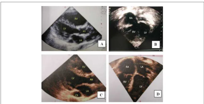
Figure 1 -Echocardiogram shows an enlargement of right cavities and a reduction in the size of left cavities especially of the left atrium in the preoperative period in a longitudinal parasternal section in A and in an apical section of four chambers in B. Images C and D, eight months after the operation, show that the cavities have been repaired. RA - right atrium, LA - left atrium, Ao - aorta, CS - coronary sinus, RV - right ventricle, LV - left ventricle.
Figure 2 -Angiography shows anomalous drainage of the right upper lobe vein (RUL) to the right superior cava vein in A, and of the right medial and lower lobe (RML and RLL) veins to the right atrium in B and C. The left upper lobe (LUL) vein and lingula (LL) drained normally into the left atrium in D. The left lower lobe vein (arrow) towards the right atrium in E. The left superior vena cava (LSVC), larger than the right one drained to the coronary sinus in F.
The sternal remained open for 36 hours, due to dilation of the right ventricle. With the use of furosemide, vasodilator (nitric oxide and captopril) and inotropics (dobutamine and adrenalin), there was stabilization of pulmonary venocapillary congestion and low cardiac output. The patient was discharged in good conditions 15 days after the procedure.
The echocardiogram performed eight months after the procedure showed normal heart cavities (LA: 29 mm; Aorta: 24 mm; RV: 20 mm; VE: 46 mm), normal biventricular function (LVEF: 65%) and competent valves. There was a 9-mm interatrial septal defect with left-to-right flow. Pulmonary systolic pressure was 23 mm Hg. There were no
Case Report
Atik et al
Anomalous drainage of 4 pulmonary veins
Arq Bras Cardiol 2007; 91(1) : e1-e3
signs of obstruction when the pulmonary venous return was redirected (fig. 1).
Discussion
The interatrial septum is intact in 20% to 33% of the cases of PAPVR1,2. Associations with other defects occur in
20% of them. Defects include cor triatriatum in 7.8%, mitral stenosis in 4.3% and aortic coarctation in 2.6% of the 139 cases reported from 1964 to 20061,2,6,7. Other defects are
sporadically associated, such as interventricular septal defect, left superior vena cava draining to the left atrium, interruption of the aortic arch, Fallot’s tetralogy, atrioventricular septum defect, in addition to genetic syndromes such as the syndromes of Turner, Jarcho Levin and Kabuki6,7. Of these 139 cases
reported, only in two of them there were three and four anomalous veins, an anatomical situation that corresponded to 1.4% of the total1-4.
It is known that interatrial septal defect (ASD) associated with PAPVR does not cause major pulmonary hypertension. However, in the absence of ASD, pulmonary venous return tends to become more intense through the anomalous pulmonary vein due to the lower resistance of right venous
structures, which causes greater right cavity volume overload and pulmonary hypertension2.
Systemic output depended exclusively on the flow from the left upper lobe vein and the lingula, and was then lower than 50% of the total pulmonary venous return, even considering the smaller size of the two anomalous upper pulmonary veins to the right. However, from a dynamic point of view, the patient behaved as if he had a classic type of ASD, with no major hemodynamic expression and without pulmonary hypertension and/or low systemic output.
This is why the existence of adaptive mechanisms is admitted in such unfavorable anatomical conditions. These mechanisms are so important that the patient was able to live with few symptoms and signs and also have normal physical growth.
The postoperative pulmonary congestion, caused by the reduced dimensions and compliance of left cavities, however, is in contrast with the preoperative adaptive picture.
Therefore, adaptive mechanisms to different stimuli, in addition to the individual response to these stimuli, remain dependent on aspects that cannot be easily explained, even from a logical point of view.
References
1. Senocak F, Ozme S, Bilgic A, Ozkutlu S, Ozer S, Saraclar M. Partial anomalous pulmonary venous return: evaluation of 51 cases. Jpn Heart J. 1994; 35: 43-50.
2. Alpert JS, Dexter L, Vieweg WV, Haynes FW, Dalen JE. Anomalous pulmonary venous return with intact atrial septum: diagnosis and pathophysiology. Circulation. 1977; 56: 870-5.
3. AboulHosn JA, Criley JM, Stringer WW. Partial anomalous pulmonary venous return: case report and review of the literature. Cathet Cardiovasc Intervent. 2003; 58: 548-52.
4. Weiman DS, Lee K, Levett JM, Replogle RL. Partial anomalous pulmonary venous return: a ten-year experience. Tex Heart Inst J. 1985; 12: 239-43.
5. Mullen JC, Waskiewich K, Bhargava R, Bentley MJ. Bilateral partial anomalous pulmonary venous return. Can J Cardiol. 1997;13: 567-9.
6. Cooley DA, Murphy MC. Cor triatriatum and anomalous pulmonary venous return: an unusual case. Tex Heart Inst J. 1990; 17: 118-21.
7. Elami A, Rein AJ, Preminger TJ, Milgalter E. Tetralogy of Fallot, absent pulmonary valve, partial anomalous pulmonary venous return and coarctation of the aorta. Int J Cardiol. 1995; 52: 203-6.