Importance of the Area of Fibrosis at the Midterm Evolution of
Patients Submitted to Ventricular Reconstruction
Gustavo Calado de Aguiar Ribeiro, Mauricio Lopes, Fernando Antoniali, Ana Nunes, Cledicyon Eloy Costa,
Juliano L Fernandes
Clinica Cardio Cirurgica Campinas, Radiologia Clinica Campinas, Hospital Samaritano de Campinas, Campinas, SP, Brazil.
Abstract
Background: Although it is acknowledged that the ventricular reconstruction surgery (VRS) can promote reverse remodeling, new studies are necessary to define the influence of the left ventricular (LV) area of fibrosis.
Objective: To evaluate whether the extension of the area of fibrosis of the LV is important in the LV functional recovery after the surgery and correlate it with clinical factors.
Methods: Prospective analysis of 82 patients with ventricular dysfunction submitted to VRS. We analyzed the importance of the clinical characteristics and the amount of fibrosis was assessed, measured by cardiac magnetic resonance (CMR) as small, medium and large.
Results: All patients were followed for 36 months, with a mortality of 6%. The amount of medium fibrosis was 25.8% ± 13.6%. There was improvement in the left ventricular ejection fraction (LVEF), from 36.9% ± 6.8% to 48.2% ± 8.2% (p < 0.001). There was an inverse association between the amount of fibrosis and the increase in LVEF (r = -0.83, p < 0.0001). There was a decrease in the LV end-systolic volume of 43.3 ± 8.2ml/m² (p < 0.001). There was an improvement in heart failure symptoms, except in patients with large areas of fibrosis (p = 0.45). The independent predictors for events were: fibrotic area (p = 0.01), age (p = 0.01), LV end-systolic volume (p = 0.03) and LVEF (p = 0.02). The event-free follow-up was different in relation to the area of fibrosis (p < 0.01).
Conclusion: In patients with ventricular dysfunction, the extension of the area of fibrosis was an independent predictor of the LV functional recovery after the VRS. The combination of cardiac MRI and clinical parameters can help in the indication for VRS. (Arq Bras Cardiol 2009; 93(6):564-570)
Key Words: Ventricular dysfunction, left; endomyocardial fibrosis; heart failure; stroke volume.
Mailing address: Gustavo Calado de Aguiar Ribeiro •
Rua Jose Teodoro de Lima 77 / 62 - Cambuí – 13015-150 – Campinas, SP, Brazil E-mail: [email protected], [email protected]
Manuscript received October 31, 2007; revised manuscript received April 14, 2008; accepted May 21, 2008.
Introduction
Approximately 70% of infarctions are the transmural type, with 54% in the anterior wall, where myocyte slippage occurs in 40% of these, resulting in wall expansion and narrowing, originating a dysfunctional akinetic or dyskinetic area1,2.
Moreover, in the remote myocardium, there are consequences in volume as well as in shape during the remodeling process, altering the normal elliptical shape to a spherical one, resulting in ventricular dysfunction3.
With the concept of ventricular reconstruction introduced by Jatene4 and modified by Dor et al5 for the correction of
the akinetic area, a new field opened for cardiac surgery. The techniques of left ventricular reconstruction surgery (VRS) result in symptom decrease, promoting the reverse remodeling and decreasing mortality6-9. However, it has been
observed, in all series, that not all patients benefit from this
type of procedure10-12, especially those with multiple infarcted
territories and high remote myocardium asynergy.
Therefore, it is crucial to develop noninvasive preoperative methods that can predict the outcome of the VRS and, ideally, that this information can be appropriate for patient selection and surgical planning. In this context, the present study determines the importance of the left ventricular (LV) area of fibrosis, correlating it with clinical characteristics at the midterm follow-up of patients submitted to VRS.
Patients and methods
Patients
A total of 82 patients (65.8% males), with a mean age of 64.2 ± 8 years, were prospectively studied for a period of 3 years; they presented surgical indication for VRS and met the following inclusion criteria at the cardiac magnetic resonance (CMR): contractile defect (akinesis and/or dyskinesis) of the LV anterior wall and LV ejection fraction (LVEF) < 45%.
viable myocardium on the anterior wall, pulmonary arterial systolic pressure > 70 mmHg; coronary artery bed that was unfavorable for myocardial revascularization, valvular lesion other than mitral insufficiency caused by the ischemic process or the remodeling.
The main indication for VRS was heart failure (HF) in 59 patients (70.9%), angina in 17 patients (20.7%), ventricular arrhythmia in 4 patients (4.8%) and 31 patients (37.8%) presented more than one criterion. All patients were submitted to VRS between 2002 and 2004 and to the concomitant myocardial revascularization and/or mitral valve plasty when indicated. The indication for myocardial revascularization was obstructive coronary artery lesion at the angiography > 50% and 70%, for left coronary trunk and artery branches, respectively. The indication for mitral valve correction was mitral regurgitation grade 3 and 4 and for grade 2, mitral annulus dimension > 40 mm.
The present study was formally approved by the Ethics Committee in Research and all patients received and signed the Free and Informed Consent Form.
Cardiac magnetic resonance imaging
Analyses of the myocardial viability, definition of the infarcted area and percentage of fibrosis of the LV were performed by cardiac magnetic resonance (CMR). The myocardial segments with previous infarction were determined according to the coronary artery irrigation territory: antero-septal (anterior interventricular artery), lateral (circumflex artery) and inferior (right coronary artery).
The infarcted area was characterized through the images obtained from a 1.5 Tesla equipment (Symphony, Siemens, Erlangen, Germany), using cardiac software. The images were acquired 10 minutes after the injection of 0.02 mmol/ kg of contrast containing gadolinium. The technique used was the late-enhancement with images obtained from 8 segments of the short axis view of the heart, performed during respiratory pauses of 6 to 10 seconds. The technique consists in a segmented pulse sequence, with FLASH (fast low-angle shot) inversion recovery pulse, with the following characteristics: TR/TE of 8/4; 8 to 12 heart beats; 8-mm thickness slices; matrix of 208 X 256; inversion recovery pulse of 180°; time of inversion ranging from 230 to 300 msec; FOV of 350 X 280 mm, with spatial resolution of 1.7 X 1.1mm, flip angle = 30°. The semiquantitative visual analysis was performed, with division of the eight slices: four divisions for slices 1 and 2 of the apex, six divisions for the slices 3 to 6 in the middle-third and 8 divisions for slices 7 and 8 of the base of the heart. In the late-enhancement areas, the myocardial thicknesses were divided: absent enhancement (0 point); enhancement from 1% to 25% f the myocardial thickness (1 point); enhancement from 26% to 75% of the thickness (2 points); enhancement > 75% of the myocardial thickness (3 points). The percentage of points is obtained by the addition of the points divided by 144 (maximum score) and multiplied by 100. The accepted margin of error is ± 4 (± 2.7%)13. The amount of fibrosis was divided and classified
in thirds: (I, small: 0% to 25%; II, moderate: 25% to 41%; III, large: 42% to 56%).
Symptoms and evolution
Heart failure (HF) was measured by functional class according to the criteria of the New York Heart Association (NYHA). Interviews and clinical assessments were carried out by the same physician during the evolution every three months up to 36 months and whenever necessary. The unfavorable evolution was defined as an event. The events included: death (cardiac and non-cardiac), worsening in functional class (NYHA), hospitalization and the need for new cardiac therapy (angiography, cardiac surgery, pacemaker implantation, multisite resynchronization, arrhythmia ablation and implantable defibrillator.
Analyses of the reverse remodeling and ventricular function during the evolution were performed by sequential echocardiograms (preoperative to 36 months), in a Vivid 3 equipment (General Electric), equipped with a second harmonic field and a 1.8 to 3.6 MHz transducer. The standard LV images were obtained at rest. The volumes and the LVEF were measured using the biplane Simpson’s method. The LV end-systolic and diastolic volumes (LESV and LEDV, respectively) were indexed by the body surface area (ESVI and EDVI).
Statistical analysis
Continuous data were expressed as mean ± standard deviation and compared by Analysis of Variance (ANOVA). Fisher’s test or Chi-square test were used for the categorical data. The multivariate analysis by logistic regression, used to identify independent factors for events, was carried out with the inclusion criterion of identified variables (p < 0.2) of the univariate analysis or variables considered to be clinically significant. The following clinical variables were analyzed during the evolution: age < or > 75 years; chronic pulmonary disease; renal failure; incomplete myocardial revascularization; diabetes mellitus; two or three coronary vascular territories; functional class III to IV; systolic volume < or > 120 ml/m²; mitral insufficiency; and time between acute myocardial infarction (AMI) and surgery. The correlation between two variables was carried out with Pearson’s and Spearman’s correlation test. The Kaplan-Meier method was used to analyze patients that were free of events.
Results
The 36-month follow-up, or to the death, was carried out in 100% of the patients. The interval between the myocardial infarction on the anterior wall and the VRS was 16.2 ± 8.5 months (ranging from 1 to 123 months).
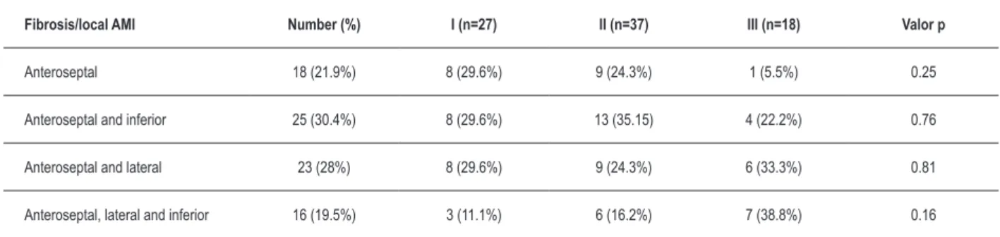
The distribution by infarcted ventricular territories was: antero-septal infarction (n = 18 patients); antero-septal and inferior (n = 25 patients); antero-septal and lateral (n = 23 patients); and antero-septal, inferior and lateral (n = 16 patients). Table 2 shows the distribution of patients by area of fibrosis and the number of infarcted territories.
Surgical data are shown in Table 3. There were no intraoperative deaths and the in-hospital mortality consisted of 3 patients (3.6%), due to cardiogenic shock, sepsis and pancreatitis, respectively.
The myocardium revascularization concomitant to the VRS was carried out in 79 patients (96.3%) and it was considered incomplete in 3 of them (3.6%). The mitral insufficiency correction with mitral valve plasty was performed in 17 patients (20.7%), with 9 patients from the group of fibrosis area grade III (p = 0.031)(Table 1) and in another patient (1.2%), from the fibrosis area grade II, six months after the VRS. The mitral valve plasty was more frequent in patients with LVEF < 30% (p = 0,021) and ESV > 120ml/m²) (p = 0.023).
Figure 1 shows the comparison of the distribution of patients with areas of fibrosis and infarcted ventricular territories in relation to NYHA –FC III to IV, with no difference in the preoperative period (p=0.51). In the postoperative period, it can be observed that patients with fibrosis area grade I had a decrease in HF symptoms in comparison to the preoperative period (p=0.03), with the same occurring with fibrosis area grade II (p=0.001), as well as regarding the distribution by infarcted vascular territories 1 (p = 0.02), 2 (p<0.001) and 3 (p=0.042). However, in patients with fibrosis area grade III, there was no regression of the HF symptoms (p = 0.45).
After 36 months, there was an improvement in LVEF from 36.9% ± 6.8% to 48.2% ±8.2% (p < 0.001). Figure 2 shows that the amount of fibrosis in the preoperative period was negatively correlated with the increase in LVEF at the end of the 36-month follow-up (r = -0.83, p < 0.0001).
Table 4 shows that the amount of fibrosis definitely influenced the 36-month evolution, as patients with fibrosis area grade III presented larger ventricular diameters and higher ESV, lower increase in LVEF and less reverse remodeling. Two
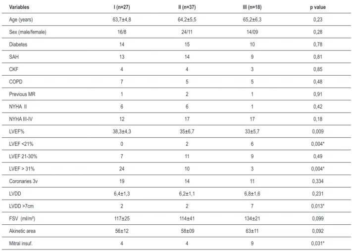
Table 1 – Preoperative data.
Variables I (n=27) II (n=37) III (n=18) p value
Age (years) 63,7±4,8 64,2±5,5 65,2±6,3 0,23
Sex (male/female) 16/8 24/11 14/09 0,28
Diabetes 14 15 10 0,78
SAH 13 14 9 0,81
CKF 4 4 3 0,85
COPD 7 5 5 0,48
Previous MR 1 2 1 0,91
NYHA II 6 6 1 0,42
NYHA III-IV 12 17 17 0,18
LVEF% 38,3±4,3 35±6,7 33±5,7 0,009
LVEF <21% 0 2 6 0,004*
LVEF 21-30% 7 11 9 0,49
LVEF > 31% 24 10 3 0,004*
Coronaries 3v 19 14 11 0,334
LVDD 6,4±1,3 6,2±1,1 6,8±1,6 0,231
LVDD >7cm 2 2 7 0,013*
FSV (ml/m²) 117±25 114±41 134±21 0,099
Akinetic area 56±12 58±09 63±11 0,092
Mitral insuf. 4 4 9 0,031*
SAH: systemic arterial hypertension; NYHA: New York Heart Association; Uremia: urea. 100mg/dl; LVEF: left ventricular ejection fraction; COPD: chronic obstructive pulmonary
Table 2 – Distribution of patients by area of ibrosis and the number of myocardial infarctions.
Fibrosis/local AMI Number (%) I (n=27) II (n=37) III (n=18) Valor p
Anteroseptal 18 (21.9%) 8 (29.6%) 9 (24.3%) 1 (5.5%) 0.25
Anteroseptal and inferior 25 (30.4%) 8 (29.6%) 13 (35.15) 4 (22.2%) 0.76
Anteroseptal and lateral 23 (28%) 8 (29.6%) 9 (24.3%) 6 (33.3%) 0.81
Anteroseptal, lateral and inferior 16 (19.5%) 3 (11.1%) 6 (16.2%) 7 (38.8%) 0.16
AMI - acute myocardial infarction.
Table 3 – Operative data according to the preoperative area of ibrosis.
Variables I (n=27) II (n=37) III (n=18) Valor p
Anas. AD 26 36 17 0.99
Distal Anas. 2.1±1.2 2.8±1.3 3.2±1.3 0.014
Vasoactive >24hrs 5 8 12 0.039
IAB 0 1 2 0.17
ECC time 85 ±09 91 ±24 88 ±16 0.44
Anox. time 52 ±11 48 ±16 47±10 0.37
Mortality 0 1 2 0.71
Anas. DA: anastomosis of vascular graft in the anterior descending artery; Distal Anas.: number of anastomoses in the coronary bed; Vasoactive > 24 hrs: need for vasoactive drugs for more than 24 hours; IAB: intra-aortic balloon; ECC time: time of extra-corporeal circulation; Anox. time: time of myocardial anoxia through total
clamping of the aorta; Variables I, II, III: small, medium and large areas of ibrosis, respectively. n: number of patients.
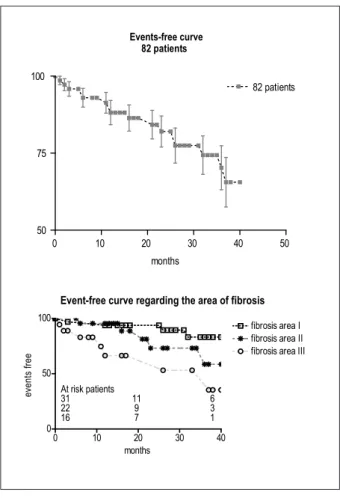
late deaths (2.4%) occurred, being one case (1.2%) due to cardiovascular cause (sudden death). Vasoactive drug support was more necessary (p = 0.039) in patients with fibrosis area grade III. Figure 3 shows the percentage of patients free of events, divided by area of fibrosis, being significant the number of events for patients with a large area of fibrosis (p < 0.01), whereas there is no difference for small and moderate areas of fibrosis (p = ns). Among the clinical events, it was necessary to implant three implantable cardiac defibrillators and four multisite pacemakers for cardiac resynchronization. Eight patients underwent 11 hospitalizations due to worsening in the HF picture and a new cardiac surgery was necessary in one patient.
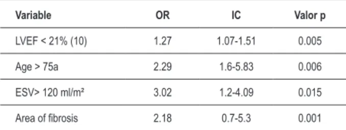
At the multivariate analysis with regression to determine independent predictors of events, fibrosis area, LVEF < 21%, systolic volume > 120 ml/m² and age > 75 years were identified (Table 5).
Discussion
The ventricular reconstruction surgery (VRS) is well established as a therapeutic option for patients with akinetic and dyskinetic areas, improving symptoms, ejection fraction and decreasing the ventricular cavity, although the myocardial 0
5 10 15 20 25 30 35 40 45 50
fib rosis 1 0 18 6 0 6 0 2 0
fib rosis 2 0 31 6 2 10 1 5 0
fib rosis 3 0 14 1 7 12 2 15 2
ter 1 0 13 3 2 8 1 6 1
ter 2 0 43 9 2 21 1 7 0
ter 3 0 12 2 2 5 1 9 1
FC1 Pre
FC1 Post
FC 2 Pre
FC 2 Post
FC3 Pre
FC 3 Post
FC 4 Pre
FC 4 Post
Figure 1 – Comparison by NYHA functional class (NYHA-FC) between the
number of infarcted vascular territories and the area of ibrosis in the pre and
Table 4 – Evolution data after 36 months, according to the area of preoperative ibrosis.
Variables I (n=27) II (n=35) III (n=15) Valor p
LVDD 4.3±0.73 4.99±0.43 5.16±0.72 <0.0001
ESV (ml/m²) 68±14 76±25 89±21 0.011
∆ ESV -43±4.2 - 46±5.2 -38±6.7 0.0001
∆ EF% 16.8±2.6 13.8±2.6 10.3±3.4 <0.0001
LVDD: post-surgical left ventricular-end diastolic diameter; ESV: LV end-systolic volume; ∆EF%: difference between the preoperative LV ejection fraction and after 36 months; ∆ESV: difference between the preoperative LV inal systolic volume and after 36 months. Variables I, II, III: small, medium and large areas of ibrosis,
respectively.
revascularization is performed concomitantly in most patients6,7.
The present study analyzes patients with multiple infarcted ventricular territories and the current literature is still controversial regarding the surgical indication for this type of patient, due to the high surgical risk11,14 and the exiguity of
data concerning the evolution in the middle and long term. Moreover, in the present study, most patients present akinetic areas and their evolution is more unfavorable than those with dyskinetic areas, as shown in the RESTORE study6, where the
5-year survival was higher in patients with dyskinesis (80%
versus 65%, p < 0.001).
Recently, Mickleborough et al11 reported that large
ventricular volumes and extensive dysfunctional areas are nor contraindications for VRS. In the present study, although many patients presented high surgical risk (low LVEF, large ventricular volume and mitral insufficiency), we obtained a low in-hospital mortality, similar to the one observed in the RESTORE study6.
We found the same results observed in other series regarding the risk factors for events at the multivariate analysis: LVEF<
21%, age > 75 years, preoperative systolic volume and fibrosis area. Systolic volume reduction was observed in the three groups of fibrosis, although the group with the higher grade of fibrosis presented a significantly higher ESV at the end of the 36 months. In the series reported by Patel et al14, some
patients did not present clinical improvement and needed other therapies. This also occurred in the present series and other types of therapy, such as resynchronization by multisite pacemaker and ventricular arrhythmia ablation were necessary. Dor et al15, reported on a large long-term series, the need for
re-interventions, arrhythmia-related mortality and the recurrence
Figure 2 – Inverse correlation between the extension of the preoperative
ibrosis area and the LV ejection fraction variation between the pre and the
postoperative periods after a 36-month period.
Events-free curve 82 patients
0 10 20 30 40 50
50 75 100
82 patients
months
Event-free curve regarding the area of fibrosis
0 10 20 30 40
0 50 100
fibrosis area I fibrosis area II fibrosis area III
months
e
ve
n
ts
f
re
e
At risk patients
31 11 6 22 9 3 16 7 1
Figure 3 – Event-free percentage for all patients and with different areas of
1. McKay RG, Pfeffer MA, Pasternak RC, Markis JE, Come PC, Nakao S, et al. Left ventricular remodeling after myocardial infarction: a corollary to infarct expansion. Circulation. 1986; 74 (4): 693-702.
2. Stone PH, Raabe DS, Jaffe AS, Gustafson N, Muller JE, Turi ZG, et al. for the MILIS Group. Prognostic significance of location and type of myocardial infarction: independent adverse outcome associated with nterior location. J Am Coll Cardiol. 1988; 11: 453-63.
3. White HD, Norris RM, Brown MA, Brandt PW, Whitlock RM, Wild CJ. Left ventricular end-systolic volume as the major determinant of survival after recovery from myocardial infarction. Circulation. 1988; 76: 44-51.
4 Jatene AD. Left ventricular aneurysmectomy: resection or reconstruction. J Thorac Cardiovasc Surg. 1985; 89: 321-31.
5 Dor V, Saab M, Coste P, Kornaszewski M, Montiglio F. Left ventricular aneurysm: a new surgical approach. J Thorac Cardiovasc Surg. 1989; 37: 11-9.
6 Athanasuleas CA, Buckberg G, Stanley GH, Siler W, Dor V, Di Donato M, et al. Surgical ventricular restoration in the treatment of congestive heart failure due to post-infarction ventricular dilatation. J Am Coll Cardiol. 2004; 44 (7): 1439-45.
References
of the adverse cardiac remodeling process15. The cardiac
magnetic resonance (CMR) allows the observation, with a high degree of reliability, the myocardial viability, the magnitude of the changes in the transversal and longitudinal axes, the infarction location and compliance, and also the analysis of the ventricular functions and the mitral insufficiency, when present. In the present study, the patients were systematically investigated by CMR concerning the extension of the infarcted area and the number of affected ventricular territories and there is no association between these factors and mortality.
Few data in the literature express the role of the remote muscle in the in the evolution post-VRS. In the present study, there was a correlation between the LV fibrosis area and evolution, ventricular function and symptoms at the end of the 36-month follow-up. Those with large areas of fibrosis presented less reverse remodeling, lower increase in EF and less relief of the heart failure. Athanasuleas et al6 suggested
that the viability of the remote muscle is determinant for
the indication or not of the surgical procedure. Patel et al14
reported that further studies are necessary to determine the importance of the lateral wall in this type of procedure. In the present study, the area of fibrosis was a more important determinant than the number of affected vascular territories. Although good results were demonstrated16,17 with patients
with multiple infarctions and/or large areas of fibrosis, the amount of muscle necessary to guarantee a favorable evolution has yet to be determined. The quantification of the area of fibrosis for each vascular territory can provide additional data and that is one limitation of the present study.
Conclusion
The extension of the area of fibrosis was inversely proportional to the LV functional recovery after the VRS, as well as to the HF symptom relief and the combination of clinical factors can help in the indication for the surgical procedure.
However, further studies are necessary to determine which patients with extensive dysfunctional LV area can benefit from this type of procedure.
Potential Conflict of Interest
No potential conflict of interest relevant to this article was reported.
Sources of Funding
There were no external funding sources for this study.
Study Association
This study is not associated with any post-graduation program.
Table 5 – Multivariate analysis with logistic regression for event-related factors.
Variable OR IC Valor p
LVEF < 21% (10) 1.27 1.07-1.51 0.005
Age > 75a 2.29 1.6-5.83 0.006
ESV> 120 ml/m² 3.02 1.2-4.09 0.015
Area of ibrosis 2.18 0.7-5.3 0.001
LVEF - left-ventricular ejection fraction Left ventricle ejection fraction (tested with
EF< 21, between 21-30, and EF between 31-40%; Age - tested every 10 years and > 75 years; ESV - left ventricular end-systolic volume (ESV was tested < 100 ml/m², ESV was tested between 100-120 ml/m² and ESV was tested > 120 ml/m²).
7 Dor V, Sabatier M, Di Donato M, Maioli M, Toso A, Montiglio F. Late hemodynamic results after left ventricular patch repair associated with coronary grafting in patients with postinfarction akinetic or dyskinetic aneurysm of the left ventricle. J Thorac Cardiovasc Surg. 1995; 110: 1291-301.
8 Maxey TS, Reece TB, Ellman PI, Kern JA, Tribble CG, Kron IL. The beating heart approach is not necessary for the Dor procedure. Ann Thorac Surg. 2003; 76: 1571-5.
9 Schenk S, McCarthy PM, Starling RC, Hoercher KJ, Hail MD, Ootaki Y, et al. Neurohormonal response to left ventricular reconstruction surgery in ischemic cardiomyopathy. J Thorac Cardiovasc Surg. 2004; 128: 38-43.
10. Di Donato M, Sabatier M, Dor V, Gensini GF, Toso A, Maioli M, et al. Effects of the Dor procedure on left ventricular dimension and shape and geometric correlates of mitral regurgitation one year after surgery. J Thorac Cardiovasc Surg. 2001; 121: 91-6.
11. Mickleborough L, Merchant N, Ivanov N, Rao V. Left ventricular reconstruction: early and late results. J Thorac Cardiovasc Surg. 2004; 128: 27-37.
15.Dor V. Left ventricular reconstruction: the aim and the reality after twenty years. J Thorac Cardiovasc Surg. 2004; 128: 1: 17-9.
16. Maxey T, Reece T, Ellman P, Butler P, Kern J, Tribble C, et al. Coronary artery bypass with ventricular restoration is superior to coronary artery bypass alone in patients with ischemic cardiomyopathy. J Thorac Cardiovasc Surg. 2004; 128: 27-37.
17. Aguiar Ribeiro G, Costa CE, Lopes MM, Albuquerque A, Antoniali A, Reinert GA, et al. Left ventricular reconstruction benefits patients with ischemic cardiomyopathy and non-viable myocardium.Eur J Cardiothorac Surg. 2006; 29: 196-201.
dysfunction: comparison with a series of large dyskinetic scars. J Thorac Cariovasc Surg. 1998; 116: 50-9.
13. Azevedo Filho CF, Hadlich M, Petriz JlF, Mendonça LA, Moll JF, Rochitte CE. Quantificação da massa infartada do ventrículo esquerdo pela ressonancia magnetica cardiaca: comparação entre a planimetria e o método de escore visual semi-quatitativo. Arq Bras Cardiol. 2004; 83: 111-7.