Calibragem de tonômetros em Brasília, Brasil
Trabalho realizado no UNIPLAC, School of Medicine - Brasília (DF) - Brazil.
1Acadêmica do 6º ano de Medicina da Escola Superior de Ciências da Saúde - ESCS - Brasília (DF) - Brazil. 2Acadêmico do 6º ano de Medicina da ESCS - Brasília
(DF) - Brazil.
3Doutor, Professor de Oftalmologia da Faculdade de Medicina do Planalto Central - FAMEPLAC - Brasília (DF) - Brazil.
Address for correspondence: Geraldo Magela Vieira. SQS 306 - Bl. G - Apto. 405 - Brasília (DF) CEP 70353-070
E-mail: [email protected] Recebido para publicação em 08.12.2008 Aprovação em 04.03.2009
Fernanda Pires da Silva Abrão1 Warley Alves Araújo2
Geraldo Magela Vieira3
Keywords: Intraocular pressure; Tonometry, ocular/instrumentation; Calibration; Equipment failure analysis; Glaucoma; Quality control
Purpose: To determine calibration errors of Goldmann applanation tonometers in ophthalmic clinics of Brasília, Brazil, and correlate the findings with variables related to tonometers model and utilization. Methods: Tonometers from ophthalmic clinics in Brasília, Brazil, were checked for calibration errors. A standard Goldmann applanation to-nometer checking tool was used to asses the calibration error. Only one trained individual made all verifications, with a masked reading of the results. Data on the model, age, daily use, frequency of calibration checking and the nature of the ophthalmic department - private or public - were collected and correlated with the observed errors. Results: One hundred tonometers were checked for calibration. Forty seven percent (47/100) were out of 1 mmHg range at least at one point checking. Tonometers mounted to slit lamp, with less than 5 years, used in less than 20 patients daily, that had a calibration check on a yearly basis, and those from private office exhibit a lower rate of inaccuracy, but only the first variable was statistically significant. Sixty one percent of to-nometers on public hospitals were out of calibration. Conclusion: Calibration of tonometers in the capital of Brazil is poor; those from general hospitals are worst, and this fact can lead to inaccurate detection and assessment of glaucoma patients, overall in the population under government assistance.
ABSTRACT
INTRODUCTION
The relevant role played by intraocular pressure (IOP) in glaucoma pathophysiology is being more and more emphasized(1-4) generating the
need to obtain its precise measurements both for diagnosis and follow-up of glaucomatous patients.
Since its introduction in the 1950s, Goldmann’s applanation tonometry (GAT) has been considered the gold standard to check IOP(5). Several
factors influence IOP evaluation by GAT, such as time and number of checks(6), amount of fluorescein drops(7), corneal curvature(8-9), central
cor-neal thickness(10-11), among others. However, before questioning any other
interfering factor, one should pay attention to instrument calibration. Recent studies reported a high prevalence of calibration errors GAT(12-13)
which may confound the ophthalmologist regarding both the diagnosis and follow-up of glaucoma suspects and glaucoma patients.
inhabitants is located), we heard of the low demand for tono-meter calibration service in the region. We believe that despite of recent publications having emphasized the need for regular GAT checks, this problem may still have an important dimen-sion in the Federal District.
The aim of this study is to verify GAT calibration in diffe-rent clinics and hospitals of the Federal District, as well as possible factors related to calibration errors.
METHODS
A hundred Haag Streit® GATs from several public or
pri-vate health care services of the Federal District were randomly tested in this transversal study.
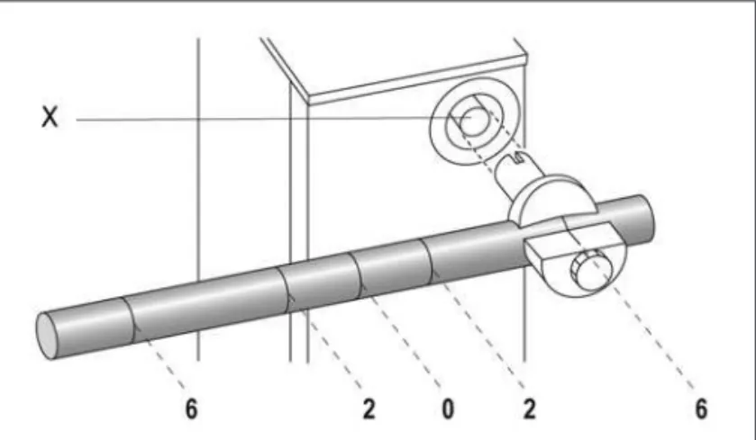
Tonometer calibration was checked in position “0” (0 mmHg), “2” (20 mmHg) and “6” (60 mmHg) using a standard checking cylinder(14) (Figure 1).
The highest and lowest reading in each of the checking points were identified according to the manufacturer’s instruc-tions manual(14). The difference between the highest and lowest
reading was considered the measure of the degree of the ins-trument play. The mid-point between the highest and the lowest reading was considered the true reading at the checked point.
Since the revolving drum had its movement limited in the position corresponding to -2.0 mmHg, tonometers with dings less than -2.0 mmHg were considered to present a rea-ding of -2.0 mmHg. Tonometers with a checking error greater than or equal to 1.0 mmHg in at least one of the three mea-suring points were considered as having a calibration error.
The following variables were also analyzed regarding their influences on calibration errors: model (mounted to slit lamp or removable), time of manufacture (≤5 years or >5 years), number of daily attended patients (≤20 patients or >20 pa-tients), calibration check frequency (at least annually or more than annually) and the nature of the health care faculty which used the tonometer (public or private).
Data were collected from January to February 2007, all checks and interviews being performed by only one examiner who was trained previously for the task (FPSA). Reading of the checking measurements was masked and made by an independent observer (WAA).
Data regarding the age of the tonometer, calibration fre-quency and daily use were obtained from the administration staff of clinics or directly from the doctor who used the tono-meter. Interviews were made always by the same examiner (WAA).
For the statistical analyses the chi-square test of the SPSS 14.0 program (SPSS Inc, Chicago, IL, USA) was used and p<0.05 was considered a statistically significant association.
RESULTS
A hundred tonometers were assessed for calibration errors. Forty seven per cent (47/100) presented with calibration error regarding at least at one of the three checked reference points. Twenty-four percent (24/100) of the instruments presented a calibration error at position “0”; 27.0% (27/100) at position “2”, and 36.0% (36/100) at position “6”.
Figure 2 shows the frequency of calibration error of the sample at each of the reference checking positions.
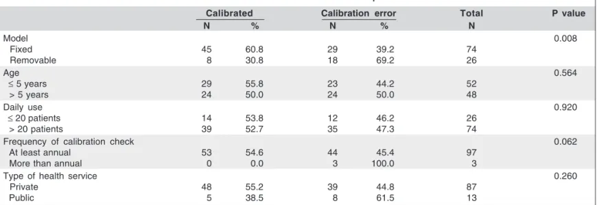
The relationship between calibration error and model, age, daily use, frequency of calibration error checks, and nature of service where the tonometer is used are shown in Table 1.
Among the evaluated associations, only the tonometer mo-del is correlated in a statistically significant way with the fre-quency of calibration error (p=0.008). The other variables do not present a statistically significant association with the occurrence of calibration error (Table 1).
Means, respective standard deviations (SD) and variation coefficients (VC) of the measurements at positions “0”, “2” and “6” in slit lamp mounted and removable tonometers are described in Table 2. The greatest variation coefficient found was 5 mmHg at position “0” and 10 mmHg at both positions “2” an “6”.
Figure 1 - Key for calibration check of Goldmann’s applanation
tonom-e t e r s
with 0 (0 mmHg), 2 (20 mmHg) e 6 (60 mmHg) markers*
*= the key should be coupled to the tonometer orifice indicated by X Adapted from Applanation Tonometer to Slit Lamp - Instruction Manual
DISCUSSION
Goldmann applanation tonometer mechanism comprises a checking prism made of an organic lens whose extremity turned towards the eyeball is plane with rounded borders in order to avoid corneal lesions. In contact with cornea, the prism’s plane surface exerts a pressure controlled by the exa-miner by turning a graded drumwhich makes the tonometer shaft move, thus determining the pressure of the prism to the patient’s eye. The drum is turned until an 7.354 mm2 area of
corneal surface is flattened. The force needed to obtain this applanation is converted into pressure and IOP value may then be determined(14).
Calibration error of a tonometer leads to an error in IOP reading. The diagnostic classification of a patient as normal, glaucomatous or glaucoma suspect, as well as the determina-tion of the target IOP, may be impaired to a certain degree(15-17).
In dealing with a mechanical system, GAT needs periodical calibration. The manufacturer’s manual recommends the fre-quency of calibration check to be monthly at the measurement points corresponding to 0 mmHg, 20 mmHg and 60 mmHg. For this purpose, a specific key supplied by the manufacturer is used to verify whether the instrument is calibrated or not (Figure 1)(14).
The manufacturer considers 0.5 mmHg as an acceptable error margin at 0 mmHg and 20 mmHg measurements, and
1.0 mmHg at 60 mmHg measurement(14). The present study
considers a tonometer with calibration error those with check error greater than or equal to 1.0 mmHg in at least one of the three measurement points.
In our study, 47.0% (47/100) presented a calibration error in at least one of the three check points. In addition, 40 GATs presented a degree of the instrument play over 1.0 mmHg at position “2”, exceeding 4.0 mmHg in five of these instru-ments. At position “6”, there were five instruments with a calibration error greater than 4.0 mmHg, one of which pre-sented 10.0 mmHg as a degree of the instrument play. On the other hand, only one tonometer showed an error greater than 4.0 mmHg at position “0”.
Our data demonstrate that calibration errors did not pre-sent an internal consistency among the three studied check points, showing variation in magnitude among them. Conver-sely, the observed high variation coefficients reveal a quite heterogeneous sample, with many reading variation between tonometers at the same check point.
Errors less than 1.0 mmHg may not have a great influence on clinical decision(18), but we found several instruments with
variation over this value, which may probably lead to an error in IOP readings of patients, with serious consequences, ove-rall in those with advanced glaucoma(19-20).
Wessels & Oh in a sample of 175 applanation tonometers mounted to a slit lamp, and checking the calibration only at position “2”, found 18.3% of instruments with calibration error with a mean error of 0.43 mmHg (varying from -0.29 to 1.44 mmHg)(12). The numbers found by these authors are
clearly inferior to those observed in our study.
In a similar investigation conducted in Recife, PE, in nor-theastern Brazil, which used the same criteria for calibration error as that of our study, Costa et al. found 12.7%, 25.3% and 10.1% tonometers with calibration errors at positions “0”, “2”, and “6”, respectively(13). In that study, it was observed that
25.3% of tonometers presented a calibration error, a quite Table 1. Characteristics of tonometers and their relationship with calibration error
Calibrated Calibration error Total P value
N % N % N
Model 0.008
Fixed 45 60.8 29 39.2 74
Removable 8 30.8 18 69.2 26
Age 0.564
≤ 5 years 29 55.8 23 44.2 52
> 5 years 24 50.0 24 50.0 48
Daily use 0.920
≤ 20 patients 14 53.8 12 46.2 26
> 20 patients 39 52.7 35 47.3 74
Frequency of calibration check 0.062
At least annual 53 54.6 44 45.4 97
More than annual 0 0.0 3 100.0 3
Type of health service 0.260
Private 48 55.2 39 44.8 87
Public 5 38.5 8 61.5 13
Table 2. Difference between fixed and removable tonometers regading degree of the instrument play of readings at 0 mmHg, 20 mmHg and 60 mmHg and their respective standard deviations
Reading Fixed Removable
Mean ± SD*; VC† Mean ± SD; VC
0 mmHg 1.54 ± 1.10; 0.71 1.54 ± 1.17; 0.76
20 mmHg 1.51 ± 1.93; 1.28 1.62 ± 2.08; 1.28
60 mmHg 1.05 ± 1.52; 1.45 1.73 ± 2.09; 1.21
lower number than that found in our research. In an Ophthal-mologic Center of Vancouver, Canada, with a sample of 25 instruments, calibration errors up to 1.0 mmHg were classified as clinically irrelevant. Even so, the obtained percentage of tonometers with calibration error was 24%(21), still a quite
smaller number than that found in the present study.
Of the removable tonometers, 69.2% had calibration error, while this index was 39.2% in slit lamp mounted instruments. This numbers are higher compared to that obtained by Costa et al., where percentages were 39.0% and 10.5%, respective-ly(13). In the same way, Wessels & Oh observed 33.0% and
12.6%, respectively, confirming a significant relationship between the tonometer model and calibration(12).
Means of degrees of the instrument play and the respec-tive standard deviations were greater in removable tono-meters when compared to fixed ones, a fact also verified in Wessels & Oh’s study who observed error and standard de-viation means of 0.36 ± 0.65 for slit lamp mounted models and 0.60 ± 0.84 for removable ones. This demonstrates that more calibration errors occur and this tends to be greater in remo-vable tonometers. This fact, as suggested, may be explained by the handling and shocks to which the removable tonome-ters are exposed(12).
Despite the fact that half of tonometers over 5 years old were calibrated, they presented proportionally more calibra-tion errors when compared to those with less than 5 years, which is in agreement with Wessels & Oh(12). This may be
linked to the fact that, due to a longer exposure time, older tonometers are more subject to dust and other debris accumu-lation in their mechanism along time, determining increase in resistance to movement and culminating in loss of precision(12).
Little difference was observed between calibration of to-nometers used for twenty or less patients per day as compared to those used for a higher number of patients. These data agrees with those obtained by some authors(13), but contradict others(12).
Most tonometers are checked at least annually, however, all three tonometers which were checked with a frequency greater than annual presented calibration error, with two of them showing inaccuracy at the three evaluated positions. Our interview regarding calibration frequency was conducted by one of the researchers by personal communication, which can cause some constraint and therefore is subjected to a bias. This is represented by the great number of tonometers with calibration error in contrast to the high calibration check frequency informed in our interview. In the United Kingdom, a study conducted using questionnaires sent by mail to oph-thalmologists showed that 85% of them never tested the calibration of their tonometers(22).
It can be seen that there is a greater proportion of tonome-ters with calibration error in public health services when com-pared to private ones. This probably occurs due to the preca-rious conditions of instrument maintenance in Brazilian public health service and, naturally, to the excessive use. Unfortuna-tely, the sample of this last group was too small to obtain a possible statistically significant association.
In consonance with the manual of the instrument manu-facturer(14), some authors(21-22) suggested that calibration check
frequency of tonometers should be monthly. According to data of the present research, we believe that a better aware-ness by ophthalmologists is needed regarding the importance of performing the simple procedure of periodically checking the tonometers, especially the models not fixed to the slit lamp. The great errors observed overall in public hospital of Federal District demonstrate the need to establish routine procedures in ophthalmic services of Brasília, determining the time when tonometer check should be performed and the per-son responsible for this task.
CONCLUSION
It was observed that removable tonometers with over five years of use, attending more than twenty patients daily, checked at time intervals over one year and which are used in public health services are proportionally those with more cali-bration errors.
The high prevalence of tonometers with calibration error in health services of the Federal District shows the importance of instruments frequent calibration checks to guarantee a correct follow-up of the patients, specially those with glaucoma.
RESUMO
Descritores: Pressão intraocular; Tonometria ocular/instru-mentação; Calibragem; Análise de falha de equipamento; Glau-coma; Controle de qualidade
REFERENCES
1. Hollows FC, Graham PA. Intraocular pressure, glaucoma, and glaucoma suspects in a defined population. Br J Ophthalmol. 1966;50(10):570-86. 2. Bengtsson B. The prevalence of glaucoma. Br J Ophthalmol. 1981;65(1):46-9. 3. Klein BE, Klein R, Sponsel WE, Franke T, Cantor LB, Martone J, Menage MJ. Prevalence of glaucoma. The Beaver Dam Eye Study. Ophthalmology. 1992;99(10):1499-504.
4. Heijl A, Leske MC, Bengtsson B, Hyman L, Bengtsson B, Hussein M; Early Manifest Glaucoma Trial Group. Reduction of intraocular pressure and glauco-ma progression: results from the Early Manifest Glaucoglauco-ma Trial. Arch Ophthalmol. 2002;120(10):1268-79. Comment in: Arch Ophthalmol. 2002;120(10):1371-2. JAMA. 2002;288(20):2607-8. Optom Vis Sci. 2002;79(12):741-2.
5. Goldmann H, Schmidt T. Über applanationstonometrie. Ophthalmologica. 1957; 134(4):221-42.
6. Moses RA. The Goldmann applanation tonometer. Am J Ophthalmol. 1958; 46(6):865-9.
7. Grant WM. Fluorescein for applanation tonometry: more convenient and uni-form application. Am J Ophthalmol. 1963;55:1252-3.
8. Mark HH. Corneal curvature in applanation tonometry. Am J Ophthalmol. 1973;76(2):223-4.
9. Holladay JT, Allison ME, Prager TC. Goldmann applanation tonometry in patients with regular corneal astigmatism. Am J Ophthalmol. 1983;96(1):90-3. 10. Ehlers N, Bramsen T, Sperling S. Applanation tonometry and central cornea
thickness. Acta Ophthalmol (Copenh). 1975;53(1):34-43.
11. Wolfs RC, Klaver CC, Vingerling JR, Grobbee DE, Hofman A, de Jong PT.
Distribution of central corneal thickness and its association with intraocular pressure: The Rotterdam Study. Am J Ophthalmol. 1997;123(6):767-72. 12. Wessels IF, Oh Y. Tonometer utilization, accuracy, and calibration under field
conditions. Arch Ophthalmol. 1990;108(12):1709-12.
13. Costa RT, Barreto RBP, Lopes FP, Lira RPC. Análise da aferição dos tonômetros de aplanação de Goldmann em serviços oftalmológicos de Recife.
Arq Bras Oftalmol. 2004;67(2):267-70.
14. Haag-Streit International. Applanation Tonometer to Slit Lamp. Instruction Manual. Koeniz: Haag-Streit Ag Ophthalmic Instruments; 2005.
15. The Advanced Glaucoma Intervention Study (AGIS): 7. The relationship between control of intraocular pressure and visual field deterioration. The AGIS Investigators. Am J Ophthalmol. 2000;130(4):429-40. Comment in: Am J Ophthalmol. 2000;130(4):490-1.
16. Gordon MO, Beiser JA, Brandt JD, Heuer DK, Higginbotham EJ, Johnson CA, et al. The Ocular Hypertension Treatment Study: baseline factors that predict the onset of primary open-angle glaucoma. Arch Ophthalmol. 2002;120(6):714-20; discussion 829-30. Comment in: Arch Ophthalmol. 2004;122(7):1088-9; author reply 1089.
17. Seddon JM, Schwartz B, Flowerdew G. Case-control study of ocular hyperten-sion. Arch Ophthalmol. 1983;101(6):891-4.
18. Sandhu SS, Chattopadhyay S, Birch MK, Ray-Chaudhuri N. Frequency of Goldmann applanation tonometer calibration error checks. J Glaucoma. 2005; 14(3):215-8.
19. Le A, Mukesh BN, McCarty CA, Taylor HR. Risk factors associated with the incidence of open-angle glaucoma: the visual impairment project. Invest Oph-thalmol Vis Sci. 2003;44(9):3783-9.
20. Medeiros FA, Pinheiro A, Moura FC, Leal BC, Susanna R Jr. Intraocular pressure fluctuations in medical versus surgically treated glaucomatous patients.
J Ocul Pharmacol Ther. 2002;18(6):489-98.
21. Chuo JY, Mikelberg FS. Calibration errors of Goldmann tonometers in a tertiary eye care centre. Can J Ophthalmol. 2007;42(5):12-4.