Randomized Comparative Study of Diamond-like Carbon Coated
Stainless Steel Stent
versus
Uncoated Stent Implantation in Patients
with Coronary Artery Disease
George César Ximenes Meireles, Luciano Mauricio de Abreu, Antonio Artur da Cruz Forte, Marcos Kiyoshi Sumita, Jorge
Hideki Sumita, Jose Del Carmen Solano Aliaga
Hospital Stella Maris – São Paulo, SP - Brazil
Summary
Objective: To compare restenosis and major cardiac event rates at one and six months after DLC-coated stent implantation with those of uncoated stents.
Méthods: Randomized, prospective, comparative study of 180 patients with coronary insufficiency undergoing DLC coated stent (Phytis™) or uncoated stent (Penta™) implantation, from January, 2003 to July, 2004. Inclusion criteria
were: de novo lesion with >50% diameter stenosis in a coronary artery with reference diameter ≥ 2,5 mm and ≤ 4 mm,
and length < 20 mm. Exclusion criteria were: left main coronary artery and bifurcation lesions, chronic total occlusion, and in-stent restenosis.
Results: Clinical and angiographic baseline characteristics of the groups were similar. Procedural success was achieved in 98.9% of the patients in both groups. One cardiac death occurred in each group during hospitalization. Reference diameter and acute gain were greater in the Penta™ group (3.21±0.37 mm vs 3.34±0.8 mm, p=0.02 and 2.3±0.5 vs
2.49±0.5, p=0.009, respectively). Angiographic follow-up at six months showed similar rates of restenosis (24.3% vs
21.8%, p=0.84) and of major cardiac events (16.8% vs 17.5%, p=1).
Conclusion: DLC coated stents did not provide better outcomes in relation to uncoated stents.
Key words: Coronary disease, coronary restenosis, angioplasty transluminal percutaneous coronary.
Mailing Address: George César Ximenes Meireles •
Rua Sena Madureira, 1265/102 - 04021-051 – São Paulo, SP - Brazil E-mail: [email protected]
Manuscript received June 19, 2006; revised manuscript received September 25, 2006; accepted November 24, 2006.
Introduction
The efficacy and safety of drug-eluting stents has been confirmed by several studies published in prestigious medical journals1-6, and these stents are used in more than 80% of the
percutaneous procedures in the United States of America, but the utilization rate is much lower in some countries in Europe and Latin America (<30%) due to the high cost of the procedure7. Most of the standard and drug-eluting stents
are made of stainless steel. Therefore, new information on stainless steel stents is of great importance.
Stainless steel stents may release heavy metal ions such as nickel, chromium, and molybdenum, and these ions may cause allergic and hypersensitivity reactions resulting in a stimulus for proliferation and migration of smooth muscle cells, and, consequently, in restenosis. Stent surface coating with hemocompatible and biocompatible material may reduce thrombogenicity by reducing platelet adhesion and activation, and inflammatory response by reducing the release of cytotoxic metal ions8-14.
Stent coating may be divided into two categories: biocompatible materials and drug-eluting coatings (ex.:
sirolimus and paclitaxel). Biocompatible materials include inert coatings such as carbon, gold and silicon carbide15.
Diamond-like carbon is essentially diamond with a small percentage of hydrogen (4%). Because of the carbon-carbon bond resistance, diamond is, along with boron nitride, the hardest substance known, and if used in stent coating it would result in inflexible endoprostheses. The introduction of hydrogen into the carbon structure of diamond adds the benefits of diamond (resistance) to the coating flexibility provided by the presence of hydrocarbon bonds within the carbon atom clusters. Some of the properties of diamond-like carbon are: low friction coefficient, hydrophilic surface, inertness, flexibility, and hemocompatibility and biocompatibility10-12.
The objective of this study was to determine whether diamond-like carbon coated stainless steel stents may reduce restenosis rates more efficiently than uncoated stainless steel stents.
Methods
infarction and target vessel revascularization). Acute lumen gain was defined as the difference between the minimum lumen diameter (MLD) immediately after implantation and baseline MLD. Late lumen loss was defined as the difference between MLD immediately after and MLD at six months after implantation. Binary restenosis was defined as a percent diameter stenosis > 50% in angiography performed at six months after implantation in the stent segment, and up to 5mm proximal and distal to the edges. Control angiographies were performed at six months after implantation or earlier, if clinically indicated.
Secondary endpoints were major cardiac event rates at one and six months after implantation. The patients were seen in the hospital’s outpatient service at one and six months after implantation. Myocardial infarction (MI) was defined as the occurrence of a new Q wave > 0.04 seconds in two contiguous leads with elevation of CK-MB levels. Non-ST elevation MI was defined as the absence of new Q waves and elevation of CK-MB levels at more than three times the upper normal limit.
Calculation of a sample of 180 patients divided into two equal groups was based on the supposition of a 0.05 type I error, with a power of test of 80%, and an angiographic restenosis rate of 30% for Penta stents17 and of 12% for Phytis
stents18. In order to test the hypothesis of equality of means
in the groups, the Student’s t test was used for independent samples. For comparison between the proportions, the chi-square test or Fisher test was used when expected frequencies below 5 occurred. To assess the correlations (univariate) between restenosis and clinical and angiographic variables, the Pearson’s correlation coefficient was used; and to study these correlations in a multivariate fashion, the multivariate linear regression model with stepwise variable selection was used. The significance level used for the tests was 5%. Data were expressed as mean ± SD for continuous variables, and as frequency for categorical variables. Statistical analyses were performed with the Statistical Analysis System (SAS) version 6.12.
Results
From January, 2003 to July, 2004, 90 patients were randomly assigned to Phytis™ stent implantation and 90 to Penta™ stent implantation.
The clinical characteristics of the patients are listed in Table 1. Both groups were similar as regards all variables analyzed. Characteristics and location of the lesions treated are shown in Table 2, and no statistically significant differences were observed.
Procedure variables were comparable in the two groups, except for stent length which was shorter in the Penta™ group (Table 3). No difference was observed between the groups with respect to procedural success (98.9% in both groups). One death occurred in each group due to MI with cardiogenic shock and, except for these, no elevations in enzyme levels above the established criteria were observed after the procedure.
Three patients in the Phytis™ group and 12 in the Penta™ implantation in patients with coronary artery disease. The
study protocol was approved by the institution’s Research Ethics Committee and was conducted according to the principles of the Declaration of Helsinki. Written informed consent was obtained from all patients.
Patients with single or multivessel coronary artery disease, stable angina, or acute coronary syndrome were included in the study. In patients with multivessel disease only one lesion was treated with one of the protocol stents. Angiographic inclusion criteria were the presence of de novo lesion with > 50% diameter stenosis in native coronary arteries, with reference diameter ≥ 2.5 mm and ≤ 4 mm and length < 20 mm.
Patients with contraindication to the use of aspirin and clopidogrel, those undergoing primary angioplasty, and those with a chronic systemic disease that could influence survival (chronic hepatic or renal failure, malignant neoplasm, cerebrovascular disease and connective tissue diseases) were excluded. Angiographic exclusion criteria were in-stent restenosis, lesions in unprotected left main coronary artery and bifurcations, and chronic total occlusions (more than three months).
After patient eligibility was evaluated and written informed consent was obtained, patients were randomly assigned to Phytis™ stent or Penta™ stent implantation in a ratio of 1:1, and only one stent was implanted per procedure. A table with random numbers was used to assign the patients to the groups.
Phytis™ stent is a laser-cut tubular stent mounted in a low-compliance balloon; it has a 90-µm-thickness strut with a diamond-like carbon film coating, and is available in five different lengths (9, 12, 16, 20, and 25mm) and four different diameters (2.5, 3, 3.5, and 4mm). Penta™ stent is also a laser-cut tubular stent mounted in a low-compliance balloon, and has a 91-µm-diameter strut. It is available in seven different lengths (8, 13, 15, 18, 23, 28 and 33 mm) and four different diameters (2, 5, 3, 3.5 and 4 mm)16.
The lesions were treated with standard stent implantation procedures, via a femoral artery approach, and the choice between predilatation with a balloon catheter 0.5mm smaller than the reference diameter and post-dilatation with a balloon catheter shorter than the stent with increasing diameter (balloon/ artery ratio ≤ 1.1/1) was left to the investigator’s discretion. When stent implantation in other vessels was necessary, another procedure was performed after hospital discharge. All patients received antiplatelet therapy with aspirin (200 mg/day), which was maintained indefinitely, and clopidogrel at a dose of 300 mg at least six hours prior to the procedure and 75 mg/day for 30 days. Use of glycoprotein IIb/IIIa inhibitors was left to the investigator’s discretion. Quantitative angiographic analysis was performed by an observer who was unaware of the type of stent implanted using on-line analysis with the Philips Integris 5.000 (Philips Medical System, The Netherlands) automatic edge detection system.
group dropped out of the protocol, so that 86 patients (95.5%) and 77 patients (85.5%) remained in each group, respectively. Clinical assessment at six months after implantation was performed for all patients who remained in the protocol,
showing 75 asymptomatic patients (87.2%) in the Phytis™ group and 67 (87%) in the Penta™ group. Control coronary angiography of the patients who remained in the protocol was performed in 70 patients of the Phytis™ group and in 55 patients of the Penta™ group (81.4% vs 71.5%, p=0.63), at 208.4±54.88 days and 203.97±45.77 days (p=0.26), respectively. Angiographic analysis at six months and restenosis rate were based on the coronary angiography performed in these patients.
Results of the quantitative analysis of the angiographies performed before, immediately after, and at six months after Phytis™ and Penta™ stent implantation are listed in Table 4. Reference diameter was larger in the Penta™ group. Percent diameter stenosis and minimum lumen diameter immediately after implantation and acute gain were higher in the Penta™ group. Cumulative distribution curves of minimum lumen diameter before, immediately after, and at six months after stent implantation are shown in Figure 1. Late loss (1.06 ± 0.73 vs 1.08 ± 0.89, respectively, p=0.02) and restenosis rate (24.5% vs 21.8%, respectively, p=0.84) did not show statistically significant differences.
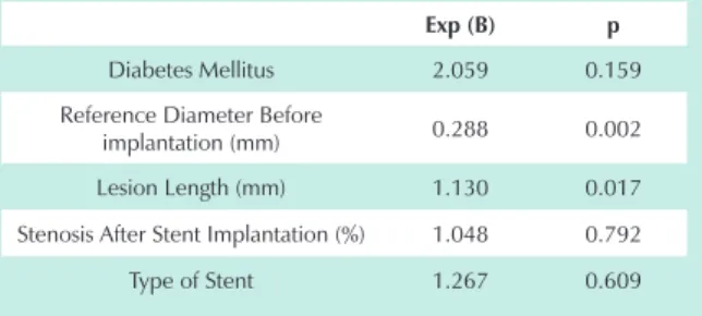
Univariate analysis showed that diabetes mellitus, post-implantation reference diameter, lesion length and stent type had a statistically significant association withrestenosis. However, only the pre-implantation reference diameter and lesion length were independently associated with restenosis (Table 5).
Cumulative rates of major cardiac events at six months were similar in both groups (16.8% vs 17.5%, respectively, p=1). Target-lesion revascularization rates were similar in both groups (15.7% vs 16.4%, respectively, p=1).
Discussion
This randomized, prospective, comparative study between diamond-like carbon coated stainless steel stents and uncoated stainless steel stents did not show statistically significant differences in restenosis rates at six months after implantation between the two groups.
Few results of diamond-like carbon coated stent implantation in human coronary arteries are published in the medical literature, and only one randomized comparative study with stents of similar design, differing only as to the diamond-like carbon coating was published, showing no
Table 1 - Clinical characteristics of the patients
Phytis™ Penta™ p
n=90 n=90
Age 60.3±9.8 66.7±33.3 0.86 Male gender 55 (61.1%) 60(66.7%) 0.64 Current smoker 30(33.3%) 35(38.9%) 0.33
High blood pressure 66(73.3%) 68(75.6%) 0.73 Hypercholesterolemia
(>200 mg/dL) 52(57.8%) 49(54.4%) 0.88 Diabetes mellitus (>125
mg/dL) 20(22.2%) 21(23.3%) 0.98 Previous myocardial
infarction 9(10%) 13(14.4%) 0.51 Previous angioplasty
(different vessel) 4(4.4%) 12(13.3%) 0.06 Previous CABG 3(3.3%) 5(5.6%) 0.72 Stable angina 35(38.9%) 45(50%) 0.18 Non-ST elevation acute
coronary syndrome 31(34.4%) 26(28.9%) 0.64 ST elevation acute
coronary syndrome 24(26.7%) 19(21.1%) 0.50 Left ventricular ejection
fraction 0.65±0.14 0.65±0.13 1
*Total cholesterol > 200 mg/dL.
Table 2 - Location and characteristics of the lesions
Phytis™ Penta™ p
n=90 n=90 Artery Treated
Anterior Descending
Artery 64(71.1%) 59(65.6%) 0.52 Right Coronary Artery 8(8.9%) 11(12.2%) 0.62
Circumflex Artery 8(8.9%) 15(16.7%) 0.18 Ramus Diagonalis 1(1.1%) 1(1.1%) 1
Diagonal Artery 1(1.1%) - -Left Marginal Branch 7(7.8%) 4(4.4%) 0.53 Posterior Descending
Branch 1(1.1%) -
-Type of Lesion
A 2(2.2%) 1(1.1%) 1
B1 31(34.4%) 40(44.5%) 0.40 B2 41(45.6%) 38(42.2%) 0.79 C 16(17.8%) 11(12.2%) 0.41
Table 3 - Characteristics of the procedure
Phytis™ Penta™ p
n=90 n=90
Stent length/lesion ratio 1.11±0.30 1.09±0.32 0.62 Mean stent length (mm) 14.57±4.17 12.88±3.71 0.002
Maximum balloon
pressure (atm) 12.26±1.29 12.53±1.55 0.19
the influence of the design of diamond-like carbon coated or uncoated stainless steel stents on restenosis rates and late loss at six months after implantation.
Phytis™ and Penta™ are stainless steel stents with equal diameters and different lengths (9, 12, 16, 20 and 25 mm vs. 8, 13, 15, 18, 23, 28 and 33 mm, respectively), a difference that did not influence the immediate post-implantation outcomes as assessed by the stent/lesion length ratio (1.11 ± 0.30 vs. 1.09 ± 0.32, respectively, p=0.62). These stents have different designs with similar strut diameters (90 and 91 µm, respectively)17, and are considered thin-strut stents, a
characteristic that has a favorable impact on late outcomes20-23.
Therefore, we believe that the stents used in the two groups are adequately comparable.
In the present study, stent reference diameter, minimum lumen diameter, and acute gain were higher in the Penta™ stent group. Univariate and multivariate analyses revealed the reference diameter as an independent predictor of restenosis. This higher diameter may have favored Penta™ stents, because a wider vessel could accommodate intimal hyperplasia more adequately. Nevertheless, if we focus on in-stent late loss, which is considered the measurement that best reflects the real and pure biological effect ofcoronary stent performance24,
we will observe that late loss was similar between the two stents, of approximately 1 mm, a value that is comparable to that observed in previous studies with diamond-like carbon18,
silicon carbide25 and carbon-coated26,27 stainless steel stents.
One of the theoretical advantages of diamond-like carbon coating is the lower incidence of acute or subacute thrombosis because of a better biocompatibility with low platelet activation10,13,26,27. The present study used the same
antiplatelet therapy in both groups (aspirin and clopidogrel) for the same period (30 days). Despite the predominance of acute coronary syndrome in the Phytis™ group (61.1% of the patients) and lesion complexity (approximately 64% of the lesions in both groups were B2 and C types), acute/subacute thrombosis and MI rates were low (1.1% for both stents), and no advantages were observed in diamond-like carbon coated over uncoated stents.
Clinical8 and laboratory9 studies suggest that stent surface
coating with hemocompatible and biocompatible material reduces the inflammatory response by decreasing the release of cytotoxic metal ions. However, when the concentration of inflammation markers (C-reactive protein and cytokines) was comparatively assessed after carbon-coated or uncoated stent implantation28, the inflammatory response was not affected. It
differences in restenosis rates and late loss between the two stents at six months after implantation. The limitation of this study was that the findings could not be extended to other types of stents with different designs or different carbon coatings19. Thus, the present study adds new information on
Fig. 1 - Cumulative distribution curves of minimum lumen diameter after, immediately after, and at six months after Phytis™ and Penta™ stent implantation.
Cumulative frequency curve of minimum lumen diameter before, immediately after, and six months after implantation
%
L
e
si
o
n
s
Before
Before After
After
Minimum lumen diameter (mm)
0.5 1.5 2.5 3.5
Table 4 - Angiographic measurements before, immediately after and six months after implantation and restenosis and target-lesion
revascularization rates
Phytis™ Penta™ p
n=90 n=90 Before implantation
Reference diameter (mm) 3.21±0.37 3.34±0.8 0.02 Minimum Lumen
Diameter (mm) 0.83±0.44 0.84±0.45 0.88 Percent Stenosis (%) 73.77±13.73 75.36±13.05 0.42
Length (mm) 12.79±5.17 12.15±3.50 0.33 After implantation
Reference Diameter (mm) 3.21±0.33 3.35±0.38 0.01 Minimum Lumen
Diameter (mm) 3.13±0.36 3.34±0.39 0.0003 Percent Stenosis (%) 0.98±0.06 0.99±0.06 0.02
Acute Gain 2.30±0.49 2.49±0.50 0.009 Six Months After
implantation n=70 n=55
Reference Diameter (mm) 3.16±0.33 3.37±0.40 0.002 Minimum Lumen
Diameter (mm) 2.07±0.79 2.25±1.02 0.27 Percent Stenosis (%) 35.68±22.45 34.73±26.99 0.83 Late loss (mm) 1.06±0.73 1.08±0.89 0.82 Angiographic restenosis 24.3% 21.8% 0.84
Target-lesion
Revascularization 15.7% 16.4% 1
Table 5 - Multivariate analysis (logistic regression) for restenosis at 6 months
Exp (B) p
Diabetes Mellitus 2.059 0.159 Reference Diameter Before
the lack of evidence that patients treated with coated stent present better results than those treated with uncoated stents may indicate that these differences do not exist or that they could not be detected in this study.
Conclusions
The results of this study did not show any differences in restenosis and major cardiac event rates at one and six months after diamond-like carbon coated stainless steel stent implantation in comparison with uncoated stainless steel stents.
is possible that only patients allergic to nickel or molybdenum benefit from stent coating with inert material7.
The present study had the following limitations: 1) subgroups of patients with more complex coronary artery disease were not included because patients with longer lesions (> 20 mm), lesions in unprotected left main coronary artery lesions and in bifurcations, chronic total stenosis, and restenotic lesions were excluded; 2) control coronary angiography rates for both groups were lower than those recommended (<80%) to assess the presence of restenosis as the main endpoint, which may create a selection bias; thus,
References
1. Sousa JE, Costa MA, Abizaid AC, Rensig BJ, Abizaid AS, Tanajura LF, et al. Sustained suppression of neointimal proliferation by sirolimus-eluting stents: one-year angiographic and intravascular ultrasound follow-up. Circulation. 2001; 104 (17): 2007-11.
2. Morice MC, Serruys PW, Sousa JE, Fajadet J, Ban Hayashi E, Perin M, et al. RAVEL Study Group. A randomized comparison of a sirolimus-eluting stent with a standard stent for coronary revascularization. N Engl J Med. 2002; 346: 1773-80.
3. Moses JW, Leon MB, Popma JJ, Fitzgerald PJ, Holmes DR, O’Shaughnessy C, et al; SIRIUS Investigators. Sirolimus eluting stents versus standard stents in patients with stenosis in a native coronary artery. N Engl J Med. 2003; 349(14): 1315-23.
4. Grube E, Silber S, Hauptman KE, Mueller R, Buellesfeld L, Gerckens U, et al. TAXUS I: six and twelve-month results from a randomized, double-blind trial on a slow-release paclitaxel-eluting stent for de novo coronary lesions. Circulation. 2003; 107(1): 38-42.
5. Sousa JE, Costa MA, Sousa AG, Abzaid AC, Seicas AC, Abizaid AS, et al. Two-year angiographic and intravascular ultrasound follow-up after implantation of sirolimus-eluting stents in human coronary arteries. Circulation. 2003; 107: 381-3.
6. Lemos PA, Lee C, Degertekin M, Saia F, Tanabe K, Arampatzis CH, et al. Early outcome after sirolimus-eluting stent evalued at Roterdam Cardiology Hospital (RESEARCH) Registry. J Am Coll Cardiol. 2003; 41: 2093-9. 7. Lemos PA, Magalhães MA. Aplicação dos stents farmacológicos baseada em
evidências; sim, quando possível, para todos. Rev Bras Cardiol Invas. 2005; 13(1): 37-42.
8. Koster R, Vieluf D, kiehn M, Sommerauer M, Kahler J, Baldus S, et al. Nickel and Molybdenum contact allergies in patients with coronary in-stent restenosis. Lancet. 2000; 356: 1895-7.
9. Gutensohn K, Beythien C, Bau J, Fenner T, Grewe P, Koester R, et al. In vitro analyses of diamond-like carbon coated stents: reduction of metal ion release, platelet activation, and thrombogenicity. Thromb Res. 2000; 99: 577-85. 10. Alanazi A, Nojiri C, Kido T, Noguchi T, Ohgoe Y, Matsuda T, et al. Engineering
analysis of diamond-like carbon coated polymeric materials for biomedical applications. Artif Organs. 2000; 24: 624-7.
11. De Scheerder I, Szilard M, Yanming H, Ping XB, Verbeken E, Neerinck D, et al. Evaluation of the biocompatibility of two new diamond-like stent coatings (Dylon) in a porcine coronary stent model. J Invasive Cardiol. 2000; 12: 389-94. 12. Linder S, Pinkowski W, Aepfelbacher M. Adhesion, cytoskeletal architecture
and activation status of primary human macrophages on a diamond-like carbon coated surface. Biomaterials. 2002; 23: 767-73.
13. Monnink SH, van Boven AJ, Peels HO, Tigchelaar I, de Kan PJ, Crijns HJ, et al. Silicon-carbide coated coronary stents have low platelet and leukocyte adhesion during platelet activation. J Invest Med. 1999; 47: 304-10. 14. Founier AF, Calabuig J, Marchan A, Augé JM, Melgares R, Colman T, et al.
Resultados iniciales y seguimiento clínico a 6 meses tras el implante de un stent coronario recubierto de carburo de silicio. Rev Esp Cardiol. 2001; 54: 567-72.
15. Babapulle MN, Eisenberg MJ. Coated stents for the prevention of restenosis: Part I. Circulation. 2002; 106: 2734-40.
16. Colombo A, Stankovic G, Moses JW. Selection of coronary stents. J Am Coll Cardiol. 2002; 40: 1021-33.
17. Cutlip DE, Chauhan MS, Baim DS, Ho KKL, Popma JJ, Corozza JP, et al. Clinical restenosis after coronary stenting: perspectives from multicenter trials. J Am Coll Cardiol. 2002; 40: 2082-9.
18. Sass N, Fedosenko G, Hoge J, Margaris N, Salachas A. The phytis DLC (Diamond-Like Carbon) coated stents. In: Serruys PW, Resing B, ed. Handbook of coronary stents. London: Taylor & Francis; 2001. p. 327-42. 19. Airoldi F, Colombo A, Tavano D, Stankovic G, Klugman S, Paolito V, et al.
Comparison of diamond-like carbon-coated stents versus uncoated stainless steel stents in coronary artery disease. Am J Cardiol. 2004; 93: 474-7. 20. Kastrati A, Dirschinger J, Boekstegers P, Elezi S, Schulen H, Pache J, et al.
Influence of stent design on 1-year outcome after coronary stent placement: a randomized comparison of five stent types in 1,147 unselected patients. Catheter Cardiovasc Interv. 2000; 50: 290-7.
21. Meireles GCX, Lemos PA, Ambrose JÁ, Ribeiro E, Horta PE, Perin M, et al. Luminal recovery from six to twelve months after implantation of thicker strut coronary stents. Am J Cardiol. 2004; 93: 210-3.
22. Kastrati A, Mehilli J, Dirschinger J, Dotzer F, Schuhlen H, Neumann FJ, et al. Intracoronary stenting and angiographic results: strut thickness effect on restenosis outcome (ISAR-STEREO) trial. Circulation. 2001; 103 (23): 2816-21. 23. Rittersma ZH, Winter RJ, Koch KT, Bax M, Schotborgh CE, Mulder KJ, et
al. Impact of strut thickness on late luminal loss after coronary artery stent placement. Am J Cardiol. 2004; 93: 477-80.
24. Sousa AGMR, Sousa JEMR. CYPHER ou TAXUS: diferentes ou semelhantes? Rev Bras Cardiol Invas. 2004; 12: 1-3.
25. Tanajura LFL, Sousa JEMR, Sousa AGMR, Abizaid A, Paula JET, Albertal M, et al. Estudo prospectivo e randomizado de pacientes tratados com e sem stents revestidos com carbeto de silício amorfo para a prevenção da reestenose coronária: avaliação ultra-sonográfica. Arq Bras Cardiol. 2004; 83 (Nº. especial): 59-63.
26. Antoniucci D, Bartorelli A, Valenti R, Montorsi P, Santoro GM, Fabbiocchi F, et al. Clinical and angiographic outcome after coronary stenting with the carbostent. Am J Cardiol. 2000; 85: 821-5.
27. Antoniucci D, Valenti R, Migliorini A, Moschi G, Trapani M, Bolognese L, et al. Clinical and angiographic outcomes following elective implantation of the carbostent in patients at high risk of restenosis and target vessel failure. Cathet Cardiovasc Interv. 2001; 54 (4): 420-6.