PS-HEMA Latex Fractionation by Sedimentation and Colloidal
Crystallization
André H. Cardoso, Carlos A.P. Leite, and Fernando Galembeck*
Instituto de Química, Universidade Estadual de Campinas,
130803-970 Campinas - SP, Brazil
Látex de poli(estireno-co-hidroxiacrilato de metila) separa-se em três camadas visualmente distinguíveis, das quais a inferior é opalescente e contém cristais coloidais. Alíquotas do látex foram coletadas em diferentes alturas, e as partículas foram caracterizadas, por espalhamento de luz dinâmico, microeletroforese, IV e microscopia eletrônica analítica. A fração inferior contém a maior parte do polímero, sendo formada por partículas de dimensões e composição química uniformes. As partículas coletadas das duas outras frações são diferentes das que formam os cristais coloidais, em praticamente todos os aspectos. A secagem da fração opalescente produz macrocristais de alta qualidade, com baixa frequência de defeitos, mostrando que a homogeneidade química das partículas é um fator importante, na sua auto-organização.
A poly(styrene-co-hydroxyethylmethacrylate) latex underwent sedimentation under gravity followed by an spontaneous and extensive colloidal crystallization. It was then fractionated in three visually distinguishable layers. Latex aliquots layers were sampled at different heigths and the particles were characterized by PCS, microelectrophoresis, infrared spectra and analytical electron microscopy. The major fraction was opalescent and contained the colloidal crystals settled in the bottom of the liquid. Two other latex fractions were obtained, which differed in their chemical compositions, particle sizes and topochemical features from the self-arraying particles. Macrocrys-tallization of the fractionated latex yielded high quality crystals with a low frequency of defects, which confirms that particle chemical homogeneity is an important factor for particle self-arraying.
Keywords: latex fractionation, colloidal crystallization, latex sedimentation,
core-and-shell latex, macrocrystal, latex
Introduction
Self-organized latex particle arrays1,2, either (dry) macrocrystals or (liquid) colloidal crystals, are often ob-served in latexes, and they have attracted the attention of many researchers. Many devices have been proposed to help building high quality macrocrystals, but the success rate is still low3-7 considering both the number of macro-crystalline domains obtained and the extent of correlated crystalline planes. On the other hand, many colloidal crys-tal-forming systems are known, and their equilibria with the colloidal “gas” phase8 have been studied but the formation of high quality colloidal crystals is still an exception rather than the rule. Practical applications of these structures call for a high degree of control over the processibility as well as the structure and composition of the latex9.
Two conditions are usually fulfilled, in successful at-tempts of latex colloidal crystallization: the particle diame-ters are monodisperse and the ionic strength of the initial dispersion liquid is very low10-11. The importance of hydro-philic particle surfaces and capillary adhesion in the forma-tion of dry macrocrystals is well acknowledged, following the work of Denkov, Nagayama et al.12, Distler and Koenig13, Whitesides and collab.3. In a paper on disorder-to-order in settling suspensions of colloidal silica, Davis et al.7 observed that ordered particle arrays were obtained at higher solid volume fractions (φ > 0.5), but only when the rate of particle sedimentation is lower than the rate of particle crystallization.
We have recently examined in detail the case of poly(styrene-co-hydroxyethylmethacrylate) latex, in
Article
which the particle polidispersity as well as the presence of salt in the initial dispersion does not preclude macrocrys-tallization14. In another recent work, we have obtained microchemical information on this latex, by energy-loss imaging (ESI)15. This showed that negative charges are distributed throughout the dry latex particles, while the positive charges make a particle shell in the dry particles, thus imparting to each particle a multipolar charge distri-bution which is relevant for particle-particle interaction and self-arraying. We confirmed that these particles are dipoles or multipoles, by observing backscattered-electron images in a field-emission electron microscope as well as by show-ing that the charge-bearshow-ing groups in the particles are asymmetrically distributed16.
Latex particle chemical heterogeneity is now well es-tablished17,18, and techniques are available to observe
dif-ferences in the chemical composition from one to another particle in a population, as well as for the microchemical characterization of domains within individual particles.
In this work, we report on the fractionation of PS-HEMA latex, and we show that this actually leads to high-quality macrocrystal formation.
Experimental
Latex preparation and characterization
The latex was prepared14 by batch surfactant-free emul-sion copolymerization of styrene (S) and hydroxyethyl methacrylate (HEMA) following procedures similar to those developed by Okubo19 and Suzawa20. The amounts
of reagents used are as follows: water 210.2 g, styrene 31.2 g, 2-hydroxyethyl methacrylate 4.5 g, potassium per-sulfate 0.1086 g.
The polymerization was carried out in a 500 mL glass kettle reactor fitted with condenser, thermometer, glass paddle-type stirrer and a gas inlet providing a constant flow of nitrogen gas. The kettle was kept at the required constant temperature (±2 °C) using a thermostated water bath. The reaction vessel was loaded with water and the monomers, and heated to 70 °C. After 30 min of N2 purging and stirring
the system, the initiator potassium persulfate dissolved in 4.5 cm3 of water was added to the reaction mixture. The polymerization reaction was carried out at 70 °C for 10 h under constant 300-350 rpm stirring. The product was filtered with a 200 mesh steel sieve, to remove coagulated latex. In order to remove unreacted monomer, oxidation products and unwanted electrolyte, the resulting latex was dialyzed against water with daily changes over a two-month period. The dialysate conductivity reached 2 µS/cm, and remained unchanged for 48 h. The dialysis tubing (a visking
membrane from Sigma) was boiled in several quantities of distilled water, prior to use.
After dialysis, part of the latex sample was lyophilized using a bench-top glass apparatus, to recover the solid polymer for spectral characterization. The remainder of the sample was dispersed in water, as required for reaching the desired concentration. Monomer conversion was 94.4%, as determined gravimetrically right after the reaction14.
Transmission electron microscopy and elemental spectroscopy imaging
Brightfield pictures and the elemental distribution within latex particles were obtained using a Carl Zeiss CEM 902 transmission electron microscope, equipped with a Castaing-Henry-Ottensmeyer energy filter spectrometer within the column. When the electron beam passes through the sample, interaction with electrons of different elements results in characteristic energy losses. A prism-mirror sys-tem deflects electrons with different energies to different angles so that only electrons with a well defined energy are selected. If elastic electrons only are chosen (∆E = 0 eV) a transmission image with reduced chromatic aberration is obtained. When monochromatic inelastically scattered electrons are selected, electron spectroscopic images (ESI) are formed, in which contrast is dependent on the local concentration fluctuations of a particular chosen element. Clear areas correspond to element-rich domains.
For individual latex particle examination, one drop of the latex dispersion (1% solids content) was deposited on carbon-coated parlodion films supported in 400 mesh cop-per grids (Ted Pella). To make sure that the whole particles were not excessively thick, they were first observed using
∆E = 0 eV electrons, then observed again at ∆E = 20-50 eV. Image contrast inversion was always obtained, showing that a significant number of electrons were transmitted throughout the particles21. This observation is understood, considering that the 80 keV electrons mean free path within these latex particles is greater than 160 nm for elastic scattering22, and is estimated as many hundreds of nanome-ters, for inelastic scattering.
technique was used to perform the background subtraction, for each elemental image23.
Image processing was performed in an IBM PC micro-computer using the Image-Pro Plus 3.0 image analyzer program (Media Cybernetics).
FESEM images
Secondary electron images were obtained in an ultra high resolution “semi-in-lens” JEOL JSM-6340F field emission scanning electron microscope, operating at 15 kV, which corresponds to a 1.2 nm nominal resolution. The films were placed on a metal stub and sputter coated with carbon prior to examination.
Infrared spectra
Infrared spectra were obtained from polymer films cast on NaCl windows. The latex fractions were centrifuged, the latex was dried and dissolved in toluene, to obtain the casting solutions.
Results
Latex sedimentation under gravity
Soon after its synthesis, ca. 200 mL of the latex disper-sion were stored in a 250-mL glass vial with a screw-cap (ca. 63 mm i.d.; initial liquid height was 115 mm). After 5 months storage, an intensely opalescent, 15-mm high layer was observed at the bottom of the container. Visual obser-vation at any given angle showed the existence of domains, presenting iridescent colors extending for a few centimeters within this bottom layer, which changed with the angle of observation. This proves that there was a high degree of ordering in this layer, characterized by macroscopic colloi-dal crystals. Many bright, millimeter-sized spots of differ-ent colors were also observed, evidencing the coexistence of colloidal polycrystals. There is a well-defined interface between this crystalline layer and an opaque milky layer above it, and another upper, translucent layer, 1-mm thick just beneath the latex surface.
Three aliquots were carefully collected with syringes fitted with long glass tips, to avoid cross-contamination. The fractions were drawn: i) from the opalescent layer, ii) from the milky layer, 30 mm above the bottom and iii) 50 mm above the bottom.
Properties of the particles in the separated latex fractions
The solids contents in the three layers were determined, as well the particle sizes and zeta potentials. The results are in Table 1.
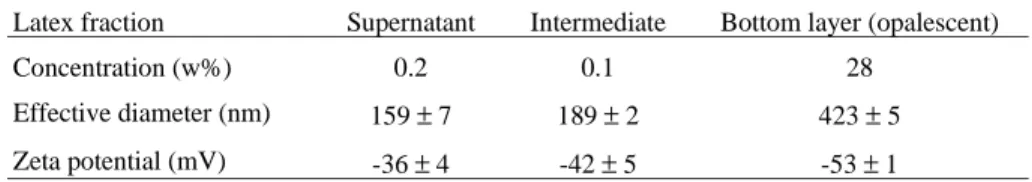
Transmission brightfield micrographs are in Fig. 1. The two upper fractions contain large numbers of small (< 200 nm) particles, which are not seen in the lower frac-tion. There are also important differences among the larger particles in the three fractions: in the lower fraction, the particles in the microscope grid are well separated, and they do not show any strong trend towards coalescence, as evidenced by necking. On the other hand, larger particles in the upper fractions coalesce with the smaller particles and with larger particles (not shown), as well. This demon-strates significant differences in larger particle surface properties, even though their diameters are very close, in the different fractions.
The smaller particles are very different from the larger ones, also considering their smooth surfaces, departure from spherical shapes and their easy coalescence as evi-denced by the pronounced neck formation.
Consequently, the coexistence of ordered and disor-dered material within this latex is not the result of an order-disorder phase equilibrium, but instead it is the result of latex particle fractionation.
Electron spectroscopy imaging of the disordered fractions
Figures 2-5 present micrographs and elemental (C, O, S and K) distribution maps, for particles in the three latex aliquots. In the bottom fraction, the particles are all similar, in the different maps. This demonstrates a rather uniform overall chemical composition, for these particles. On the other hand, the opposite is observed in the intermediate fraction, particularly among the smaller particles. Some specific observations are the following:
i) Some small particles appear brighter than others in the top fraction sulfur map (Fig. 4), but these same particles are not distinguished in the carbon map (Fig. 2). Conse-quently, these particles have an above-than-average sul-fur/carbon atom ratio, probably associated with a lower MW of the polymer chains.
Table 1. Concentration, particle effective diameters and zeta potentials for the three latex fractions.
Latex fraction Supernatant Intermediate Bottom layer (opalescent)
Concentration (w%) 0.2 0.1 28
Effective diameter (nm) 159 ± 7 189 ± 2 423 ± 5
ii) The oxygen map in Fig. 3 also shows some darker particles, which are nevertheless very clear in the carbon and potassium maps. The conclusion is the existence of a significant variation in the O/C ratio, and the existence of styrene-richer (and acrylate-poorer) small particles.
iii) Even in the very uniform particles in the bottom fraction, a few brighter points are observed (e.g., in the
particle to the right, ca. 5 o’clock in Fig. 3c), showing domains in which C content is higher than average.
iv) In the top fraction, potassium is distributed rather uniformly (Fig. 5) throughout the coalesced particles. In the
intermediate fraction this ion tends to concentrate at the outer shell of either larger or smaller particles, and in the
Figure 2. C elemental distribution maps of the upper, medium and bottom
fractions collected from the latex.
Figure 1. Brightfield transmission electron micrographs of particles from
lower fraction potassium concentration at the particle sur-face is very marked.
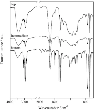
Infrared spectra
Infrared spectra for films obtained for the three aliquots are in Fig. 6. The spectra for the two upper fractions are
rather similar, but they show large differences with the bottom fraction, particularly in the 1300-1050 cm-1 range. The spectrum of the bottom fraction in this range
corre-sponds closely to the sum of the homopolymers spectra. Consequently, these particles are predominantly formed by
block copolymer chains, or by a mixture of homopolymers.
Figure 4. S elemental distribution maps of the upper, medium and bottom
fractions collected from the latex.
Figure 3. O elemental distribution maps of the upper, medium and bottom
The poor resolution of the bands in this same region, for the two upper fractions, is an evidence that the acrylic mers are interspersed with styrene, forming random copolymer chains24.
The ratios between the absorbances at 1727 (assigned to C=O acrylic groups) and 700 (from out-of-plane
aro-matic ring deformation) cm-1 in the three fractions are respectively 11.1, 4.8 and 0.77, while this ratio for the unfractionated latex is 0.83. The particles in the upper fractions are thus much richer in acrylic monomer (which confirms the information from ESI imaging) than the pre-dominating population.
Scanning electron microscopy
A scanning electron micrograph of a fractured macro-crystal prepared by drying an aliquot of the bottom fraction is presented in Fig. 7. We note the perfection of the particle ordering in this image, which demonstrates the usefulness of particle fractionation, to prepare high-quality macrocrys-tals.
Discussion
The fractionation of the PS-HEMA latex revealed that even the as-prepared latex is already rather homogeneous. This is probably related to the specific synthesis protocol used in this work; in a previous work, we found significant differences in the degree of heterogeneity of three styrene-butyl methacrylate latexes, depending on the specific syn-thesis procedure24.
The lower, opalescent, fraction is by far much more concentrated than the others, and this is probably the main factor for the easy self-arraying of this latex, even prior to fractionation. On the other hand, latex fractionation elimi-nates particles of many different sizes and chemical com-positions, which can only impair crystallization.
Figure 6. Infrared spectra of films prepared with the three latex fractions. Figure 5. K elemental distribution maps of the upper, medium and bottom
The present results show that latex particles undergo an spontaneous sedimentation-driven crystallization, produc-ing a fraction with improved chemical uniformity. This fraction has a great ability for colloidal and macrocrystal formation.
In a previous work16 we have shown that PS-HEMA latex particles are dipoles (or multipoles), and we proposed that this feature is relevant for self-arraying. In the present paper we have described the extensive spontaneous forma-tion of colloidal crystals at rather low (28%) volume frac-tion, in an undialyzed sample, and we suggest that the polar nature of the particles contributes to this behavior.
The present findings give us an explanation for the difficulties in observing and reproducing self-arrayed latex structures, even in size-monodisperse latex: this is probably due to a large heterogeneity of particle chemical composi-tions, which poses difficulties for the encounters of identi-cal particles, with good associating characteristics.
On the other hand, we observe that colloidal crystal-lization is an effective tool for latex purification, which is understood here as the process yielding a highly uniform fraction, concerning size, morphology as well as particle chemical composition.
In the present case this was done just by sedimentation under gravity, but other fractionation techniques (centrifu-gation in density gradients, osmocentrifu(centrifu-gation25) are likely to be helpful, as well.
Conclusion
The PS-HEMA latex sediments under gravity and it undergoes spontaneous fractional colloidal crystallization, yielding a large fraction of chemically and morphologically uniform particles. Minor fractions contain particles with
large chemical differences, in a range of diameters. The predominance of chemically uniform particles in the as-prepared latex explains the unusually easy macrocrystalli-zation of this latex.
Acknowledgments
F G a c kn ow ledg es t h e s u pp or t o f Fap es p , Pronex/Finep/MCT and CNPq. During this work, AHC was a Capes pre-doctoral fellow.
References
1. Alfrey Jr., T.; Bradford, E.B.; Vanderhoff, J.W.; Os-ter, G. J. Opt. Soc. Am. 1954, 44, 603.
2. Hachisu, S.; Kobayashi, Y.; Kose, A.J. J. Colloid Interface Sci. 1973, 42, 342.
3. Kim, E.; Xia, Y.; Whitesides, G. Adv. Mater. 1996, 8, 245.
4. Michelleto, R.; Fukuda, H.; Ohtsu, M. Langmuir 1995, 11, 3333.
5. Trau, M.; Saville, D.A.; Aksay, I.A. Science 1996, 272, 706.
6. Burmeister, F.; Schäfle, C.; Matthes, T.; Böhmisch, M.; Boneberg, J.; Leiderer, P. Langmuir 1997, 13, 2983.
7. Davis, K.E.; Russel, W.B.; Glantschnig, W.J. Science 1989, 245, 507.
8. Böhmer, M. Langmuir 1996, 12, 5747.
9. Meier, W. Curr. Op. Colloid Interface Sci. 1999, 4, 6. 10. Joanicot, M.; Wong, K.; Maquet, J.; Chevalier, Y.; Pichot, C.; Graillat, C.; Lindner, P.L.; Rios, Cabane B. Progr. Colloid Polym. Sci. 1990, 81, 175.
11. Keddie, J.L. Materials Science and Engineering 1997, R21, 101.
12. Denkov, N.D.; Velev, O.D.; Kralchevsky, P.A.; Ivanov, I.B.; Yoshimura H.; Nagayama, K. Nature 1993, 361, 26.
13. Distler, D.; Kanig, G. Colloid Polym. Sci. 1978, 256, 1060.
14. Cardoso, A.H.; Leite, C.A.P.; Galembeck, F. Colloids Surfaces A 1998, 144, 207.
15. Cardoso, A.H.; Leite, C.A.P.; Galembeck, F. Lang-muir 1998, 14, 3187.
16. Cardoso, A.H.; Leite, C.A.P.; Galembeck, F. Lang-muir 1999, 15, 4453.
17. Moita Neto, J.M.; Cardoso, A.H.; Testa, A.P.; Galem-beck, F. Langmuir 1994, 10, 2095.
18. Galembeck, F.; Souza, E.F. In Polymer Interfaces and Emulsions, Esumi, K., ed., M. Dekker, New York, p. 119, 1999.
Figure 7. A scanning micrograph of a fractured PS-HEMA macrocrystal.
19. Kamei, S.; Okubo, M.; Matsuda, T.; Matsumoto, T. Colloid Polym. Sci. 1986, 264, 743.
20. Tamai, H.; Fujii, A.; Suzawa, T. J. Colloid Interface Sci. 1987, 116, 37.
21. We are grateful to Dr. W. Probst (LEO-Zeiss Elek-tronenmikroskopie Gmbh) for this valuable private communication.
22. Newbury, D.E. In “Principles of Analytical Electron Microscopy” Joy, D.C.; Romig Jr., A.D.;Goldstein, J.I., eds., Plenum Press, New York, 1986.
23. Reimer, L.; Zepke, U.; Moesch, J.; Schulze-Hillert, St.; Ross-Messemer, M.; Probst, W.; Weimer, E. “EELS Spectroscopy: A Reference Handbook of Standard Data for Identification and Interpretation of Electron Energy Loss Spectra and for Generation of Electron Spectroscopic Images” Carl Zeiss, Oberko-chen, 1992.
24. Cardoso, A.H.;.Moita Neto, J.M; Leite, C.A.P.; Galembeck, F. Colloid Polymer Sci. 1997, 275, 244. 25. Galembeck, F.; Robilotta, P.R.; Pinheiro, E.A.;
Joekes, I.; Bernardes, N. J. Phys. Chem. 1980, 84, 112.
Received: August 6, 1999