Article
J. Braz. Chem. Soc., Vol. 25, No. 8, 1455-1461, 2014. Printed in Brazil - ©2014 Sociedade Brasileira de Química 0103 - 5053 $6.00+0.00
A
*e-mail: [email protected]
Behaviour of Pseudoisocyanine in Macromolecular and Hydrotropic Solutions
Lukese R. Menegussi, Silvano R. Valandro, Alessandra L. Poli, Carla C. Schmitt and Miguel G. Neumann*
Instituto de Química de São Carlos, Universidade de São Paulo, CP 780, 13560-970 São Carlos-SP, Brazil
O comportamento fotofísico do corante pseudoisocianina (PIC) foi estudado em solução macromolecular (alginato de sódio) e hidrotrópica (toluenossulfonato e estirenossulfonato de sódio). A localização do corante nestes meios modifica as propriedades espectroscópicas e os tempos de vida dos estados excitados. Os efeitos são atribuídos à interação do corante com as macromoléculas ou sua localização nos agregados hidrotrópicos, que induzem a formação de agregados J ou constringem suas movimentações internas reduzindo a velocidade da conversão interna. Os tempos de vida do estado triplete do corante em alginato foram de 20-30 ns em soluções recém preparadas, enquanto que, deixando a solução repousar por algumas horas o decaimento vira biexponencial com a formação de uma nova espécie com tempo de vida ao redor de 150 ns. Esta última é atribuída às moléculas do corante localizadas no ambiente do alginato.
The photophysical behaviour of the dye pseudoisocyanine (PIC) was studied in macromolecular (sodium alginate) and hydrotropic (sodium toluenesulphonate and sodium styrenesulphonate) solutions. It was found that the placement of the dye in these media affected the spectroscopic properties and the lifetimes of its excited states. The effects are traced down to the interactions of the dye molecules with the macromolecules or their placement in the hydrotropic aggregates, inducing the formation of J-aggregates of the dye or constraining its internal movements reducing the rate of internal conversion. The lifetimes of the triplet state of the dye in alginate were of the order of 20-30 ns in freshly prepared solutions, whereas when leaving the solution to rest for some hours the decay became biexponential with the growing of a species with lifetime around 150 ns. The latter is assigned to dye molecules placed in the alginate network.
Keywords: pseudoisocyanine, hydrotropes, alginate, J-aggregates
Introduction
The photophysics of the cyanine known as pseudoisocyanine (PIC) has a relative long history due to its early use as a spectral sensitizer in photography.1 Its
photophysics is well known.2-4 The monomer band appears
around 520 nm with a vibronic shoulder at 490 nm and a smaller one at 460 nm.3 The decrease of the monomer
band with increasing dye concentration is accompanied by the appearance of a band at ca. 485 nm on the vibronic shoulder of the monomer band, characterizing the formation of dimers.3 Under some conditions, the dye can also form
J-aggregates characterized by a narrow band around 570 nm.5-13
PIC presents a very short singlet excited-state lifetime (12-18 ps)14,15 and low fluorescence quantum yield
(6.8 × 10⁻⁵) in low viscosity media.16,17 These facts can be
related to the losses of energy by the deactivation of the singlet state by a non-radiative process due to the rotation of the quinolinic rings around the methine bridge. Indeed, the dye 1,1’-methylene-2,2’-cyanine, whose structure differs from PIC only by the rigid linkage of the aromatic rings, has high emission intensity even in fluid media at room temperature.18
As a result of the efficient non-radiative decay of its singlet excited-state, the triplet state of PIC cannot be detected by direct excitation. However, formation of the PIC triplet can be sensitized by benzophenone.2
(alginate) and hydrotropes (p-toluenesulphonate and styrenesulphonate) and compared with the behaviour in aqueous and organic solvent solutions.
Experimental
Chemicals
Pseudoisocyanine chloride (PICCl, Sigma-Aldrich, 100%), and pseudoisocyanine iodide (PICI, Sigma-Aldrich, 97%), sodium styrenesulphonate (StyS, Sigma-Aldrich,
≥ 99%), sodium p-toluenesulphonate (TS, Sigma-Aldrich, 95%), benzophenone (Vetec, 99%), and sodium alginate (Alg, Sigma-Aldrich) were used as received. Ethanol (Mallinckrodt), methanol (Tedia), butanol (Mallinckrodt), ethyleneglycol (Mallinckrodt), acetonitrile (Tedia) and Milli-Q purified water were used to prepare the solutions. The chemical structure of PIC and the similar 1,1’-methylene-2,2’-cyanine (MEC) are given in Scheme 1.
Methods
Absorption spectra were recorded on a Shimadzu UV-2550 spectrophotometer and fluorescence spectra were obtained using a Hitachi F-4500 spectrofluorimeter at room temperature. For emission studies, PIC was excited at 490 nm. Laser flash photolysis measurements were made with a Luzchem LFP-112/122 system combined with a Quantel Brilliant B Nd:YAG laser delivering 5.2 ns pulses of the second harmonic (532 nm). For transient studies
ethyleneglycol. Working solutions of the hydrotropes were prepared by diluting 0.8 and 2.0 mol L–1 stock solutions of
StyS and TS, respectively. The stock solution of sodium alginate was 2.8 g L–1.
For flash photolysis measurements, the samples were deaerated by five freeze/thaw cycles under vacuum.
Results and Discussion
The absorption spectra of PIC in aqueous solution and other solvents are shown in Figure 1a. Except for the difference in the extinction coefficients, all spectra are similar with a small shift to the red (2-4 nm) for the peaks in organic solvents. These effects may be due to the solvent-dye interactions in organic solvents, as already observed for other dyes.
On the other hand, as expected, the emission (Figure 1b) increases with solvent viscosity, due to the increasing medium rigidity that reduces the rotation of the aromatic rings around the methine bridge, thus diminishing the internal conversion deactivation of the excited singlet state. This effect is confirmed by the extremely high emission of MEC, that has a similar structure, but an additional ring structure that prevents the rotation around the methine bond.18
The absorption and emission spectra for PIC in hydrotropic solutions of sodium styrenesulphonate (StyS) and in sodium toluenesulphonate (TS) were investigated in the concentration range 0.03 to 0.6 mol L–1 and 0.2 to
1.8 mol L–1, respectively, as shown in Figures 2 and 3. The
selected concentration range encompasses the MHC regions for these hydrotropes, which is around 1.0 mol L–1 for TS
and 0.1 mol L–1 for StyS.23
As can be seen in Figure 2, the absorption spectra of the dye present a bathocromic shift when in aqueous solution of hydrotropes, together with a moderate increase in the absorption intensity. This behaviour is similar to that observed for PIC in aqueous solutions of poly(methacrylic acid) (PMAA) at low pH and in organic solvents. The
changes can be traced to the interactions between PIC and the hydrotropes that are essentially hydrophobic (between the dye and the hydrotrope aggregates above the MHC), and electrostatic (between the sulphonate group of the hydrotrope and the cationic dye). Similar variations of the spectra of PIC in PMAA where assigned to hydrophobic
interaction between PIC and the polyelectrolyte in a hypercoiled conformation.22
The emission of the dye (Figure 3) also increases in hydrotropic medium as observed for PIC in PMAA solution at low pH.21 This behaviour is similar to that found for the
dye in other rigid media: the dye-hydrotrope interaction
Figure 1. Absorption (a) and emission (b) spectra of PIC in water and organic solvents. [PIC] = 1 × 10⁻⁵ mol L–1; λ
ex = 490 nm.
Figure 2. Absorption spectra of PIC in aqueous solutions of: (a) sodium styrenesulphonate (StyS) and (b) sodium toluenesulphonate (TS). [PIC] = 5 × 10⁻⁶ mol L–1.
Figure 3. Emission spectra of PIC in aqueous solution of: (a) StyS and (b) TS. [PIC] = 5 × 10⁻⁶ mol L–1; λ
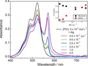
The absorption of PIC in alginate solutions showed two bands, corresponding to monomers at 525 nm (with the corresponding vibronic band at 490 nm) and J-aggregates at 570 nm (Figure 4). It can be observed that, upon addition of increasing amounts of alginate, the increase in the absorbance of the J-aggregates parallels the decrease of the monomer (inset in Figure 4). The excitation spectra of PIC in alginate solutions (not shown), measured around the emission maximum (575 nm) shows the same two peaks. The slight variation in the relative absorption of the peak at 490 nm, assigned by some authors to the formation of H-aggregates,31 has to be
attributed to the modification of the vibrational modes when PIC is in the alginate environment, which should affect the vibronic band.
J-aggregation band increases. This effect is depicted in Figure 6b and can be observed in alginate concentration up to approximately 0.2 g L–1. In this range, the emission
intensity of the dye also increases steadily. At higher alginate concentrations, the alginate aggregates start to approach each other allowing the interaction of the already bound to the -COO– dye molecules to interact between
them forming induced non-emitting aggregates (Figure 6d). Hence, the emission intensity decreases, whereas the J-band increases. Similar results were found for PIC in solutions of sodium polyacrylate (MM 30,000 D), potassium polyvinyl sulphonate, and sodium poly(styrenesulphonate).32
The transient spectra resulting from 532 nm excitation of PIC 5.7 × 10⁻⁶ mol L–1 in an aqueous solution of
TS 1.8 mol L–1 is shown in Figure 7.
The decay of the transient at 740 nm, shown in Figure 8, presents a lifetime of around 7.5 µs. This lifetime is the same found for the decay of triplet PIC sensitized by benzophenone.2 Therefore, it can be assigned to the triplet
state of PIC.
Longer decay times (around 20-30 µs) were found for the transient absorptions in the 640 and 400 nm regions. The absorption at these wavelengths did not return to the baseline. The same behaviour was also found for the dye in PMAA solution.2 This behaviour can be due to a stable
(or long-lived) species formed from the triplet. The dye safranine was found to have the same behaviour in TS and PMAA solutions.33 In this case, the residual absorbance was
assigned to the formation of semioxidized or semireduced radical species.
The transient spectra resulting from 532 nm excitation of PIC 5 × 10⁻⁶ mol L–1 in an aqueous solution of Alg
2.5 g L–1 is shown in Figure 9. The transient at 740 nm
showed a biexponential decay with lifetimes around 170 ns (68%) and 30 ns (32%). Similar values were found for the transient absorption at 400 nm.
The experiment was performed again with another solution of PIC in Alg in the same concentrations. The transient lifetimes in 740 nm and in 400 nm were about
Figure 4. Absorption of PIC 5 × 10⁻⁶ mol L–1 in alginate solutions.
Inset: Maximum absorbance of the monomer (520 nm) and J-aggregate (570 nm).
Figure 5. Emission of PIC 5 × 10⁻⁶ mol L–1 in alginate solutions. Inset:
Figure 6. PIC-alginate interactions in solutions with growing alginate concentrations. A: free PIC monomers, B: free monomers, and monomers and J-aggregates bound to alginate sites; C: J-aggregates formed by PIC molecules bound to different alginate chains; D: redistribution of PIC monomers at alginate sites.
Figure 7. Transient spectra of PIC in aqueous solution of TS.
λexc = 532 nm; [PIC] = 5.7 × 10⁻⁶ mol L–1; [TS] = 1.8 mol L–1.
Figure 8. Transient decay of PIC at 740 nm in aqueous solution of TS.
λex = 532 nm; [PIC] = 5.7 × 10⁻⁶ mol L–1; [TS] = 1.8 mol L–1.
Figure 9. Transient spectra of PIC in aqueous solution of Alg.
λexc = 532 nm; [PIC] = 5 × 10⁻⁶ mol L–1; [Alg] = 2.5 g L–1.
Figure 10. Transient decay of PIC at 740 nm in aqueous solution of Alg with double-exponential fit. λexc = 532 nm; [PIC] = 5 × 10⁻⁶ mol L–1;
transient lifetimes between 15 to 30 ns were found for solutions examined immediately after preparation, whereas the 150 ns component was only observed when the solutions were allowed to rest for about 24 h or longer. The 15-30 ns lifetime is assigned to the J-aggregate. When this aggregate stabilizes in alginate medium, its lifetime grows up to about 150 ns.
Conclusions
Absorption and emission
Organic solvent solutions compared to water solutions
No shifts of the peaks of the absorption spectra of PIC were found when increasing the chain length of alcohols. The PIC emission band also remained at the same maximum for all solvents, but the intensity increased with the viscosity of the solvent. This effect is because rigid media prevent the rotation of the aromatic rings around the methine bridge reducing the non-radiative deactivation.
Hydrotropic solutions
In the presence of hydrotropes (StyS and TS), the PIC absorption spectra present a bathocromic shift and an increase in the emission. This behaviour is similar to that for PIC in aqueous solution of PMAA at low pH. The dye-hydrotrope interactions diminish the mobility of the quinolinic rings and, thus, the emission increases.
Alginate solutions
A band in 570 nm is found when increasing the amount of alginate in aqueous solutions containing PIC. This band was assigned to the formation of J-aggregates of the dye. The PIC fluorescence emission increases with alginate concentration up to 0.2 g L–1. After this concentration,
the emission decreases until reaching the same intensity than that of a 2.5 g L–1 aqueous solution. This behaviour
can be explained by the initial PIC-alginate interactions diminishing the mobility of the PIC-quinolinic rings, further
lifetime between 15 to 30 ns. When the solution is left standing for a couple of hours, a new lifetime component can be identified with a lifetime around 150 ns. The 15-30 ns lifetime is assigned to the J-aggregate and the longer lifetime to the aggregates stabilized in the alginate microenvironment.
Acknowledgments
Financial support by FAPESP, Brazil (Proc. 2012/19656-0), is gratefully acknowledged. L. R. M. also thanks CNPq, Brazil for a graduate fellowship.
References
1. James, T. H.; Mees, C. E. K.; The Theory of the Photographic Process; Macmillan: New York, 1966.
2. Jones II, G.; Oh, C.; J. Phys. Chem. 1994, 98, 2367. 3. Horng, M. L.; Quitevis, E. L.; J. Chem. Educ. 2000, 77, 637.
4. Horng, M. L.; Quitevis, E. L.; J. Phys. Chem. 1993, 97, 12408. 5. von Berlepsch, H.; Böttcher, C.; Dähne, L.; J. Phys. Chem. B
2000, 104, 8792.
6. Möbius, D.; Adv. Mater. 1995,7, 437. 7. Cooper, W.; Chem. Phys. Lett. 1970,7, 73.
8. Yu, Z. X.; Lu, Y.; Alfano, R. R.; Chem. Phys. 1983,79, 289. 9. Iwasaki, M.; Kita, M.; Ito, K.; Kohno, A.; Fukunishi, K.; Clays
Clay Miner. 2000,48, 392.
10. Peyratout, C.; Donath, E.; Daehne, L.; J. Photochem. Photobiol. Chem. A 2001,142, 51.
11. Tani, T.; Yamaguchi, Y.; Saeki, M.; Oda, M.; Vacha, M.; J. Lumin. 2003,102-103, 27.
12. Sluch, M. I.; Vitukhnovsky, A. G.; Yonezawa, Y.; Sato, T.; Kunisawa, T.; Opt. Mater. 1996,6, 261.
13. Bujdák, J.; Iyi, N.; J. Colloid Interface Sci. 2008,326, 426. 14. Rentsch, S. K.; Danielius, R.; Gadonas, R. A.; Piskarskas, A.;
Chem. Phys. Lett. 1981,84, 446.
15. Kopainsky, B.; Kaiser, W.; Chem. Phys. Lett. 1982,88, 357. 16. Stiel, H.; Teuchner, K.; Becker, W.; Freyer, W.; Dähne, S.;
17. Buettner, A.; J. Chem. Phys. 1967, 46, 1398. 18. Cooper, W.; Photogr. Sci. Eng. 1973, 17, 3.
19. Hodgdon, T. K.; Kaler, E. W.; Curr. Opin. Colloid Interface Sci. 2007,12, 121.
20. Bauduin, P.; Renoncourt, A.; Kopf, A.; Touraud, D.; Kunz, W.; Langmuir 2005,21, 6769.
21. González, G.; Nassar, E. J.; Zaniquelli, M. E. D.; J. Colloid Interface Sci. 2000, 230, 223.
22. Maheshwari, R. K.; Saxena, M.; Gahlot, M.; Chaki, R.; Kinariwala, M.; Jagwani, Y.; Indian J. Pharm. Sci. 2010,72, 649.
23. Neumann, M. G.; Schmitt, C. C.; Prieto, K. R.; Goi, B. E.; J. Colloid Interface Sci. 2007, 315, 810.
24. Liu, Z.; Jiao, Y.; Wang, Y.; Zhou, C.; Zhang, Z.; Adv. Drug Delivery Rev. 2008,60, 1650.
25. Baldursdóttir, S. G.; Kjøniksen, A. L.; Karlsen, J.; Nyström, B.; Roots, J.; Tønnesen, H. H.; Biomacromolecules 2003,4, 429. 26. Neumann, M. G.; Schmitt, C. C.; Iamazaki, E. T.; Carbohydr.
Res. 2003,338, 1109.
27. Rehm, B. H. A.; Alginates: Biology and Applications; Springer: London, 2009.
28. Kenawy, E. R.; Sakran, M. A.; Ind. Eng. Chem. Res. 1996,35, 3726.
29. Dvir, T.; Timko, B.; Kohane, D. S.; Langer, R.; Nat. Nanotechnol. 2011, 6, 13.
30. Eiselt, P.; Yeh, J.; Latvala, R. K.; Shea, L. D.; Mooney, D. J.; Biomaterials 2000,21, 1921.
31. Eisfeld, A.; Briggs, J. S.; Chem. Phys. 2006, 324, 376. 32. Liu, M.; Kira, A.; Thin Solid Films 2000,359, 104.
33. Borsarelli, C. D.; Bertolotti, S. G.; Previtali, C. M.; Photochem. Photobiol. Sci. 2002,1, 574.
Submitted on: March 26, 2014
Published online: June 3, 2014
![Figure 3. Emission spectra of PIC in aqueous solution of: (a) StyS and (b) TS. [PIC] = 5 × 10 –6 mol L –1 ; λ ex = 490 nm](https://thumb-eu.123doks.com/thumbv2/123dok_br/18998245.462887/3.892.103.820.831.1080/figure-emission-spectra-pic-aqueous-solution-stys-pic.webp)