Rev Odontol UNESP. 2016 Jan-Feb; 45(1): 59-64 © 2016 - ISSN 1807-2577
ORIGINAL ARTICLE
Doi: http://dx.doi.org/10.1590/1807-2577.05915
Assessment of the efects of decontamination and storage
methods on the structural integrity of human enamel
Avaliação dos efeitos de métodos de descontaminação e armazenamento sobre a
integridade estrutural do esmalte humano
Adriana Rodrigues de FREITAS
a, Fábio Duarte da Costa AZNAR
a, André Luís da SILVA
b,
Arsenio SALES-PERES
a, Silvia Helena de Carvalho SALES-PERES
a*
aFaculdade de Odontologia de Bauru, USP – Universidade de São Paulo, Bauru, SP, Brasil
Resumo
Introdução: O armazenamento de dentes para utilização em pesquisas é uma questão controvérsia e não há consenso sobre o método de tratamento mais apropriado para esta finalidade. Objetivo: O objetivo deste estudo foi analisar a efetividade e a influência de diferentes métodos de descontaminação e armazenamento do esmalte dentário humano, a fim de manter sua integridade. Material e método: A amostra foi constituída por 124 molares distribuídos aleatoriamente em três grupos, de acordo com o método: controle – água destilada, soluções de timol a 0,1% e de azida de sódio a 0,02%. Os testes realizados foram fluorescência a laser, microdureza de superfície e análise de perfilometria (0, 15 e 30 dias) e teste microbiológico (7, 15 e 30 dias). Os dados foram analisados por meio dos testes ANOVA e Tukey (p<0,05). Resultado: No teste de fluorescência a laser, a solução de timol a 0,1% demonstrou ser mais viável na manutenção da integridade do órgão dentário, uma vez que este não apresentou variações significativas nos valores entre os períodos avaliados (p<0,05). A análise de microdureza de superfície evidenciou perda de estrutura dentária em todos os métodos, sendo que a azida de sódio promoveu menor perda dentária. Na análise de perfilometria observou-se perda de estrutura em todos os grupos, com maior perda no grupo azida de sódio. Nenhum dos métodos
conseguiu inibir o crescimento bacteriano. Conclusão: Dentre os métodos de processamento analisados nenhum
foi capaz de aliar a efetividade na descontaminação ao armazenamento com manutenção da integridade estrutural do esmalte dentário humano.
Descritores: Esmalte dentário; infecção; dureza; fluorescência.
Abstract
Introduction: The storage of teeth for use in research is a controversial issue with no consensus on the most appropriate treatment method for this purpose. Objective: The aim of this study was analyze the effectiveness and the influence of different methods of decontamination and storage of human enamel samples, in order to maintain their integrity.
Material and method: The sample consisted of 124 molars distributed randomly into three groups according to the method: control - distilled water, 0.1% thymol and 0.02% sodium azide. The tests performed were laser fluorescence, surface microhardness and profilometry analysis (0, 15 and 30 days) and Microbiological test (7, 15 and 30 days). Data were analyzed by the ANOVA and Tukey tests (P <0.05). Result: In the laser fluorescence test, thymol proved to be more feasible for maintaining the integrity of the dental organ, since it did not show significant variations in values among the analyzed periods (P> 0.05). The surface microhardness analysis showed loss of tooth structure in all methods, and sodium azide led to a lower level of tooth loss. Profilometry analysis showed loss of mass in all groups whereas sodium azide showed the greatest loss. None of the methods was able to inhibit bacterial growth.
Conclusion: Among the processing methods analyzed none was able to combine effective decontamination and storage with maintenance of the structural integrity of the human enamel.
Descriptors: Dental enamel; infection; hardness; fluorescence.
INTRODUCTION
he use of extracted teeth in in vitro and in situ experiments contributes to the development of new techniques and dental materials1,2.
Teeth can be considered a potential source of cross infection and contamination3,4, and eicient methods of decontamination should
that allow the maintenance of their physical properties5-7. In order
to preserve teeth, a variety of storage methods have been used, including chloramines, formalin, sodium hypochlorite, thymol, alcohol, glutaraldehyde8 and the autoclaving process3,4.
he majority of cariogenicity and erosive tests are conducted intraorally in an efort to use the natural environment of the tooth and in situ conditions9-11, consequently, in many experiments enamel
specimens from human or bovine sources ind their way into the volunteer’s mouth5.
Enamel, the hardest tissue in nature, is a highly mineralized non-regenerative tissue with a well-organized microstructure12.
he decontamination method must not afect the structural integrity of dental enamel and hard tissue specimens, in order to maintain the baseline condition of specimens13.
he method of decontamination, conditions and time of storage of teeth seem to be important variables in studies that use this type of substrate4,14,15. Elucidating this question is crucial to
understanding the maintenance of the structural integrity of tooth enamel under storage conditions.
he aim of this study was to analyze the efectiveness and the inluence of diferent methods of decontamination and storage of human teeth, in order to maintain their properties and the structural integrity of the enamel.
MATERIAL AND METHOD
One hundred and twenty-four freshly extracted and non-carious human third molars were selected for this study. Teeth were provided by a Human Tooth Bank, which collects, cleans and stores teeth in iltered water under refrigeration until their use. he study protocol was approved by the Research Ethics Commission of the
Institution and was conducted in compliance with the norms of the Declaration of Helsinki.
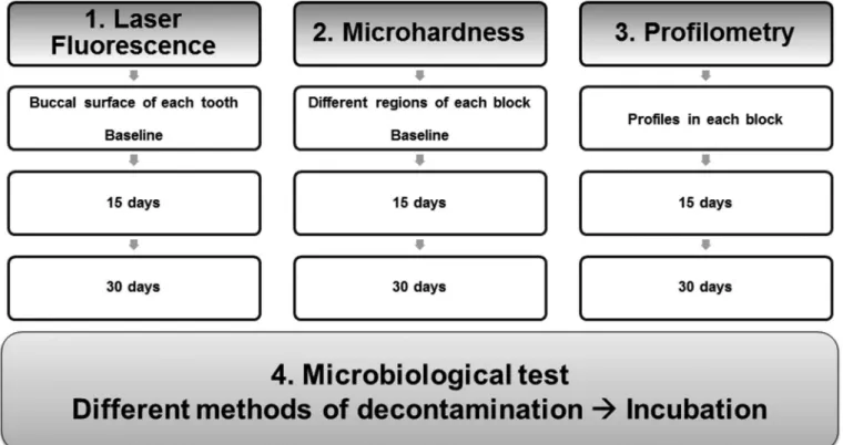
An in vitro study was conducted to assess the efectiveness and inluence of diferent methods of decontamination and storage of human teeth, in order to maintain their properties and structural integrity. he teeth were immersed in solutions of 0.1% thymol, 0.02% sodium azide and distilled water (control) (solutions previously prepared by the biochemical laboratory of BSD-USP) and subjected to a microbiological test, laser luorescence, microhardness and proilometry analyses (Figure 1).
For the Laser Fluorescence analysis, we used 30 molars, ten for each group (distilled water – control, thymol and sodium azide). he teeth were analyzed by means of the laser luorescence device DIAGNOdent ® (KAVO, Biberach, Germany). According to the manufacturer’s instructions, the instrument was calibrated to its ceramic standard before the measurement of each tooth, and the teeth were let at room temperature for 5 minutes. Ater this, they were immersed in deionized water and dried on sterile gauze. Five sites on the buccal surface of each tooth were selected and identiied for analysis, acquisition and recording of the values, which were subsequently used to calculate the mean value for each tooth. Ater these initial analyses, the teeth were randomly distributed, placed in jars with lid, immersed in the test solutions and stored under refrigeration (4 °C). New records of laser luorescence were obtained for each group ater 15 and 30 days of storage.
For the Surface Microhardness analysis, enamel fragments obtained from 12 molars. he crowns were sectioned from the roots with a diamond disk (Isomet 1000; Buehler, Lake Bluf, IL, USA). he enamel surface of the blocks was ground lat with water-cooled carborundum discs (320, 600 and 1200 grit Al2O3 papers; Buehler, Lake Bluf, IL, USA), and polished with felt pads with diamond
spray (1 lm; Buehler, Lake Bluf, IL, USA). To standardize the blocks, specimens were previously selected to measure the initial KNOOP microhardness (Five indentations made in diferent regions of the blocks, 25 g, 10 s, HMV-2000; Shimadzu Corporation, Tokyo, Japan). Blocks that presented 10% below and 10% above the mean value were excluded from the sample. Twelve specimens with a mean surface microhardness of between 417.40 and 464.80 KHN (mean=441.10) were randomly divided into groups, and immersed in the following solutions: distilled water- control, thymol and sodium azide. Ater time intervals of 15 and 30 days of storage, new microhardness analyses were performed in each group.
For proilometry analysis, enamel fragments obtained from 12 molars and prepared in the same way as those used for the microhardness analysis were used. he purpose of this analysis was to highlight enamel loss, thus it was not performed at time interval 0 (full surface), only in the subsequent periods. In order to maintain reference surfaces for determining lesion depth, two layers of nail varnish (Risque, Niasi, Taboão da Serra, São Paulo, Brazil) were applied on half of the specimen surfaces. Enamel blocks were randomly divided into groups and immersed in the following solutions: distilled water- control, thymol and sodium azide. At the time interval of 15 days the blocks were remove from the solutions, washed with deionized water and dried with gauze. Surface proiles of the enamel specimens were obtained with a contact proilometer (Hommel Tester T 1000, Hommelwerke, VS, Schwenningen, Germany). To determine enamel loss, the nail varnish was removed from the specimens and ive proiles were recorded. he proile scans were performed in the centre of each specimen at intervals of 250 µm. Treatment scans was superimposed and the average depth of the area under the curve in the eroded area was calculated with a specially designed sotware program (Turbo Datawin-NT, Version 1.34, Copyright© 2001). he results of the ive scans were averaged for each specimen. hen the nail varnish was applied again on half of the specimen surfaces. he blocks were stored for the time of 30 days, when new proilometry analyses were performed at exactly the same sites as for the measurement at the time interval of 15 days.
Prior to the Microbiological test, a pilot study was conducted, using a laminar low chamber to assess the initial contamination of the teeth. For this purpose, 10 molars were selected (5 sterilized-control and 5 test), and placed in two sterilized jars with lid containing 40 mL of Brain Heart Infusion (BHI) each. he test group was inoculated with 40 µL of broth containing 3.5 × 10⁴ spores of Bacillus subtilis
(Microbiology Laboratory of BSD-USP), while the other group remained uncontaminated (control). Both samples were stored at 37 °C for 24 hours. Ater the incubation period the samples were removed from the broths, the contents deposited on glass slides, and submitted to Gram stain. he slides were examined under an optical microscope and strains of microorganisms identiied for each group.
To conduct the experiment 60 molars were used, and were divided into the groups: 15 distilled water (positive control), 15 thymol, 15 sodium azide and 15 autoclave (negative control). All teeth were immersed in sterilized test tubes containing 40 ml of BHI broth, kept in an incubator at 37 °C, for 24 hours. Ater this
stage the teeth from the Autoclave group were wrapped in gauze, placed in self-sealing envelopes, and submitted to a 15-minute sterilization cycle at 121 °C, with the drying process of the device being excluded.
In the next step all teeth were immersed in their solutions: distilled water (positive and negative controls), thymol and sodium azide. Each group was stored under refrigeration (4 °C) for time intervals of 7, 15 and 30 days (5 teeth in each group and t each time). Ater the mentioned time intervals, the tubes were removed from the refrigerator and let for 5 minutes at room temperature. hen the teeth were removed from solution, dried on sterile gauze and put into new tubes containing BHI broth, which were stored in an incubator at 37 °C for 6 hours.
he tubes containing broth + tooth were initially analyzed by the turbidity of the medium. Originally the culture medium was shown to be yellow and clean; this condition was considered a negative result for bacterial growth, whereas the presence of turbidity in samples was considered positive for bacterial growth. In order to conirm the results of this analysis (positive or negative) serial decimal dilutions (10–1 to 10–3) of the broth contained in the
tubes was performed, using 9 mL of sterile saline solution in each tube. Aliquots of 50µl of the 10–2 and 10–3 dilutions were seeded on
the surface of culture plates containing BHI agar, by means of an automatic pipette, always working from the more diluted solution to the less diluted. he plates were incubated in an incubator at 37 °C for 24 hours. Ater this period the culture plates were analyzed for the presence/absence of Colony Forming Units (CFU).
Data were entered into Excel® spreadsheets (Microsot, Redmond, WA, USA, 2010), and analyzed using the sotware program STATISTICA® statistical package version 10.0 (Statsot, Tulsa, OK, USA, 2011). he laser luorescence, surface microhardness and proilometry analysis data were checked for the assumptions of equality of variances and normal distribution of errors. Since the assumptions were satisied, data were analyzed by two and one-way analysis of variance, ANOVA. For individual comparisons among the groups, the Tukey test was used. he level of signiicance was set at 5% (P<0.05) and Interval of Conidence of 95%. he microbiological test was analyzed qualitatively, according to the bacterial growth.
RESULT
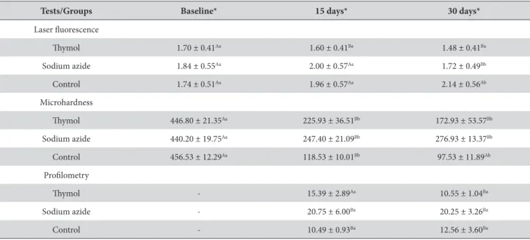
he main results of the laser luorescence, surface microhardness and proilometry analysis are described in Table 1. In all tests performed we observed signiicant relations between the groups, storage time and interaction of these two variables (P>0.05).
In the pilot study of the microbiological test, no diferences were observed in morphological forms of bacterial strain that were found in the both control and experimental group.
he culture plates produced from dilutions of the contents of the tubes (culture medium and teeth) were qualitatively analyzed. Since there was great proliferation of microorganisms on the plates, Colony Forming Unit counts proved to be unfeasible. he positive control, thymol and sodium azide plates showed bacterial growth in all the time intervals analyzed. he autoclave culture plates (negative control) showed negative results in this analysis, however ater 24 hours of incubation there was turbidity in 40% of the samples.
DISCUSSION
In laboratory conditions, many variables such as temperature, humidity and storing solutions need to be considered14. he storage
method can be used to prevent dehydration of the specimens, but may also incorporate anti-microbial substances in order to prevent the growth of microorganisms4,16. he use of extracted teeth requires
the adoption of measures for cross contamination prevention, such as the use of personal protective equipment3,4.
he 0.1% thymol solution was investigated and showed decline in comparison with the laser luorescence values during the time intervals evaluated (0, 15 and 30 days), however without signiicant diferences (P<0.05). Storage solutions may reduce the concentration of luorophores in the samples by the dilution, resulting in decreased response to laser luorescence17. In this case the thymol solution
may have favored a decrease in concentration of luorophores by its bacteriostatic potential. In the surface microhardness and proilometry analysis this group presented loss of enamel structure (Table 1) with signiicant diferences between the analyzed time intervals (P<0.05). he microbiological test showed the ineiciency of this solution for the inhibition of bacterial growth in samples of teeth stored for 7, 15 and 30 days. hymol solution has been largely used as storage method in in vitro18 and in situ studies9,19,20.
he use of this solution has not determined signiicant inluence on shear resistance21 and microleakage22 tests, however studies
have described the use of thymol solution without standardization, probably due to insuicient parameters to enable the choice of the most appropriate method23. However, this agent did not appear
to ofer useful properties, since it promoted signiicant loss of enamel structure and it was not able to inhibit bacterial growth in the samples analyzed.
he 0.02% sodium azide solution presented an increase in laser luorescence values at 15 days of storage, and later showed signiicant decrease in these values between 15 and 30 days (P=0.045). Two hypotheses can be raised, the irst being the bacteriostatic efect ater 15 days of storage, and the second that it allowed dispersal of the mineral content of the teeth17. In the microhardness analysis
this solution presented the lowest loss of structure (–37.09%), however in the proilometry analysis it showed the highest loss of mass values (Table 1). It was observed that sodium azide promoted loss of tooth structure from enamel specimens in the time interval of 15 days, and hardened these structures ater 30 days of storage (Table 1). Sodium azide presented no activity in the inhibition of bacterial growth in all samples and periods analyzed. his solution showed some instability in the tests and diferent storage times, which seems to agree with the indings shown in the scientiic literature14,15. Gamma irradiation is an efective method for tooth
decontamination5,24 but has an unfavorable cost-beneit ratio for
its application in this type of research. Bovine enamel specimens were disinfected by microwave irradiation without prejudice to the property of hardness25 and other properties, such as laser luorescence
and the proilometry should be analyzed in future studies.
he control group, distilled water showed linear performance throughout the laser luorescence test (Table 1), with a signiicant increase in these values between the time intervals (P=0.002).
Table 1. Mean, standard deviation and association among processing method and time of storage in Laser luorescence, Microhardness and Proilometry analysis
Tests/Groups Baseline* 15 days* 30 days*
Laser luorescence
hymol 1.70 ± 0.41Aa 1.60 ± 0.41Ba 1.48 ± 0.41Ba
Sodium azide 1.84 ± 0.55Aa 2.00 ± 0.57Aa 1.72 ± 0.49Bb
Control 1.74 ± 0.51Aa 1.96 ± 0.57Aa 2.14 ± 0.56Ab
Microhardness
hymol 446.80 ± 21.35Aa 225.93 ± 36.51Bb 172.93 ± 53.57Bb
Sodium azide 440.20 ± 19.75Aa 247.40 ± 21.09Bb 276.93 ± 13.37Bb
Control 456.53 ± 12.29Aa 118.53 ± 10.01Bb 97.53 ± 11.89Ab
Proilometry
hymol - 15.39 ± 2.89Aa 10.55 ± 1.04Ba
Sodium azide - 20.75 ± 6.00Ba 20.25 ± 3.26Ba
Control - 10.49 ± 0.93Ba 12.56 ± 3.60Ba
With regard to the surface microhardness and proilometry analyses it promoted the loss of structure on the enamel block in both cases, with the greatest loss of hardness (–78.58%). Distilled water was used as control in this study and somehow favored the loss of structure of the enamel specimens. In view of this inding, this matter should be investigated in the future, which refers to the possibility of replacing the deionized water with luoridated water to store enamel specimens, in an attempt to minimize these losses.
Storage in distilled water was used as positive control in the microbiological test, while the sterilization of teeth in an autoclave was used as negative control. he autoclave is described as a simple, inexpensive and accessible method for decontamination of dental elements used for didactic and scientiic purposes3. As expected
distilled water allowed bacterial growth in all samples, while the autoclave sterilization showed the opposite performance. Although the sample of tubes for the autoclave group did not show bacterial growth in the stipulated period of 6 hours ater the initial analysis of these samples, they were incubated for a period of 24 hours in order to assess possible changes in an extended period, which was observed in 40% of the tubes. his late contamination of the samples can be attributed to the presence of viable microorganisms in dentinal tubules or due to the presence of cracks on the tooth surface26.
In this study, the storage of specimens in diferent conditions was shown to be capable of inluencing the results, and the time was less favorable in the storage of 30 days in all groups. he chemical and optical changes in dental properties are important factors to be considered when choosing the solution and the storage time14,15,27,28.
he limitation of this study was due to the previous contamination of the teeth used. he results of pilot test showed no diference between the groups. An alternative to this bias would be the prior sterilization of the teeth by immersion in 10% formalin for seven days3,4 or processing in an autoclave3. Nonetheless, this procedure
would not meet the requirements of laboratory analysis, since these processes could inluence the results of tests to which the teeth would be submitted.
CONCLUSION
he processing methods analyzed showed no efectiveness in the decontamination and the maintenance of the structural integrity of the human enamel. he performance of sodium azide solution seems to invalidate its use in laboratory tests that require the use of tooth structures, due to the perceived changes it causes in these structures. he 0.1% thymol solution seems to be the storage method most accepted, however it needs to be applied in combination with another efective decontamination method.
ACKNOWLEDGEMENTS
he authors wish to thank the Microbiology Laboratory of the Bauru School of Dentistry, University of São Paulo; Professor Fausto Mendes Medeiros from the Department of Orthodontics and Pediatric Dentistry, Faculty of Dentistry, University of São Paulo, for the loan of an equipment used in this study and CAPES - Brazilian Federal Agency For Support and Evaluation of Graduate Education, for inancial support.
REFERENCES
1. Farah R, Drummond B, Swain M, Williams S. Linking the clinical presentation of molar-incisor hypomineralisation to its mineral density. Int J Paediatr Dent. 2010 Sep 1;20(5):353-60. http://dx.doi.org/10.1111/j.1365-263X.2010.01061.x. PMid: 20642473.
2. Manton DJ, Cai F, Yuan Y, Walker GD, Cochrane NJ, Reynolds C, et al. Effect of casein phosphopeptide-amorphous calcium phosphate added to acidic beverages on enamel erosion in vitro. Aust Dent J. 2010 Sep;55(3):275-9. http://dx.doi.org/10.1111/j.1834-7819.2010.01234.x. PMid: 20887514.
3. Dominici JT, Eleazer PD, Clark SJ, Staat RH, Scheetz JP. Disinfection/sterilization of extracted teeth for dental student use. J Dent Educ. 2001 Nov;65(11):1278-80. PMid:11765875.
4. Kumar M, Sequeira PS, Peter S, Bhat GK. Sterilisation of extracted human teeth for educational use. Indian J Med Microbiol. 2005 Oct;23(4):256-8. PMid:16327123.
5. Amaechi BT, Higham SM, Edgar WM. Efficacy of sterilisation methods and their effect on enamel demineralisation. Caries Res. 1998; 32(6):441-6. http://dx.doi.org/10.1159/000016485. PMid: 9745118.
6. Toledano M, Osorio R, Osorio E, Aguilera FS, Yamauti M, Pashley DH, et al. Durability of resin-dentin bonds: effects of direct/indirect exposure and storage media. Dent Mater. 2007 Jul;23(7):885-92. http://dx.doi.org/10.1016/j.dental.2006.06.030. PMid: 16949659.
7. Lee JJ, Nettey-Marbell A, Cook A Jr, Pimenta LA, Leonard R, Ritter AV. Using extracted teeth for research: the effect of storage medium and sterilization on dentin bond strengths. J Am Dent Assoc. 2007 Dec;138(12):1599-603. http://dx.doi.org/10.14219/jada.archive.2007.0110. PMid: 18056105.
8. Titley KC, Chernecky R, Rossouw PE, Kulkarni GV. The effect of various storage methods and media on shear-bond strengths of dental composite resin to bovine dentine. Arch Oral Biol. 1998 Apr;43(4):305-11. http://dx.doi.org/10.1016/S0003-9969(97)00112-X. PMid: 9839706. 9. Lippert F, Lynch RJ, Eckert GJ, Kelly SA, Hara AT, Zero DT. In situ Fluoride response of caries lesions with different mineral distributions
at baseline. Caries Res. 2011;45(1):47-55. http://dx.doi.org/10.1159/000323846. PMid: 21293122.
10. Grazziotin GB, Rios D, Honorio HM, Silva SMB, Lima JEO. In situ investigation of the remineralizing effect of saliva and fluoride on enamel following prophylaxis using sodium bicarbonate. Eur J Dent. 2011 Jan;5(1):40-6. PMid:21228955.
12. Xie Z, Swain MV, Hoffman MJ. Structural integrity of enamel: experimental and modeling. J Dent Res. 2009 Jun;88(6):529-33. http://dx.doi. org/10.1177/0022034509337130. PMid: 19587157.
13. Shellis RP, Ganss C, Ren Y, Zero DT, Lussi A. Methodology and models in erosion research: discussion and conclusions. Caries Res. 2011;45 Suppl 1:69-77. http://dx.doi.org/10.1159/000325971. PMid: 21625135.
14. Komabayashi T, Ahn C, Zhang S, Zhu Q, Spangberg LS. Chronological comparison of root dentin moisture in extracted human teeth stored in formalin, sodium azide, and distilled water. Oral Surg Oral Med Oral Pathol Oral Radiol Endod. 2009 Jul; 108(1): e50-e54. http://dx.doi. org/10.1016/j.tripleo.2009.03.022. PMid: 19540443. PMCID: PMC2746056.
15. Li HP, Burrow MF, Tyas MJ. The effect of long-term storage on nanoleakage. Oper Dent. 2001 Nov-Dec;26(6):609-16. PMid:11699186. 16. Ziskind D, Gleitman J, Rotstein I, Friedman M. Evaluation of cetylpydinium chloride for infection control in storage solution. J Oral Rehabil.
2003 May;30(5):477-81. http://dx.doi.org/10.1046/j.1365-2842.2003.01091.x. PMid:12752926.
17. Francescut P, Zimmerli B, Lussi A. Influence of different storage methods on laser fluorescence values: a two-year study. Caries Res. 2006; 40(3):181-5. http://dx.doi.org/10.1159/000092223. PMid: 16707864.
18. Bishara SE, Soliman M, Laffoon JF, Warren J. Shear bond strength of a new high fluoride release glass ionomer adhesive. Angle Orthod. 2008 Jan;78(1):125-8. http://dx.doi.org/10.2319/100405-347.1. PMid: 18193962.
19. Mathews MS, Amaechi BT, Ramalingam K, Ccahuana-Vasquez RA, Chedjieu IP, Mackey AC, et al. In situ remineralisation of eroded enamel lesions by NaF rinses. Arch Oral Biol. 2012 May;57(5):525-30. http://dx.doi.org/10.1016/j.archoralbio.2011.10.010. PMid: 22041022. 20. Sales-Peres SH, Pessan JP, Buzalaf MA. Effect of an iron mouthrinse on enamel and dentine erosion subjected or not to abrasion: an in situ/
ex vivo study. Arch Oral Biol. 2007 Feb;52(2):128-32. http://dx.doi.org/10.1016/j.archoralbio.2006.08.010. PMid: 17045952.
21. Farret MM, Gonçalves TS, Lima EMS, Menezes LM, Oshima HMS, Kochenborger R, et al. Influência de variáveis metodológicas na resistência de união ao cisalhamento. Dental Press J Orthod. 2010 Jan-Feb;15(1):80-8. http://dx.doi.org/10.1590/S2176-94512010000100010. 22. Haller B, Hofmann N, Klaiber B, Bloching U. Effect of storage media on microleakage of five dentin bonding agents. Dent Mater. 1993
May;9(3):191-7. http://dx.doi.org/10.1016/0109-5641(93)90119-B. PMid:8056175.
23. Campregher UB, Arruda FZ, Samuel SMW. Meios utilizados para armazenagem de dentes em pesquisas odontológicas de impacto: uma revisão sistemática. RPG Rev Pos-Grad. 2007 Abr-Jun;14(2):107-12.
24. White JM, Goodis HE, Marshall SJ, Marshall GW. Sterilization of teeth by gamma radiation. J Dent Res. 1994 Sep;73(9):1560-7. PMid:7929992. 25. Viana PS, Machado AL, Giampaolo ET, Pavarina AC, Vergani CE. Disinfection of bovine enamel by microwave irradiation: effect on the
surface microhardness and demineralization/remineralization processes. Caries Res. 2010;44(4):349-57. http://dx.doi.org/10.1159/000318528. PMid: 20616552.
26. Zapata RO, Bramante CM, de Moraes IG, Bernardineli N, Gasparoto TH, Graeff MS, et al. Confocal laser scanning microscopy is appropriate to detect viability of Enterococcus faecalis in infected dentin. J Endod. 2008 Oct;34(10):1198-201. http://dx.doi.org/10.1016/j.joen.2008.07.001. PMid: 18793919.
27. Strawn SE, White JM, Marshall GW, Gee L, Goodis HE, Marshall SJ. Spectroscopic changes in human dentine exposed to various storage solutions: short term. J Dent. 1996 Nov;24(6):417-23. http://dx.doi.org/10.1016/0300-5712(95)00106-9. PMid: 8990687.
28. Goodis HE, Marshall GW Jr, White JM, Gee L, Hornberger B, Marshall SJ. Storage effects on dentin permeability and shear bond strengths. Dent Mater. 1993 Mar;9(2):79-84. http://dx.doi.org/10.1016/0109-5641(93)90079-6. PMid: 8595846.
CONFLICTS OF INTERESTS
he authors declare no conlicts of interest.
*CORRESPONDING AUTHOR
Silvia Helena de Carvalho Sales Peres, Departamento de Odontopediatria, Ortodontia e Saúde Coletiva, Faculdade de Odontologia de Bauru, USP - Universidade de São Paulo, Al. Octávio Pinheiro Brisolla, 9-75, 17012-901 Bauru - SP, Brasil, e-mail: [email protected]