91
REVISTA BRASILEIRA DE OTORRINOLARINGOLOGIA 71 (1) PARTE 1 JANEIRO/FEVEREIRO 2005 http://www.sborl.org.br / e-mail: [email protected]
Cholesteatoma of external
auditory canal: a case report
Summary
Fá bio D. Za nini1, Ever ton S. Ameno2,
Sidney O. Ma ga ldi3, R uben A. La ma r4
1 Otorhinolaryngologist, Hospital Regional de São José Homero de Miranda Gomes – São José/SC; Hospital Infantil Joana de Gusmão – Florianópolis/SC; Fundação Catarinense de Educação Especial São José/SC.
2 Otorhinolaryngologist, Head of the Service of Otorhinolaryngology, Hospital da Lagoa – Rio de Janeiro/RJ. 3 Physician, Service of Otorhinolaryngology, Hospital da Lagoa – Rio de Janeiro/RJ.
4 Physician, Service of Otorhinolaryngology, Hospital da Lagoa – Rio de Janeiro/RJ. Affiliation: Hospital da Lagoa – Rio de Janeiro/RJ.
Address correspondence to: Fábio Duro Zanini– Rua Lauro Linhares, 2123 Torre A Sala 612 Agronômica Florianópolis SC 88036-002. Tel (55 48) 334-9045/ 9962-7485/ 9962-3577 – Fax (55 48) 228-8228 – E-mail: [email protected]
Article submited on December 30, 2002. Article accepted on April 29, 2003.
T
he authors present a case of cholesteatoma of external auditory canal (CEAC) with extensive invasion of mastoid; ossicle chain and tympanic membrane remained intact. The only symptom was chronic otorrhea. Diagnosis was based on clinical elements and CT scan was used to measure pathology and program surgery. Treatment was modified radical mastoidectomy associated with meatoplasty. Due to the insidious character of CEAC and the proximity with important structures of the external auditory canal, it must be always considered in differential diagnosis for lesions of external auditory canal. This case report intended to review clinical and surgical aspects of treatment of CEAC and present our approach in a case with severe lesions.Rev Bras Otorrinolaringol. V.71, n.1, 91-3, jan./feb. 2005
Key words: cholesteatoma, external auditory canal, otorrhea. C ASE REPORT
92
REVISTA BRASILEIRA DE OTORRINOLARINGOLOGIA 71 (1) PARTE 1 JANEIRO/FEVEREIRO 2005 http://www.sborl.org.br / e-mail: [email protected]
INTRODUCTION
Cholesteatoma of the external auditory canal (CEAC) is a rare condition affecting especially elderly people. The progress of the disease is slow and the symptoms are not too evident. This leads to late diagnosis, with progressive bone lysis affecting important circumjacent structures1,2.
The authors present a case of CEAC with extensive invasion of the mastoid, exposing the lateral sinus and the dura. Hearing acuity and structures of the tympanic cavity were unaffected.
We discuss the possible causes and treatment of choice.
LITERATURE REVIEW
In clinical practice of ear diseases, cholesteatoma occurs on the tympanum-mastoid segment. It rarely originates from the external auditory canal (EAC), whose incidence is about 0.1-0.5% in new patients with ear problems1-3.
In 18502,4, Toynbee was the first author describing
that cholesteatoma originates from the external auditory canal as “epidermal sheets”. Up to 1980, CEAC and keratosis obturans (KO) were considered different presentations of the same disease. Pipergerdes et al.2 described CEAC and
keratosis obturans (KO) as two distinct clinical-pathological processes: KO as keratin accumulation in the EAC; and CEAC as bone erosion resulting from squamous tissue on a specific spot of the EAC2.
In CEAC, lateral epithelium desquamation is affected. Keratin debris are blocked, causing local erosion and bone lysis, which can be severe2,5. Holt2,3,6,7 listed situations in
which the disease can be diagnosed: postoperative period of ear surgery; EAC trauma; EAC obstruction – due to osteoma or stenosis, for instance; idiopathic – possibly, in these cases, local periostitis may originate the process of hyperkeratosis. Diagnosis is made through physical examination. There is otalgia, otorrhea, hearing acuity is unaffected; otoscopy shows an intact tympanic membrane and erosion on a distinct spot of the external auditory canal4,5,8.
Otorhinolaryngologists must be aware that cholesteatoma may affect circumjacent structures (lateral sinus, facial nerve, posterior cranial fossa). Therefore, a CT scan is recommended for all patients1,2,4.
Differential diagnosis for CEAC should include necrotizing external otitis, tumors and KO. The latter is more frequent in young adults presenting severe otalgia and bilateral sensorial hearing loss. Differently from CEAC, which usually affects older patients, KO is unilateral, there is otorrhea, chronic pain and hearing acuity is preserved. In KO, the EAC has a keratin plug; otoscopy, following keratin plug removal, shows stenosis, redness and granulation tissue. In CEAC, there is an epidermal diverticulum on the lowest
part of the canal, with epidermal debris and otorrhea; the other parts of the EAC are normal1-3.
The treatment of CEAC may be clinical or surgical. The former is carried out through local flushing and topic antibiotics. The latter is through resection of the cholesteatoma and necrotic bone5-8.
Clinical treatment is indicated whenever the lesion is limited and there is no otalgia, in cases that surgery is contraindicated, in patients refusing surgical procedures and, when observation without treatment is an option to evaluate progression2,6.
Surgical treatment is indicated in the following situations: chronic pain (despite treatment), frequent infection (there is risk for bacterial resistance), facial palsy or chronic dizziness, progression during follow-up, involvement of hypotympanum, jugular or mastoid (showed in CT scans), diabetes mellitus or immunosuppression (predisposition to necrotizing external otitis) 2,4,6,9.
CASE REPORT
Patient A. F., male, 59 years old. Visited the otorhinolaryngological outpatient facility showing otorrhea, otalgia and ear fullness on the right for ten (10) years. Otoscopy on his right ear showed epidermal debris in the canal. Following debris removal, the tympanic membrane was intact, and there was severe erosion of the postero-inferior quadrant of the EAC. Otoscopy of the left ear was normal. Bilateral pure tone audiometry was normal.
We suspected of CEAC and requested a CT scan of temporal bones. CT scan showed erosion of the posterior wall of the EAC, mastoid involvement and exposure of the lateral sinus and meninges (Figure 1).
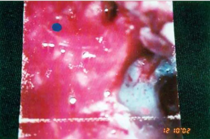
Transoperative observation revealed erosion of the entire posterior wall of the EAC, except for a thin bone band close to the tympanic ring (Figure 2). The tympanic membrane, ossicle chain and facial nerve canal were preserved.
We conducted modified radical mastoidectomy type I plus meatoplasty. The patient showed good outcomes and normal hearing, without recurrences, in a postoperative period of two years.
DISCUSSION
93
REVISTA BRASILEIRA DE OTORRINOLARINGOLOGIA 71 (1) PARTE 1 JANEIRO/FEVEREIRO 2005 http://www.sborl.org.br / e-mail: [email protected]
Mastoid invasion is one of the indications for surgery. After the patient agreed to undergo surgery, we performed modified radical mastoidectomy and wide meatoplasty; thus, we could achieve good control of the cavity during the postoperative period.
We were able to preserve the tympanic cavity and its structures; therefore, the surgical procedure was able to cure and preserve the patient’s hearing acuity. It is worth mentioning that, in spite of the increased progression of the disease, there was no involvement of the facial nerve, inner ear or nerves emerging from the jugular foramen.
CLOSING REMARKS
Otorhinolaryngologists must suspect of CEAC in cases of chronic otorrhea and intact tympanic membrane. After diagnosis of CEAC, CT scans of the temporal bone must be performed in all patients to evaluate the real severity of the disease and involvement of circumjacent structures (jugular bulb, cranial nerves and structures of the tympanic cavity). In most cases, the treatment of choice is surgery, whose main purpose is to eradicate the lesion and, if possible, preserve the patient’s hearing acuity. The modified radical
mastoidectomy is the most common surgical procedure carried out in these cases.
REFERENCES
1. Malcom PN, Francis IS, Wareing MJ, Cox TCS. CT Appearances of External Ear Canal Cholesteatoma. The British – Journal of Radiology 1997; 70: 959-60.
2. Garin P, Degols JC, Delos M. External Auditory Canal Cholesteatoma. Otolaryngol-Head-Neck-Surg 1997; 123(1): 62-5.
3. Birzgalis AR, Farrington WT, Hartley C, Hartley RH, Lyons TJ. External Ear Canal Cholesteatoma. Ann Otol Rhinol Laryngol 1995; 104(11):868-70.
4. Coelho LB, Delegido RM, Finamore CMJM, Rodrigues J, Secchi MMD. Cholesteatoma of External Auditory Canal. Rev Bras de Otorrinolaringol 2000; 66(3):285-8.
5. Segal J, Ostfeld E, Hermon R, Rabinson S. Primary Cholesteatoma of the External Auditory Meatus. Harefuah 1991 Jun 16; 120 (12):719-20.
6. Vrabec JT, Chaljub G. External Canal Cholesteatoma. Am J Otol 2000 Sep; 21(5):608-14.
7. Farrior J. Cholesteatoma of the External Ear Canal. Am J Otol 1990 Mar; 11(2):113-6.
8. Holt JJ. Ear Canal Cholesteatoma. Laryngoscope 1992 Jun; 102(6):608-13.
9. Spaçi T, Ugur G, Karavus A, Agrali N, Akbulut UG. Giant Cholesteatoma of the External Auditory Canal. Ann Otol Rhinol Laryngol 1997 Jun; 106(6):471-3.
Figure 2. Transoperative picture of modified radical mastoidectomy
showing intact TM and CEAC affecting the mastoid.
Figure 1. Temporal bone axial CT scan showing contact of the