Original Article
Artigo Original
Aline Papin Roedas da Silva1 Wanderléia Quinhoneiro Blasca1 José Roberto Pereira Lauris1 Jerusa Roberta Massola de Oliveira2
Descritores
Perda auditiva Idoso Membrana timpânica Orelha externa Meato acústico externo Audiologia Auxiliares de audição
Keywords
Hearing loss Aged Tympanic membrane Ear external Ear canal Audiology Hearing aids
Correspondence address: Aline Papin Roedas da Silva
Rua Aviador Mario Fundagem Nogueira, 3-9, Jardim América, Bauru (SP), Brasil, CEP: 17017-324.
E-mail: [email protected]
Received: 02/14/2013
Study carried out at the Department of Speech-Language Pathology and Audiology, School of Odontology of Bauru, Universidade de São Paulo – USP – Bauru (SP), Brazil.
(1) School of Odontology of Bauru, Universidade de São Paulo – USP – Bauru (SP), Brazil.
(2) Hospital de Reabilitação de Anomalias Craniofaciais, Universidade de São Paulo – USP – Bauru (SP), Brazil. Financial support: São Paulo Research Foundation (FAPESP).
Conlict of interests: nothing to declare.
Correlation between the characteristics of resonance
and aging of the external ear
Correlação entre as características da ressonância e o
envelhecimento da orelha externa
AbstRACt
Purpose: Aging causes changes in the external ear as a collapse of the external auditory canal and tympanic membrane senile. Knowing them is appropriate for the diagnosis of hearing loss and selection of hearing aids. For this reason, the study aimed to verify the inluence of the anatomical changes of the external ear resonance in the auditory canal in the elderly. Methods: The sample consisted of objective measures of the external ear of elderly with collapse (group A), senile tympanic membrane (group B) and without changing the external auditory canal or tympanic membrane (group C) and adults without changing the external ear (group D). In the retrospective/clinical study were performed comparisons of measures of individuals with and without alteration of the external ear through the gain and response external ear resonant frequency and the primary peak to the right ear. Results: In groups A, B and C was no statistically signiicant difference between Real Ear Unaided Response(REUR) and Real Ear Unaided Gain (REUG), but not for the peak frequency. For groups A and B were shown signiicant differences in REUR and REUG. Between the C and D groups were signiicant statistics to the REUR and REUG, but not for the frequency of the primary peak. Conclusion: Changes inluence the external ear resonance, decreasing its amplitude. However, the frequency of the primary peak is not affected
ResuMo
INtRoDuCtIoN
The external ear consists of the pinna and the exter-nal auditory caexter-nal (EAC). The pinna is a fibrocartilagi-nous structure with hillocks and recesses, among them are the helix, anti-helix, tragus, anti-tragus, concha, and the external auditory meatus. The lower portion, compris-ing the lobe, is the only region of the ear that has no car-tilage and composed of adipose tissue, dermis, and sub-cutaneous tissue(1,2). The EAC is slightly sinuous, being
about 2.5–3.0 cm long in adults, from its opening up to the tympanic membrane(2). It is a tube with an open end
(concha portion) and a closed end (tympanic membrane), which behaves as a resonator from a quarter-wave, with the resonance frequency being represented by the equa-tion F=v/4L, where “v” is the speed of sound and “L” the length of the EAC. The resonance of the canal occurs at a frequency range of 2,700 Hz, with amplitude between 10 and 20 dB(3,4), and these frequencies are essential for
speech recognition(4,5).
This resonance may suffer interference from anatomi-cal and physiologianatomi-cal conditions of the external ear and/ or middle ear(6–8). Resonance means the natural
ampliica-tion that the structures of the external ear (pinna, concha, and external auditory meatus) promote in the sound, that is, the external ear starts vibrating at the same frequency as the sound wave from the external source incident on the tympanic membrane, being inluenced by it.
Studies suggest that resonance is dependent on age, according to the size of the ear. It is observed that the reso-nance peak occurs at a frequency at which the wavelength is equal to one quarter(7,8). The resonance of the concha is in
the range of 5,000–6,000 Hz (amplitude of approximately 10 dB), and that of the pinna is approximately 4000 Hz (approximate amplitude of 3 dB). For the EAC, the reso-nance is approximately 2,500–2,700 Hz at 13 dB(9,10).
With aging, anatomical and structural changes in the global auditory system can be observed. In the external ear, a loss of elasticity and increased sagging occurs with con-sequent collapse of the EAC, which causes a decrease in its volume as well as a decrease in the fat layer. There is an increase in the production of earwax, in the growth of hairs, and in the growth of the pinna(10). These changes can cause
ear fullness and dizziness, relecting in conductive hearing loss and attenuating or preventing the conduction of sound to structures such as the cochlea(10,11).
The measurement of the resonance of the external ear may be performed by probe microphone measurements. In this procedure, important information is obtained on the acoustic variations of the sound incident on the tympanic membrane, caused by both the structures of the external ear itself and by the head and body of the individual(12).
With the development of miniature microphones, it was possible to record the variations in the EAC, because they can be introduced into the canal by a lexible probe tube, being placed near the tympanic membrane and hence measuring
the sound pressure level (SPL). Thus, these measurements contributed to the quick, objective, and accurate evaluation of electroacoustic characteristics of hearing aids(12,13). Also
referred to as in situ (Latin, on site), it refers to the condi-tion in which the hearing aid (HA) is evaluated in the EAC. The measurement checks the SPL achieved, considering a given SPL input. The advantage of this method is that it requires minimal cooperation from the individual, being easy to perform(13-15).
The resonance of the external ear can be quantiied by measuring the level of absolute sound pressure generated in the tympanic membrane for a given input sound, known as real-ear unaided response (REUR). However, when the level of the input sound is subtracted from REUR, it is possible to obtain the natural gain (ampliication) of the external ear, known as real-ear unaided gain (REUG)(16,17).
In REUG, little resonance is observed in the frequency range below 1,500 Hz (between 0 dB and 4), with the pres-ence of a primary ampliication peak between 2,600 and 3,000 Hz (amplitude between 14 and 18 dB) and secondary peak between 4,000 and 5,000 Hz (amplitude between 10 and 15 dB), given by the properties of the concha(18).
It is importantt to ascertain the extent of the gain of the external ear because it serves as a basis for obtaining the real-ear insertion gain (REIG), determining the ampliication provided by the HA in the EAC, being compared with REIG values preestablished by prescriptive formulas, to verify if the ampliication needs were met. The formulas that pre-scribe REIG are based on the user’s typical REUG. If this is not the case, the use of other measures, such as real-ear aided response, is required for verifying the real-ear perfor-mance of the prosthesis(18,19).
Thus, this study aims to verify the interference of ana-tomical changes of the external ear as a result of aging, in the responses of REUR and REUG.
MetHoDs
This retrospective study was conducted between August 2010 and July 2011 at the Clinic of Speech-Language Pathology and Audiology of Faculdade de Odontologia de Bauru, Universidade de São Paulo, accredited by the Brazilian Unified Health System (SUS).
The study was approved for implementation under proto-col no. 58/2010, being carried out with the inancial support from Fundação de Amparo à Pesquisa do Estado de São Paulo (FAPESP), process no. 2010/05908-2.
The sample consisted of 141 measures of individuals dis-tributed as described below:
• Group A: consisting of measures from 15 elderly subjects,
5 women and 10 men between 60 and 84 years, presenting collapse of the EAC;
• Group B: consisting of measures from 40 elderly subjects,
18 women and 22 men between 60 and 82 years, present-ing senile tympanic membrane;
• Group C: consisting of measures from 17 elderly individu -als, all females between 60 and 79 years, without changes in the EAC;
• Group D: consisting of measures from 69 adult subjects,
32 women and 37 men between 20 and 59 years, without anatomical or otological changes of the external ear. Anatomical changes of the external ear, such as collapse and senile tympanic membrane, were diagnosed considering the inspection performed by an otolaryngologist.
The following analyses were performed: measure of REUR, measure of REUG and, inally, the primary peak frequency to the right ear, chosen as the test ear, because all individuals had it registered in their medical records, which did not occur with the left ear. The measurement procedure with the probe microphone were carried out with an Unity device (Siemens), in free ield, at position 0° azimuth at a distance of approxi-mately 50 cm from the sound ield.
The REUR records the SPL according to the frequency, obtained by the probe microphone positioned at a spe-cific point in the EAC, not occluded, for a given sound field. The speech noise test signal and the intensity level of 65 dB SPL were the parameters used in the REUR and REUG measurements, obtaining, respectively, the natural resonance curve of the external ear and the gain relative to the intensity of the stimulus, according to the protocol preestablished in the literature.
For statistical analysis, analysis of variance (ANOVA) was used, and Tukey’s test and Student’s t-test were used for the groups’ correction analysis.
ResuLts
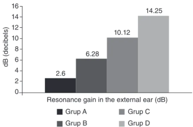
Figures 1 and 2 show, respectively, the average absolute values of the REUR and REUG measures for the four groups studied, and it is possible to visually verify the difference in these parameters. After application of ANOVA, statisti-cal signiicance between the values of samples for REUR (p=0.0000) and REUG (p=0.0000) measures was conirmed, as shown in Table 1.
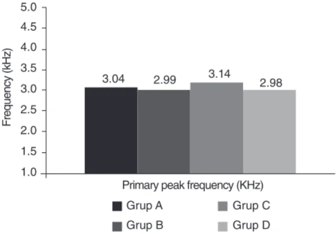
Figure 3, together with Table 1, shows the frequency in which the primary peak of resonance is registered, a variable that presented an absence of statistical signiicance (p=0.5416) in all groups.
Once the signiicance of the ANOVA was conirmed between groups A, B, and C regarding the average of the REUR and REUG measures for the right ear, Tukey’s test was used for comparison of means, to ascertain whether there was minimal signiicant difference.
Figure 1. Average of real-ear unaided response measurements of the
groups studied
Caption: Group A = elderly showing collapse of the external acoustic canal; group B = elderly showing senile tympanic membrane; group C = elderly without changes in the external auditory canal; group D = adults without anatomical or otological changes in the external ear
80
75
70
65
60
55
50
45
40
Response of resonance of the external ear (dBHPS)
Grup A
Grup B
Grup C
Grup D 67.6
71.53
76.41
79.25
Sound pressure level (d)
Figure 2. Average of real-ear unaided gain measurements of the
groups studied
Caption: Group A = elderly showing collapse of the external acoustic canal; group B = elderly showing senile tympanic membrane; group C = elderly without changes in the external auditory canal; group D = adults without anatomical or otological changes in the external ear
16 14 12 10
8 6 4 2 0
Resonance gain in the external ear (dB)
Grup A
Grup B
Grup C
Grup D 2.6
6.28
10.12
14.25
dB (decibels)
Table 1. Comparison of groups A, B, and C regarding the response of
measures of real-ear unaided response of real-ear unaided gain and of primary peak frequency
Measurement Group A Group B Group C p-value Mean±SD Mean±SD Mean±SD
Real-ear unaided
response (dB NPS) 67.60±1.06 71.53±2.61 76.41±1.73 0.0000* Real-ear unaided
gain (dB) 2.60±1.06 6.28±1.99 10.12±1.32 0.0000* Peak frequency
(kHz) 3.04±0.39 2.99±0.51 3.14±0.43 0.5416 (ns)
ANOVA = analysis of variance, *p<0.05 (statistically significant)
The results showed that the difference occurred between groups A and B in REUR and REUG measures with p=0.0001 in both cases, as can be seen in Table 2.
Table 3 shows the comparison of results between individu-als over 60 years from group C and adults from group D.
By applying Student’s t-test, it became possible to compare the average of the samples, verifying the signiicance between groups C and D for REUR and REUG measures, respectively (p=0.0043 and 0.0000), and no signiicance was observed for the primary peak frequency (p=0.1983).
DIsCussIoN
Today, worldwide, the increase in the number of elderly is already a reality, while in Brazil it becomes increasingly sig-niicant. In the age pyramid, this gradual change is enforcing a different behavior in different areas of health, and with it, new public policies to the elderly are being developed, providing improved quality of life.
In the course of aging, endogenous and exogenous factors affect humans. In this process, degenerative changes in the physi-cal, cognitive, and sensory aspects negatively affect the elderly, and hearing loss is considered one of the toughest of them in the psychosocial context.
There are many primary and secondary consequences of hearing loss in the elderly. In addition, the process of selection and adaptation to the HAs is a step that should be performed carefully, following appropriate protocols throughout the pro-cess, along with the guidance and counseling of the patient, so that problems related to acceptance and dificulty of the device’s daily maintenance are minimized. In this context, the dificulties related to the anatomical aspect of the external ear are extremely important as they may direct the response of the HA ampliication. However, a detailed study of these fac-tors was necessary, affecting the better use of ampliication. Thus, the aim of this study was to verify the inluence of the anatomical changes of the external ear as a result of aging in response to REUR and REUG.
Analyzing the results presented in Table 1, it was possible to verify a statistically signiicant difference in the comparison of groups A, B, and C in relation to measurements of the reso-nance of the external ear. Groups A and B (presenting collapse of the EAC and senile tympanic membrane) obtained results that differed from the elderly group without changes in the external ear (group C) in the procedures performed (REUR and REUG). Patients in groups A and B obtained inferior results to group C, with the lowest results being observed in group A. The difference between groups A, B, and C showed the effect of the changes caused by aging of the natural resonance of the external ear, due to loss of elasticity and increased sagging of the EAC, ratifying the consulted literature(3,6,7).
The primary peak frequency of REUR and REUG mea-sures was also investigated, with no offset being veriied in the record of the primary peak frequency for REUR or for REUG considering participating groups. Statistical tests revealed no signiicance for primary peak frequency (p=0.5416) for three groups of individuals over the age of 60. This difference was not observed because, according to the literature, the dimen-sions of the external ear among the elderly are similar, justify-ing the answers provided.
According to the results presented by the groups, a more detailed analysis of groups A and B was made necessary to verify which among the two anatomical changes presented by them would cause major impact on the response of the reso-nance of the ear (Table 2). Through this correlation, it was pos-sible to observe a statistically signiicant difference between the groups in the measures of REUR and REUG. Knowing that
Caption: Group A = elderly showing collapse of the external acoustic canal; group B = elderly showing senile tympanic membrane; group C = elderly without changes in the external auditory canal; group D = adults without anatomical or otological changes in the external ear
Figure 3. Average of primary peak frequency in the groups studied
5.0
4.5
4.0
3.5
3.0
2.5
2.0
1.5
1.0
Primary peak frequency (KHz)
Grup A
Grup B
Grup C
Grup D
3.04 2.99 3.14 2.98
Frequency (kHz)
Table 2. Comparison of groups A and B regarding the correlation of
anatomic changes in the response for real-ear unaided response and real-ear unaided gain
Measurement Group A Group B p-value
Mean±SD Mean±SD
Real-ear unaided
response (dB NPS) 67.60±1.06 71.53±2.61 0.0001* Real-ear unaided
gain (dB) 2.60±1.06 6.28±1.99 0.0001*
Tukey’s test; *p<0.05 (statistically significant) Caption: SD = standard deviation
Table 3. Comparison of groups C and D regarding real-ear unaided
response, real-ear unaided gain, and primary peak frequency
Measurement Group C Group D p-value
Mean±SD Mean±SD
Real-ear unaided
response (dB NPS) 76.41±1.73 79.25±3.87 0.0043* Real-ear unaided
gain (dB) 10.12±1.32 14.25±3.87 0.0000*
Peak frequency (kHz) 3.14±0.43 2.98±0.45 0.1983 (ns)
Student’s t-test, *p<0.05 (statistically significant)
Caption: ns = no statistically significant difference for Student’s t-test;
both changes interfere with the response of the resonance, we observed a major impact on individuals of group A.
Regarding aspects of aging, Table 3 shows that it was pos-sible to verify statistical signiicance between groups C and D for measures of REUR and REUG when comparing the aver-age of the samples, which leads us to relect on the importance of senile factors in the anatomical characteristics of the EAC.
When comparing elderly individuals (groups A, B, and C) with adults (group D), it was verified, by the statistical results, that the resonance values found, both for REUR and REUG, were lower for the elderly, which may be jus-tified by anatomical changes of the external ear due to the aging process. Therefore, changes in the natural resonance of the external ear were observed, being in accordance with the studies.
In the selection and veriication process for HAs, it is impor-tant to mention that the responses can be signiicantly affected because of changes in the anatomical characteristics of the EAC.
In the literature, authors consider that the dimensions of the EAC are different only between children and adult/elderly. In this study it was found that the resonance characteristics of adults are preserved. However, for elderly patients with ana-tomical changes in the EAC, these characteristics may be in a modiied state. In parallel with ampliication, it is known that changes in the natural resonance of the external ear directly affect the response of the HA, so it is important to consider the characteristics of the aging process for the selection and itting of HAs(8,16).
This study aimed to understand the role of aging in the natu-ral resonance of the external ear, which could aid audiological diagnostic tests in the process of selection and adaptation to HAs process, and consequently, in better use of ampliication.
CoNCLusIoN
According to the study, it is possible to conclude that the anatomical changes caused by aging interfere with measure-ments of natural resonance of the external ear and, consequently, on its resonance gain.
We believe that this study contributes to the Audiology ield, speciically in the selection and veriication of electroacoustic characteristics of HAs within the auditory rehabilitation pro-cess of elderly individuals with disabilities, aiming to maximize ampliication and its adaptation to the user’s auditory needs.
*ARPS was responsible for the project, study design, collection, and tabulation of data; WQB directed, and was responsible for, the project, study design and orientation of execution steps, and collaborated with the supervision of data collection; JRPL monitored the collection of data and collaborated with its analysis; JRMO was responsible for drafting the manuscript.
ReFeReNCes
1. Ballachanda BB. Theoretical and applied external ear acoustics. J Am Acad Audiol.1997;8(6):411-20.
2. Shaw EAG. Transformation of sound pressure from the free ield to eardrum in the horizontal plane. J Acoustical Society of America. 1974;56:1848-61.
3. Bonaldi LV. Bases anatômicas da audição e do equilíbrio. São Paulo: Livraria Santos; 2004.
4. Chisolm TH, Willott JF, Lister JJ. The aging auditory system: anatomic and physiologic changes and implications for rehabilitation. Int J Audiol. 2003;42(Suppl 2):2S3-10.
5. Couto CM, Carvalho RMM. O efeito das orelhas externa e média nas emissões otoacústicas. Rev Bras Otorrinolaringol. 2009;75(1):15-23. 6. Dirks DD, Ahlstrom JB, Eisenberg LS. Comparison of probe insertion methods on estimates of ear canal SPL. J Am Acad Audiol.1996;7(1):31-8.
7. Wiener FM, Ross DA. The pressure distribution in the auditory canal in a progressive sound ield. J Acoust Soc Am. 1946;18(2):401-8. 8. Bortholuzzi SMF. Ressonância da orelha em indivíduos com idades de
dois a 14 anos [monograia]. Santa Maria: Universidade Federal de Santa Maria; 1992.
9. Howarth A, Shone GR. Ageing and the auditory system. Postgrad Med J. 2006;82(965):166-71.
10. Navarro MR. Reducing feedback during the hearing aid evaluation. Audiology & Hearing Educ. 1978;4(6):15.
11. Liu TC, Chen YS. Aging and external ear resonance. Audiology. 2000;39(5):235-7.
12. Roland PS, Marple BF. Disorders of the external auditory canal. J Am Acad Audiol. 1997;8(6):367-78.
13. McSpaden JB. Basic tympanometry in the dispensing ofice. Hear Rev. 2006;13(12):28.
14. Byrne D, Upfold G. Implications of ear canal resonance for hearing aid itting. 1991;12:34-41.
15. Upfold G, Byrne D. Variability of ear canal resonance and its implications for the design of hearing aids and earplugs. Australian J. Audiol. 1988;10(2):97-102.
16. Oliveira RJ, Hoeker G. Ear canal anatomy and activity. Semin Hear. 2003;24(4):265-76.
17. Oliveira RJ. The active earcanal. J Am Acad Audiol. 1997;8(6):401-10. 18. Stenklev NC, Vik O, Laukli E. The aging ear: an otomicroscopic and
tympanometric study. Acta Otolaryngol. 2004;124(1):69-76.