Nº de ordem 01/D/2008
TESE DE DOUTORAMENTO
Apresentada à
Universidade da Madeira
Para obtenção do grau de Doutor
Cláudia Maria Neves Delgado
Gonad Development and Hormone Titres in Loggerhead Sea Turtles
(
Caretta caretta
) in the NE Atlantic
Júri
Reitor da Universidade da Madeira
Doutor David W. Owens, University of Charleston
Doutor Eduardo José Frias Gonçalves Crespo, Universidade de Lisboa
Doutor Adelino Vicente Mendonça Canário, Universidade do Algarve
Doutor Nuno Miguel dos Santos Ferrand de Almeida, Universidade do Porto
I
Abstract
The study proposed to describe sexual development in pelagic stage loggerhead sea turtles
Caretta caretta and compare this to hatchlings and adults. It is meant as an ontogenic approach, in order to understand reproductive development and population composition and their dynamics in the pelagic environment. The study focused on the pelagic loggerheads that are found in the waters offshore Madeira Island (Portugal) in the North-eastern Atlantic and use it as a developmental habitat.
The innovating character of this work relied on the lack of any description regarding the gonad ontogenesis and reproductive development for the pelagic stage in any of the 7 existing sea turtle species, all of them in danger of extinction.
Three methods were used to diagnose the sex of each juvenile individual and asses the level of reproductive development: (1) laparoscopy, (2) gonad biopsy and (3) the assessment of two sex steroids circulating levels, namely testosterone and estradiol.
In order to cover all life stages and compare data obtained for the juvenile stage, hatchlings and nesting female adults were sampled at the nearest nesting rookery at Boa Vista Island in the Cape Verde Archipelago. Gonads from dead hatchlings were collected for gonad histology and blood was collected from nesting females for sex steroids assessment.
Laparoscopies revealed to be a valid sexing method for the juvenile stage, since gonads are morphologically differentiated at these size classes. Moreover, laparoscopy was validated using gonad histology.
Gonad histology of juveniles showed that gonads are already completely differentiated into ovaries or testes at the size classes examined, but development seems to be quiescent.
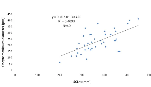
Males present already developed seminiferous tubules with spermatogonia lining the interior of the seminiferous tubule. Female gonads present oocytes at different development stages, but only oocytes up to stage III were observed. The maximum oocyte diameter in each individual correlated with body size, suggesting that reproductive development is an on-going process in juvenile females.
II No bimodal distribution was found for any of the sex steroids analysed and thus circulating hormone levels were not a reliable tool for sexing juvenile individuals with a non-invasive technique. The ratio testosterone:estradiol did not show a bimodal distribution either.
The levels of testosterone correlated with sea surface temperature. The fact that temperatures observed during this study were below 24ºC might have hindered a differential testosterone pattern between juvenile males and females.
Sex ratios for this population were generated according to laparoscopy results and compared among years and size classes. An overall sex ratio of 2 females for each male was found, but they varied among size classes but not among years. Possible causes for the sex ratios observed are discussed.
III
Resumo
O estudo propôs-se descrever o desenvolvimento sexual no estadio pelágico de tartaruga comum Caretta caretta e compará-lo com o dos neonatos e adultos da mesma espécie.
Pretendeu-se uma abordagem ontogénica de modo a compreender o desenvolvimento reprodutivo e a composição populacional e respectiva dinâmica em ambiente pelágico.
O estudo focou as tartarugas comum que se encontram nas águas da Ilha da Madeira (Portugal) no Atlântico Nordeste e que usam estas águas como habitat de desenvolvimento.
O carácter inovador deste trabalho reside na ausência de qualquer descrição da ontogénese da gónada e do desenvolvimento reprodutivo para o estado pelágico de todas as 7 espécies de tartarugas marinhas existentes, todas elas ameaçadas de extinção.
Usaram-se 3 métodos para diagnosticar o sexo de cada indivíduo juvenil e avaliar o nível de desenvolvimento reprodutivo: (1) laparoscopia, (2) biópsia da gónada e (3) medição dos níveis circulantes de duas hormonas esteróides, nomedamente de testosterona e de estradiol.
Por forma a descrever todos as fases do ciclo de vida e comparar os dados obtidos para o estadio juvenil, amostraram-se neonatos e fêmeas adultas da população nidificante geograficamente mais próxima, designadamente na Ilha da Boa Vista no Arquipélago de Cabo Verde. Foram amostradas góndas de neonatos mortos para histologia da gónada e recolheram-se amostras de sangue das fêmeas nidificantes para medição de esteróides recolheram-sexuais.
A técnica laparoscópica revelou ser um método de diagnóstico do sexo válido para o estadio juvenil, uma vez que as gónadas se encontram morfologicamente diferenciadas nestas classes etárias. Adicionalmente, a laparoscopia foi validada através da histologia da gónada.
A histologia da gónada em juvenis revelou que as gónadas estão já completamente diferenciadas em ovários e testículos nas classes etárias examinadas, mas o desenvolvimento aparenta estar quiescente.
IV correlacionado com o tamanho do indivíduo, o que sugere que o desenvolvimento reprodutivo é um processo contínuo em fêmeas juvenis.
Os níveis circulantes de testosterona e de estradiol em juvenis de ambos os sexos foram muito baixos, e consistentemente mais baixos do que os observados em fêmeas nidificantes da Ilha da Boa Vista.
Não foi encontrada uma distribuição bimodal para nenhuma das hormonas esteróides analisadas e deste modo os níveis hormonais não são fidedignos como ferramenta não invasiva para diagnóstico do sexo em indivíduos juvenis. O racio testosterona:estradiol não mostrou também uma distribuição bimodal.
Os níveis de testosterona apresentaram uma correlação com a temperatura de superfície do oceano. O facto das temperaturas observadas durante este estudo estarem abaixo dos 24ºC poderá ter impedido uma resposta hormonal distinta entre machos e fêmeas juvenis no que respeita à testosterona.
A laparoscopia permitiu a identificação dos sex ratios para esta população, os quais foram comparados entre anos e classes etárias. Encontrou-se um sex ratio de 2 fêmeas para cada macho, tendo-se observado variações entre classes etárias mas não entre anos. As causas possíveis para os sex ratios observados são discutidas.
V The water was warm. There was plenty to eat.
The turtles had everything turtles might need.
And they were all happy. Quite happy indeed.
...
And the turtles, of course... all the turtles are free
As turtles and, maybe, all creatures should be.
(from: Myrtle the Turtle by Dr. Seuss)
VII
Acknowledgments
Although I am the only author of this thesis, this is the product of the contributions of many people, which somehow influenced my work and ideas these last few years. I thank all of them, and am aware I can't name them all here.
I would like to thank my supervisors Dr. Thomas Dellinger and Dr. Adelino Canário for supporting and supervising me throughout the project time, providing me with good suggestions and advice, and particularly for encouraging me in the end and to finish the thesis writing. I also thank Dr. Thomas Dellinger for hiring me in two previous projects; it allowed me to gain an invaluable experience from field to paperwork.
Two persons deserve special recognition: Dr. Manfred Kaufmann have put aside his work to help me many times. Thank you for everything, from PC troubleshooting to lunch-breaks and wise advices. Renato Barradas, a.k.a. 'Aladino', was always there: from the boat to histology lab and for software clues, he was always helping a fellowship in despair. Thank you for Fridays dedicated to non-sense.
I am especially grateful to the members of the turtle team: Telma Ferreira, Tomás Chada, Susana Ferreira and Afonso Rocha. And Renato – again.
I thank Elsa Couto for all the patience with me at the lab doing the RIA's – I would have never been able to survive so much ether without her – and all the other fellowships who made me feel welcome in their laboratory. Acknowledgements are also due to Dr. Graça Costa for her assistance with the histology work and else.
A special thanks goes to the volunteers Nina Keul, Anísia Correia, Sandra Mendonça, Marta Oliveira and Nuno Queiroz who proved themselves exemplary assistants. Also Paulo Branco, Cláudia Moreira and Sandra Ferreira.
I thank Dr. Luís Felipe-Lopez from University of Las Palmas de Gran Canaria and Óscar Melicio from INDP (Cape Verde) for giving me the opportunity to work in Boa Vista Island.
VIII Cape Verde. And to the other two members of the 'trio equipo de investigación' – Ana Liria-Loza
'Chuchu' and Ana B. Casal – for all the delicious laughing on the most despairing situations out in a desert beach inhabited by (too) heavy and clumsy reptiles: they helped making the long patrolling nights a lot more lighter. And sorry for the bad humour sometimes: sleep deprivation! And to the shooting stars crossing the black skies of Ervatão and Ponta Cosme… they made my stay there a lot more… brighter.
I thank Cláudia Ribeiro for being the (only) closest fellow PhD student whit whom I could share my doubts, as well as Ana Luísa Valente for sharing her knowledge and special encouragement – and all the 'viagens na maionese'.
I also thank the dozens of volunteers at Madeira... and the dozens of volunteers at Boa Vista Island – can't name them all. Without them I would not be able to complete this work. Volunteers were an invaluable resource since it is impossible to work with turtles on our own, both at lab and in the sea.
Thanks to all the anonymous tourists that willingly (sometimes not so much, but definitely puzzled!) allowed me to use their cars to plug an 'high-tech' portable centrifuge and get plasma out of blood samples in the middle of nowhere.
Dr. Thomas Dellinger, Paulo Branco, Nélio Freitas and João Monteiro, among others, for the photos while general "turtling".
Funding for this project was provided by the National Science and Technology Foundation (FCT) through the fellowship SFRH/BD/8413/2002. I thank the University of Madeira and the Centre for Macaronesian Studies (CEM), who contributed with many resources, such as facilities, the boat and transportation and other equipment, as well as the University of Algarve through the Comparative and Molecular Endocrinology Laboratory.
Acknowledgements are also due to the Marine Biology Station of Funchal and all the people working there.
IX (Fundação Luso-Americana para o Desenvolvimento) for a travel grant to 25th ISTS in Savannah
and SIRAM for a travel grant to 26th ISTS in Crete. The Direcção Regional de Pescas through the
veterinary Isabel Quaresma and the veterinary clinics VetFunchal and VetMedis. And also Museu da Baleia and the tourism vessels Sea Pleasure, Ventura do Mar and Beluga.
To SANAS, who rescued BIOMAR II and its three castaways when we were already 14 nm offshore, drifting with the current on 22nd July 2005… and again Manfred and Renato who went
for help and got in trouble themselves.
My gratitude extends as well to all the sea turtle people I have met along the several ISTS's I attended. Many of them had a strong influence on my attitude and work in sea turtle research and conservation and made me feel I belong to this peculiar sea-turtle family. Special thanks go to Dr. David Owens for his patience and wisdom when teaching me how to perform laparoscopies. Dr. Nicholas Mrosovsky for his controversial opinions on sea turtle conservation and for being an inspiration for research. Dr. Thane Wibbels, Dr. Jeanette Wyneken and Dr. Marc Girondot who offered valuable advice and insights at the several ISTS's and for believing in this work. And Dr. Brendan Godley for a most welcome 'good luck' just before the oral presentation at 27th ISTS… and all the constructive comments afterwards.
To my parents, a.k.a. 'Fundação Delgado & Delgado' – always willing to fund Portuguese research through two special fellowships – for supporting and encouraging me always, even when they didn't understand the when's and why's… as well as my sister who said the right things at the right times (and for layout advice).
To some very special friends: Marla, Marta, 'Becas', Sílvia and little Inês for supporting me and sometimes being a second family. Lélia Matos, a truly 'turtle-freak'. Isabel Figueira for 'baby-catting' Mushi and then Felis whenever I had to leave the Island. And Eduarda – just because. Thank you to the many friends with whom I could not be in order to finish this: sorry for neglecting you and for general absence many times. Sorry for un-replied e-mails. Thank you for letting me work when I was not with you; and Ana Paula for the inspired and inspiring coffees.
X
List of abbreviations
AR: androgen receptor B: corticosterone
Bkm: banded krait minor CCL: Curved Carapace Length
CITES: Convention on International Trade in Endangered Species of Wild Fauna and Flora DDE: Dichlorodiphenyldichloroethylene
DHT: dihydro-testosterone E2: estradiol
EDC’s: endocrine disrupting components EEZ: Exclusive Economic Zone
ER: estrogen receptor
ESD: environmental sex determination GPS: Global Positioning System GSD: genotypic sex determination GSI: Gonadossomatic Index
IUCN: The World Conservation Union mRNA: mitochondrial ribonucleic acid mtDNA: mitochondrial deoxyribonucleic acid nDNA: nuclear deoxyribonucleic acid NE (trade winds): North-East trade winds PCB’s: polychlorinated biphenyl compounds PIT: Passive Integrated Transponder RIA: radioimmunoassay
SCL: Straight-line Carapace Length SST: sea surface temperature T: testosterone
XI
Table of Contents
CHAPTER I Sex in sea turtles ... 1
General Introduction ... 2
Loggerhead sea turtle Ecology ... 4
Species Description ... 4
Distribution ... 5
Life History/Cycle ... 6
Conservation Status and Threats ... 8
Sex Determination and Sex Identification in marine turtles ... 10
Sex determination mechanisms ... 10
TSD and Gonad Development ... 14
Sexual dimorphism during ontogeny and sex identification in sea turtles ... 19
TSD and sex-ratios: why bother? ... 22
Research objectives ... 23
Materials and Methods ... 25
Sexing sea turtles ... 26
Study Areas ... 26
Madeira Island (Portugal) ... 27
Study area description and population characterization ... 27
Sea turtle capture ... 27
Biometry and Tagging Activities ... 28
Boa Vista Island (Republic of Cape Verde) ... 30
Study area description and population characterization ... 30
Sea turtle capture ... 32
Nesting female adults ... 32
Dead hatchlings ... 33
CHAPTER II Laparoscopies ... 34
Introduction ... 35
Objectives ... 37
Materials and Methods ... 37
Results ... 40
Discussion ... 43
CHAPTER III Gonad Histology ... 45
Introduction ... 46
Objectives ... 48
Materials and Methods ... 49
Juveniles ... 49
Hatchlings ... 49
XII
Results ... 53
Juveniles ... 53
Hatchlings ... 56
Discussion ... 57
Juveniles ... 57
Hatchlings ... 60
Madeira Island juveniles and Boa Vista Island hatchlings ... 61
CHAPTER IV Identification of sex by measurement of blood plasma sex steroids ... 64
Introduction ... 65
Objectives ... 67
Materials and Methods ... 68
Juveniles ... 68
Adult females ... 68
Hormone extraction from blood plasma ... 69
Steroid radioimmunoassay ... 71
Environmental data: Sea Surface Temperature (SST) ... 71
Sex identication and oocyte size ... 72
Statistics ... 72
Results ... 73
Juveniles ... 73
Sex steroids and SST: is steroids RIA temperature-dependent? ... 76
Circulating hormones and oocyte size ... 77
Adult females ... 78
Madeira Island juveniles and Boa Vista Island nesting females ... 78
Discussion ... 80
Sex steroids levels for Madeira Island juveniles and Boa Vista Island nesting females ... 80
Sex steroids and SST: are steroids temperature-dependent? ... 84
Circulating sex steroids and oocyte size ... 84
Ratios T:E ... 85
Relevance of the Sex steroids analyzed ... 86
Sex steroids levels and population sex ratio ... 87
CHAPTER V Sexual Dimorphism and sex ratios in Juvenile Marine Turtles ... 89
Introduction ... 90
Objectives ... 91
Materials and Methods ... 92
Sexual Dimorphism ... 92
Sex ratios ... 93
Results ... 94
Sex diagnosis and correlation among methods ... 94
Biometrical parameters and correlation with sex ... 94
XIII
Sex ratio comparison between Eastern and Western North Atlantic ... 95
Sex ratio variation on a temporal scale ... 95
Sex ratio among size/age classes ... 96
Discussion ... 97
Sex identification and correlation among methods ... 97
Biometrical parameters and correlation with sex ... 98
Population sex ratio ... 98
Sex ratio on the Eastern vs. Western North Atlantic ... 99
Sex ratio variation on a temporal scale ... 100
Sex ratio among size/age classes ... 101
Causes for Dynamic Sex Ratios ... 101
Different migratory routes and/or developmental areas ... 102
Sex reversal ... 102
Differential mortality ... 104
CHAPTER VI Conclusions and Further Research Needs ... 107
Sexing techniques and gonad development ... 108
Sex ratios ... 111
TSD sex ratios and climate change ... 112
Summary ... 114
References ... 117
Appendices ... 147
List of tables ... 147
List of figures ... 147
1
CHAPTER I
2
General Introduction
The natural history and the behavioural ecology of marine turtles have received growing attention during the past 4 decades in parallel with declining populations. However there are still many gaps in our knowledge of the life history and ecology of these marine reptiles, knowledge which is essential for their management and protection.
Seven species of sea turtles representing two families, Cheloniidae and Dermochelyidae, are the only living members of what has been a large and diverse marine radiation of cryptodiran turtles. These seven species include the loggerhead (Caretta caretta), the green (Chelonia mydas), the hawksbill (Eretmochelys imbricata), the Kemp’s ridley (Lepidochelys kempi), the olive ridley (Lepidochelys olivacea), the flatback (Natator depressus) and the leatherback (Dermochelys coriacea) turtles (Meylan and Meylan, 1999).
All of them are endangered or threatened on a worldwide basis, and are protected under several national and international laws (Abreu et al., 1995; Groombridge, 1990; Márquez M., 1990). However, comparison of present population levels to historical levels are difficult to make because there is little or no information (Ross, 1995). Except for the Kemps’ ridley, each species has a large proportion of widely distributed populations, and the number of large populations with which current populations can be compared is small. A further problem is the taxonomic confusion between species, namely between Caretta and Lepidochelys which, until recently, led to many misidentifications (Ross, 1995).
Macaronesian waters comprise the seas around and between the Madeira and the Azores Archipelagos, the Selvagens Islands (Portugal) and the Canary Islands (Spain) (Brongersma, 1995). The German botanist A. Engler gave this name to the Azores, Madeira (plus Porto Santo) and the Canaries in 1879, because of similarities in the plant life of these islands. The name comes from the greek makaros and nesios, and means the blissful islands (Wirtz, 1994).
3 (Brongersma, 1995). All of these species are also reported to Madeira (Biscoito, 1987; Cabral et al., 2005; Dellinger, in prep.).
Macaronesian waters are used by juvenile loggerhead marine turtles that stay in the area for a part of their life: they get there when they are young, as post-hatchlings and juveniles, and leave oceanic waters when they become adult (Bolten et al., 1998). Thus, Madeiran loggerheads belong to the Macaronesian loggerhead aggregation which is all constituted by juveniles ranging from small, nearly hatched, juveniles to large juveniles. The loggerhead turtles found in Madeira are almost exclusively (99%) from the nesting beaches of the west coast of the United States of America and from Mexico according to mitochondrial DNA data (Bolten et al., 1998). Since loggerhead marine turtles do not reproduce here we cannot define it as a true population, but for further discussions the juveniles occurring within Madeira or Macaronesian waters will be referred as a population.
Some authors include also the Cape Verde Archipelago (Republic of Cape Verde) within the Macaronesian region owing to its bio-geographical similarities with the aforementioned European archipelagos. Recently, a fairly large breeding population of loggerheads has been reported for this archipelago, and specially for Boa Vista Island (Cejudo et al., 2000). Juveniles of green turtles seem to use those waters as a feeding area (Nuria Varo, pers. comm.).
4
Loggerhead sea turtle Ecology
Species Description
Marine turtles were common in the cretaceous, 130 MY ago, and their fossil record extends back at least 200 MY. All present day genera and species originated in the period from the early Eocene to the Pleistocene, between 60 and 10 MY ago. Together with the marine snakes, crocodiles and iguanas, they are the only surviving marine adapted reptiles, and depend entirely on land for reproduction (except for some viviparous snakes) (Márquez M., 1990). These marine reptiles have only secondarily adapted to the marine environment as they evolved from terrestrial turtles over 100 MY (Pritchard, 1997). The minimum chronologic separation of modern sea turtle lines is probably no less than 30 MY, and Dermochelys at least 50 MY (Carr, 1995). The loggerhead was originally described in 1758 as Testudo caretta by Linnaeus, but is currently known as Caretta caretta (Linnaeus, 1758). Genetic analyses from globally distributed sites have not provided support for dividing C. caretta into subspecies (Bowen, 2003; Bowen et al., 1994). Taxonomically it is placed in the
Phylum Chordata
Subphylum Vertebrata
Superclass Tetrapoda
Class Reptilia
Subclass Anapsida
Order Testudines
Suborder Cryptodira
Superfamily Chelonioidae
Family Cheloniidae
5 Individuals of the species Caretta caretta are diagnosed by the two pairs of prefrontal scales, the conspicuously large heads and the elongated carapace, thickened above the caudal region. The head is very broad and triangular in shape, with powerful jaws. They usually have five pairs of well cornified costal scutes; neural bones are usually 7 or 8. The dorsal scutes do not imbricate, except in very young specimens. The adults’ vertebral scutes are smooth and do not overlap. C. caretta has five pairs of pleural scutes, the first contacting the pre-central and usually with three or four inframarginal laminae enlarged and poreless, and 12-13 marginal scutes. Two rudimentary claws are present on each flipper but the claw on the first digit has a specialized secondary function in adult males, in which is enlarged and hooklike, and is used for clasping the anterolateral marginal area of the female during copulation. The carapace is reddish-brown and the plastron yellowish-brown. Both the carapace and plastron of the loggerhead are heavily keratinized as a protective barrier against attack and the environment (Dodd, 1988).
Juvenile vertebrals are keeled with a knob-like process on the posterior portion of each keel (most distinct on the anterior vertebrals). In juveniles the knobs generally disappear although the keels are still present, and by the end of the juvenile phase the keels also disappear (Brongersma, 1972). These are supposed to provide what may be protective armor against some oceanic predators. Larger juveniles (> 45 cm SCL) that have begun to feed in shallow coastal waters have lost their thickened shell scutes, and their carapace colouration is often masked by fouling organisms such as algae, hydroids, and barnacles (Witherington et al., 2006).
Distribution
The total range of the loggerhead turtle includes foraging areas, migration corridors, and nesting beaches distributed throughout the subtropical and temperate oceans of the world (Dodd, 1988).
Thus, loggerhead turtles are circumglobal, inhabiting continental shelfs, bays, lagoons, and estuaries in the temperate, subtropical and tropical waters of the Atlantic, Pacific, and Indian Oceans.
6 with the exception of Masirah Island, in Oman. Nesting does occur in tropical regions, and nearly all nesting occurs between 19 and 36 degrees latitude in each hemisphere (Witherington et al., 2006). Some warm temperate zone nesters are known to migrate to tropical waters in Australia and Africa after the nesting season (Dodd, 1988).
Loggerheads do not nest anywhere on the Atlantic coast of Europe and the anomalous absence of nesting grounds of Caretta in the central and western Pacific is unexplained (Ross, 1995).
Life History/cycle
All marine turtles share a common general life cycle that includes a iteroparous reproduction (Hirth, 1980), steroptyped nesting behaviour, laying of relatively large numbers of eggs several times during the reproductive season and relatively strong attachment to particular locations for nesting (i.e., philopatry), but inter- and intra-specific variation exists (Miller, 1995).
After hatching and emerging from the nest loggerhead hatchlings crawl down the beach and enter the ocean. Not much is known from this point onwards, but solitary as well as aggregations of young hatchling loggerhead sea turtles in the sargassum offshore the eastern coasts of the United States are reported (Caldwell, 1968; Carr, 1986b; Schwartz, 1988). Apparently loggerhead hatchlings use drift lines created by upwellings, downwellings, currents, and other types of convergences of different bodies of water (Carr, 1986a; Carr, 1986b; Carr, 1987). These convergence zones produce concentrations of resources that are rich in potential prey items for young turtles and floating material, such as Sargassum and debris from land sources, providing shelter for both turtles and prey.
For many years this pelagic phase out in the open ocean was known as the "lost year", and Carr (1986a; 1986b; 1987) speculated that hatchlings and juveniles would probably ride the currents and gyres in the North Atlantic between North America and Europe and go back as a subadult to the developmental habitats in the western Atlantic. Carr (1986) suggested that hatchlings born in the south-eastern USA become entrained in the “Gulf Stream-Azores” current and travel eastward to the Macaronesian area, returning to the western Atlantic in the “North Atlantic gyre”.
7 Bjorndal et al., 2001a; Bjorndal et al., 2000), during which the juvenile feeds and grows into a subadult.
Historical records of juvenile loggerheads are scarce (summarized by Carr (1986c)), but large numbers are reported from the Azores, Madeira and even between Madeira and mainland Portugal. Juvenile loggerheads are also found stranded on the coasts of northern Europe (Brongersma, 1972; Brongersma, 1982; Dellinger, in prep.).
After this pelagic or oceanic phase, where these juveniles stay in the Macaronesian region inhabiting the oceanic pelagic environment (Bolten 2003), and attain sizes up to 60 cm or more straight carapace length (personal observation) loggerheads migrate to the neritic feeding areas for a benthic phase, close to the same nesting areas where they were born. In the western Atlantic, most juveniles recruit from oceanic pelagic to neritic demersal habitats at a size 50 or more cm CCL (curved carapace length) at an estimated age of about 6 to 12 years (Bjorndal et al., 2003a; Bjorndal et al., 1999; Klinger and Musick, 1995). This ontogenetic shift in habitat utilization characterizes the passage from the juvenile to the subadult life stage.
Mating occurs during a relatively short female receptive period in the vicinity of the nesting beach (Owens, 1980) and the individual’s mating season is completed before egg laying begins. The reproductive and ovipositional cycles are triggered and regulated by changes in specific serum gonadotropins and gonadal steroids (Guillette Jr. et al., 1991; Wibbels et al., 1992). Marine turtles show a strong conservationism during the nesting process and the nesting sequence is fairly similar between species, with only minor variations. They nest nocturnally on sandy beaches of mainland shores, islands and barrier islands and dig in material that ranges from fine siliceous particles to spherical pellets of calcareous algae in beaches within tropical areas. The homing ability, also called phylopatry, has been supported by evidence of spatial population structure in Atlantic loggerheads (Bowen et al., 1993; Encalada et al., 1998), as well as for other populations.
8 often widely separated (Carr, 1995).
For reference of further discussions, the following size categories are defined as follows, adapted from Dodd (1988):
Hatchling and Post-hatchling: from hatching to the first few weeks of life; characterized by the presence of the umbilical scar.
Juvenile: the pelagic, oceanic life stage, typically referred to as “lost year”; pool of animals of a size and/or age at which little or no sexual development is occurring.
Subadult: from the end of the pelagic oceanic stage to the onset of sexual maturity, also called immature or benthic juveniles; pool of animals that move from oceanic to coastal, benthic foraging grounds.
Adult: attainment of full reproductive maturity, at different sizes, depending on population; the size at sexual maturity for males is assumed to be similar to that of females.
Conservation Status and Threats
The best assessments of marine turtles’ abundance worldwide originate from nest counts on beaches. Temporal trends for pelagic loggerheads are much less accurate or even unfeasible due to the difficulties in having reliable, standardized in-water monitoring.
The current status for loggerheads is Endangered [EN A1abd ver. 2.3 (1994)] (IUCN 2007.
2007 IUCN Red List of Threatened Species. <www.iucnredlist.org>. Downloaded on 17 October 2007), and the species is quoted in the Portuguese Regional Red Data Book (Oliveira et al., 2005). Thus, as well as all other marine turtles, loggerheads are listed under Appendix I (i.e., prohibited from international trade from or to signatory countries) of the Convention on International Trade of Endangered Species of Wild Fauna and Flora (CITES), Appendix II Bern Convention and Appendix I Bonn Convention, as well as various regional or national laws, regulations, decrees and acts. Particularly in Madeira, they are protected since 1985 (Dec. n 18/85/M) which was the first Portuguese law to protect a reptile species.
9 for meat on the nesting beaches and incidental capture by commercial fisheries worldwide have been reducing the nesting populations worldwide. Accidental mortality became an increasing issue in sea turtle conservation with increasing fishing efforts (e.g., trawling, long-lining, gill nets, purse-seines). Thus, current populations are a fraction of historic levels. The considerable loss of nesting habitats to coastal development worldwide presents another threat, as well as climate change. The small size of most populations may be a result of continued pressure (Ross, 1995). Although historically high numbers of turtles were caught for food and later for tourists as stuffed souvenirs (Brongersma, 1968; Brongersma, 1982; Dellinger, 2007), incidental capture by the commercial black scabbard-fish (Aphanopus carbo Lowe, 1839) fishery has been identified as the main source of by-catch within Madeiran waters. This fishing fleet captures over 500 loggerheads turtles every year (Dellinger and Encarnação, 1999; Ferreira, 2001). This accidental capture is the result of turtles attempting to take the bait, or becoming accidentally entangled in the line or hooked in the flippers (Lewinson et al. 2004). Measures to mitigate by-catch would allow the conservation of juvenile pelagic stage loggerhead turtles in the Madeira archipelago to be more successful, but would need a governmental backup to be implemented. Additional sources of at-sea mortality and morbidity include other fishing methods, ingestion of and entanglement in marine debris, oil spills, and other pollution sources.
In fact, this population shows heavy metals contamination such as mercury and cadmium (Dellinger et al., unpublished), but the long-term consequences of these contaminants accumulation at the individual and population levels are not known.
Turtles spend most of their lives submerged within the pelagic realm (Bjorndal, 1999), but nearly all sea turtle conservation efforts have focused on the two, easily accessible, life history stages, i.e., the eggs and the adult females on the nesting beaches. However, a species’ endangered status implies the collection of population structure data and use of population modeling to fill information gaps, a prerequisite to its management.
10 parameters, like survivorship and age-at-maturity. Thus, rookery protection is of the out most importance for the species preservation but without protection of large juveniles and adults, the conservation of sea turtles will be unsuccessful (Carreras et al., 2004).
Therefore protection and conservation of oceanic stage turtles is extremely important (Crouse et al., 1987). One of the aims of this work is to contribute to a better understanding of the pelagic phase of this endangered species.
Sex Determination and Sex Identification in marine turtles
Sex determination mechanisms
One of the most fundamental traits for any species is reproduction. In sexually reproducing organisms the sex ratio is an important factor for population growth and therefore the existence of male and female organisms. A remarkable diversity of sex determination systems can be found among different animal taxa (Ciofi and Swingland, 1997), and vertebrates have evolved several different sex determining mechanisms. These are usually classified as either genotypic sex determination (GSD) or environmental sex determination (ESD). Even though, cases of mixed sex determination, i.e. combinations of genotypic and environmental sex determination are also observed, although the extent of their relative contribution varies. In these cases, both genetic and environmental cues play a role in an individual’s sex phenotype. In fact, genotypic and environmental sex determination systems are now considered the extremes of a continuum (Sarre et al., 2004).
The most common type of GSD (or polygenic sex determination) involves sex chromosomes. In the GSD system sex chromosomes exert an ultimate control on whether the gonad will evolve to a testes or an ovary. The system of genotypic sex determination implies that the sex of an individual depends entirely on its genotype which is determined at the moment of fertilization, i.e., at the zygote stage, and is fixed from that moment on.
11 male heterogamety, and the sex chromosomes referred to as X and Y in mammals, and female heterogamety in birds, and sex chromosomes referred to as X and Z.
GSD is also present among reptiles, amphibians, and fish with either male or female heterogamety. In reptiles at least three variations of heterogamety occur for both sexes: XX/XY, XXX/XXY and pseudo XO when males are the heterogamic sex, and ZZ/ZW, ZZZ/ZZW and ZZZ/ZWW when females are the heterogamic sex (Ciofi and Swingland, 1997). Yet many fish and reptile species lack sex chromosomes altogether (Crews, 1994).
Only in the last three decades researchers have come to realize that many vertebrates also exhibit environmental sex determination (ESD). There are two basic types, behaviour-dependent and temperature-dependent, and in both instances gonadal sex is determined after fertilization (Crews, 2000). Environmental sex determination is common among reptiles and is also observed in amphibians and fish. ESD also occurs among invertebrate taxa (see Ciofi and Swingland (1997) for a review).
Environmental sex determination implies that the sex of an individual is determined irreversibly by the environment experienced during early embryonic development, i.e., post-fertilization. When the key environmental factor is temperature, it is referred as ‘temperature-dependent sex determination’ (TSD). In these cases, incubation temperature serves as the trigger to initiate the cascade of events that leads to the development of ovaries or testes. Other environmental cues are the chemical milieu, such as pH or the social status of the individual.
Temperature-dependent sex determination was first described in the lizard Agama agama
12 lizards and most turtles (Bull, 1980; Janzen and Paukstis, 1991) and the tuatara (Cree et al., 1995).
Owens and Hendrickson (1978) were the first to hypothesise that environmental clues might be the mechanism responsible for variable sex ratios observed in marine turtles. TSD was first demonstrated for loggerhead sea turtles Caretta caretta (Yntema and Mrosovsky, 1979; Yntema and Mrosovsky, 1980) and subsequently documented for all the remaining marine turtle species (Wibbels, 2003).
TSD mechanisms are not homogeneous, and at least four different patterns have been described (Fig. 1):
a) Females develop at low temperature and males at high temperature, i.e., female-male pattern (FM);
b) Males develop at low temperatures and females at high ones, i.e., male-female pattern (MF);
c) Females develop at low and high temperatures and males at intermediate ones, i.e., female-male-female pattern (FMF);
d) The hatchling sex ratio of some species is not significantly influenced by incubation temperature.
13
Fig. 1 Response of sex ratio to incubation temperature in reptiles (from Kraak and de Looze (1993)).
All marine turtles exhibit a male-female pattern of sex determination (Wibbels, 2003), i.e., temperatures below the pivotal temperature generate more males and above the pivotal temperature generate more females. Pivotal or threshold temperature is defined as the incubation temperature at which an even sex ratio is generated. For loggerhead hatchlings incubated under laboratory conditions males are produced at temperatures around 27ºC, while females form at warmer temperatures around 31ºC. A 1:1 sex ratio is produced at a pivotal temperature between the two extremes (≈ 29ºC) (Yntema and Mrosovsky, 1982). In the wild, incubation environments of approximately 29ºC have also been found to produce an equal number of male and female hatchlings (Limpus et al., 1983; Marcovaldi et al., 1997; Mrosovsky, 1988). However, pivotal temperature can vary among different populations within a species (Limpus, 1985b). For loggerheads, pivotal temperatures have been reported to vary by up to 1.0ºC in the wild (Wibbels, 2003).
14 moisture, for the whole process of embryogenesis.
As already stated, cooler temperatures produce males and warmer temperatures produce females, but within fluctuating environments such as sand beach temperature, the sex of the hatchlings is determined by the proportion of development at a given temperature, and not by the duration of exposure to that temperature (Georges et al., 1994). More precisely, sexual differentiation is not determined by temperature throughout incubation but by temperature levels prevailing during a critical period of the embryonic development, the thermosensitive period (TSP) for sexual differentiation. This sensitive period occurs during the middle third of the incubation period (Desvages et al., 1993; Maxwell et al., 1988; Yntema and Mrosovsky, 1982); until this period the gonad is considered bipotential.
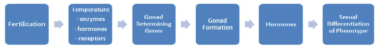
TSD and Gonad Development
Sexual differentiation can be seen as a kind of phenotypic plasticity. In vertebrate species that lack sex chromosomes, gonadal sex is fixed during embryonic development, and thus gonadal sex is plastic during a short period of embryonic development (Crews, 2000).
Many studies addressed the underlying molecular mechanisms of TSD, but the physiological, biochemical, and molecular mechanisms by which incubation temperature influence the gonad’s fate are still poorly understood, i.e., how the external signal of temperature is transduced into a signal that determines gonadal sex and channels sexual development.
The developing gonad itself does not appear to give the signal for its own differentiation into a testis or ovary, suggesting that an externally driven factor (or factors) moves into the developing gonadal tissue during a sensitive period to induce the normal differentiation, and possibly trigger the initial switch to one sex or the other (Desvages et al., 1993; Merchant-Larios and Villalpando, 1990).
15 Therefore, steroids play a pivotal role in sex determination in embryos and are a key element of sex determination due to the organizing effects of sex steroid hormones on the tissues that mediate reproduction (Crews, 1994). Only after the gonad is formed do hormones begin to exert an influence that modifies specific structures that eventually will differ between the sexes. According to Deeming and Ferguson (1988; 1991) the dose of a particular molecule determines sex, whatever the sex determining system is: in GSD the dose is genetically specified and in ESD the efficiency of gene transcription, or translation, or the stability of the mRNA or gene product, or the activity of the gene product, is determined by environmental conditions.
Research on TSD indicates that gonadal sex depends ultimately on which genes encoding for steroidogenic enzymes and hormone receptors are activated during the middle third of embryonic development by temperature – i.e., the temperature sensitive period (TSP). Therefore, incubation temperature modifies the activity as well as the temporal and spatial sequence of enzymes and hormone receptors such that sex-specific hormone milieus, created in the urogenital system of the developing embryo, determine gonad sex. Thus, estrogens are the physiologic equivalent of incubation temperature and the proximate cue that initiates female sex determination (Crews et al., 1995a). In any case, testosterone (T) serves as a precursor molecule destined for conversion to dihydro-testosterone (DHT) (via 5α-reductase) or estradiol (E2) (via
aromatase), and incubation temperature is hypothesized to activate steroid hormone receptor genes [e.g., male-producing incubation temperature upregulates androgen receptor (AR) and female-producing incubation temperature upregulates the estrogen receptor (ER)] (Crews et al., 1995a).
16 Studies using the administration of exogenous estrogens, antiestrogens and aromatase inhibitors, demonstrated the role of estrogens in sexual differentiation of the gonads in TSD species, and the activity level of the enzyme aromatase was well correlated with gonadal structure, since it increased exponentially in differentiating ovaries, whereas it remained low in differentiating testes (Pieau, 1996). Moreover, estrogens can even override the temperature effect and induce ovarian differentiation at masculinizing temperatures, as when injecting eggs with inhibitors of estrogen synthesis (aromatase) produces male offspring, even if the eggs are incubated at temperatures that usually produce females (Dorizzi et al., 1994; Rhen and Lang, 1994). In contrast, the administration of exogenous estrogen to an egg incubating at a male-producing temperature can reverse the effect of temperature and result in a female hatchling (Bull et al., 1988; Crews et al., 1991; Crews et al., 1989; Gutzke and Bull, 1986; Raynaud and Pieau, 1985; Tousignant and Crews, 1994; Wibbels et al., 1991a; Wibbels et al., 1991b). Similarly, when eggs are incubated at increasing temperatures that progressively produce a larger proportion of females, the dose of E2 required to reverse the sex of 50% of the animals
decreases significantly (Wibbels et al., 1991b). Moreover, the sensitive period for the effects of estrogens and their inhibitors coincides with the time when sex determination usually occurs (Bull et al., 1988; Gutzke and Chymiy, 1988), and there is a high correlation between the thermosensitive periods and increase in aromatase activity (Pieau, 1996).
This hormonally induced gonadal differentiation does start in the gonad itself since assays carried out on the gonads alone, i.e. separated from the adrenal/mesonephros, provided evidence that the gonads themselves respond to temperature shifts by modifying their sexual differentiation and are the site of aromatase activity and oestrogen synthesis during the thermosensitive period. Therefore, estrogens act locally on both the cortical and the medullary part of the gonad to direct ovarian differentiation (Pieau and Dorizzi, 2004).
17 destined to become Sertoli cells (Moreno-Mendoza et al., 1999; Spotila et al., 1998).
The morphological changes that occur during embryonic development of marine turtles – loggerheads included – was already described for both Cheloniidae and Dermochelyidae and is similar in morphological detail and sequence up to the stage 22 (see Miller (1985) for a detailed description of embryonic developmental stages). In general terms, development involves three main phases: 1) structural differentiation of body and organs (organogenesis), 2) functional development of organs and systems and 3) embryonic growth. Embryonic development begins immediately following fertilization, and although cleavage begins within hours after fertilization, development does not advance beyond the 6th stage while still within the female’s oviduct. After
oviposition, development resumes in a few hours (4 to 8, depending on temperature) and progresses. During stages 22-27 (which comprises the middle third of development and TSP) generic and species-specific characteristics become increasingly evident, such as the shape of the scales and the pigmentation of the carapace (Miller and Limpus, 2003).
Although the genital systems differ greatly in the two sexes, one sex frequently possesses rudiments of the structures characteristic of the other. This is due to the fact that for some time during embryonic life there is an indifferent or bipotential stage. The gonads may attain considerable size without showing specific features of either ovary or testis, and both male and female duct systems may differentiate to a considerable degree in potential members of both sexes. Eventually, however, there appears a definite sexual stage; the gonads become specifically testes or ovaries, and only the ducts and other accessory structures appropriate to one sex or the other continue their development. The nonpertinent structures characteristic of the opposite sex generally cease to develop and may be resorbed, but they are sometimes merely arrested in their growth, to persist as rudiments in the adult (Fig. 3).
18 the definitive gonad is formed toward the anterior end of the abdominal cavity; fat bodies or other structures may arise from abandoned portions of the genital ridge. The germinal epithelium of the ridge, continuous with the mesodermal lining of the rest of the coelom, forms the more important structural elements of the gonad; mesenchyme lining the epithelium forms connective tissues (see Ackerman (1997), Miller (1985) and Miller et al. (2003) for a review).
Merchant-Larios (1989) found that the ultrastructure of Lepidochelys olivacea primordial germ cells (PGC’s) is very similar to that described for Caretta caretta during the migratory stage and the early colonization of the genital crest (Fujimoto et al., 1979). Two morphogenetic events occur in the undifferentiated gonads of Lepidochelys olivacea female embryos: surface epithelium thickening and medullary cord fragmentation. The male gonads, on the other hand, keep the same histological structure as the undifferentiated gonad. Thus, they are recognisable as “testes” only because they are not differentiated ovaries (Merchant-Larios, 1989).
Working under laboratory conditions, Yntema & Mrosovsky (1980) demonstrated that gonads of
Caretta caretta reared for 1-7 weeks after hatching were similar to those of newly hatched specimens and recently Wyneken (2007) showed that captive reared hatchlings up to 120 g had perfectly distinguishable gonads.
19
Fig. 3 Diagram of female and male gonadal differentiation in amniote vertebrates (from Crews (2003)).
Sexual dimorphism during ontogeny and sex identification in sea turtles
Ecological modellers of sea turtle populations require accurate quantitative data of population structure for developing predictive models needed for management decisions. Critical demographic parameters such as growth rates, survivorship, recruitment, age at first reproduction, percent of animals reproductively active each year, age and duration of the reproductive life history and sex ratio are essential for the development of population models (Owens, 1997) and are especially important for marine turtles since all species are threatened (Casale et al., 2006). However, demographic models and management plans are limited by the lack of data on the immature pelagic phase for sea turtle populations.
20
Fig. 4 (A, B) - Adult male loggerhead showing the developed tail (A) and fore-flipper claw (B).
Marine turtles require decades to reach maturity (Bjorndal et al., 2003a; Chaloupka and Limpus, 1997; Limpus and Chaloupka, 1997), and not all individuals mature at the same size even within the same population (Limpus et al., 1994a; Limpus et al., 1994b), since sexual dimorphism in marine turtles becomes apparent very late in the life cycle.
For example, Caldwell (1962) stated that in the Pacific black turtle Chelonia mydas secondary sexual characteristics become obvious at a carapace length of about 75 cm, but at the Grand Cayman turtle farm it was possible to misidentify large immature male green sea turtles (120 kg, 80 cm carapace length) as females based on external characteristics only (Owens et al., 1978). Hughes (1974a) reported that sexual differentiation was apparent in South-African loggerhead turtles 60 cm to 67 cm SCL, but the few animals within this size ranges observed (and sexed through laparoscopy) in Madeira Island did not show any secondary sexual characteristics. Estimates from young captive loggerheads pin-pointed the age of sexual maturity at 6 to 7 years (Uchida, 1967), but these estimates from captive-reared animals are believed to be misleading (Bjorndal and Zug, 1995). Age at sexual maturity for loggerheads is now estimated to span 12-37 years (Bjorndal et al., 2000; Bjorndal et al., 2001b; Frazer, 1983; Frazer and Ehrhart, 1985; Heppell et al., 1996; Parham and Zug, 1997).
Because juvenile marine turtles lack sexual dimorphism, various methods to develop non-harmful sex diagnosing tools have been attempted in order to estimate sex ratios in species with non-dimorphic sexes.
Long before TSD was demonstrated in marine turtles, karyotyping was suggested as a method to distinguish the sexes based on heterogametic chromosomes (Makino, 1952). However, later
21 studies demonstrated that the karyotpe of Caretta caretta consists of 56 nearly identical chromosomes, and there are no obvious morphologically distinctive male and female chromosomes in this species (Bickham, 1979; Bickham et al., 1980). In fact, primary sex determining genes have not been identified in TSD species up to now (Schartl, 2004).
Several assays for H-Y antigen histocompatibility were developed for TSD species (Engel et al., 1981; Engel and Schmid, 1981; Wellins, 1987), since it is a sex-specific antigen in many vertebrates. These assays proved to be an accurate marker for sex but some conflicting results occurred (Zaborski et al., 1982; Zaborski et al., 1988).
Bkm (banded krait minor) DNA fingerprinting was also screened for sex specificity in green and Kemp’s ridley sea turtles (Demas et al., 1990). The test proved it could potentially be used as a sexing technique, but was not validated. More tests are needed in order to evaluate whether sex can be reliably identified by molecular markers in these species. In any case, if proven valid, the logistics and costs of these methods would probably hinder their widespread use for examining large numbers of turtles (Wibbels, 1999).
Therefore, given the lack of any genetic or molecular biomarkers suitable and reliable for sex identification in TSD species such as marine turtles, 3 methods are currently accepted:
a) direct visualisation of the gonad in the case of dead animals (hatchlings and juveniles included), preferentially with histological validation (Mrosovsky & Benabib, 1990) b) direct visualisation of the gonad through laparoscopy of live animals (in the case of
juveniles and sub-adults)
c) indirectly by determining hormonal titres, which correlate with sex.
22 large numbers of immature turtles (Wibbels, 1999).
New sexing technology such as the development of an assay for measuring the anti-Mullerian hormone (Wibbels et al., 2000) or assessing the female-specific vitellogenin (Roldan Valverde, pers. comm.) might prove to be feasible tools in the future for sexing juvenile turtles. Right now, a method for sexing immature sea turtles, both simple and reliable, has not yet been identified.
TSD and sex-ratios: why bother?
A general consequence of TSD is that biased sex ratios are more common in species with TSD than in species with genotypic sex determination (Bull, 1980), and these can vary widely (Mrosovsky, 1994). Biased sex ratios have obvious consequences for mating dynamics and population structure, as well as reproductive rate of natural populations (Orzack, 2002). Therefore, the importance of knowing natural sex ratios cannot be overstated regarding population dynamics for conservation and management purposes, since biased sex ratios can limit or increment a population’s reproductive rate. In these endangered marine reptiles even small changes in the incubation temperature can cause dramatic changes in the sex ratio (Bull, 1980). On the other hand, on a more theoretical perspective, knowledge on natural sex ratios provides an important background for explaining the biological importance of TSD systems (Owens, 1997), as well as on life history and evolutionary biology.
For instance, Fisher’s principle of even sex ratios applies widely to organisms where equal investment is made in male and female offspring. Since marine turtles have equal egg sizes (within the same species), do not show any parental care and mating occurs in large, effectively panmitic populations (Bowen and Karl, 2007; West et al., 2002), a 1:1 sex ratio would be expected. However, highly skewed sex ratios for natural sea turtle populations have been reported, which provides an interesting floor for debate, namely the issue of adaptively biased sex ratios.
23 TSD and sex ratios, with controlled environmental conditions, are powerful research tools but do not necessarily correspond to the situations in the field. Knowledge of the situation in the field is thus indispensable in order to assess the potential existence of biased or dynamic sex ratios in the wild.
Although significant advances have been made regarding sex and reproduction in marine turtles, gaps remain for the juvenile phase, since open-water surveys addressing sex ratios and gonad development in wild juvenile pelagic turtles are logistically difficult to obtain, owing to both the at-sea sampling requirement and the absence of an accurate sexing method without deep manipulation of the animal. In fact, most ecological studies and conservation efforts on sea turtles have focused on the two, easily accessible, life history stages found at the beach, i.e., the eggs and adult females, and most of them deal with some aspect of reproduction. As a result, the literature concerning the reproductive biology of sea turtles is immense, albeit uneven (Miller, 1997), and focus only on adult or sub-adult stages, found close to nesting beaches. Additionally, male sea turtles are less well studied than females because they do not come ashore (Owens, 1997).
In order to develop appropriate management and conservation plans, methods to assess relative population abundance and population trends and structure for the oceanic stage are needed (Bjorndal et al., 2000).
Research Objectives
Although several studies of the reproductive biology of the loggerheads have been made on the several nesting populations, few or no studies have been done on the pelagic, oceanic stage population. In fact, although sea turtles spend at most 1% of their lives in or on nesting beaches – in the form of embryos, hatchlings, and adult females that emerge to deposit their eggs – approximately 90% of the literature on sea turtle biology is based on nesting beach studies (Bjorndal, 1999).
24 gonadal sex differentiation in loggerhead sea turtles and how it correlates with sexual hormone levels. On the other hand, the generation of sex ratios for this population is a keystone objective for demographic models, and 3 different sexing methods were investigated (laparoscopy, gonad histology and sex steroid hormones).
Sex ratios from dead turtles stranded or accidentally caught in fishing gears have been provided for this same population, but sex-ratios provided by the analysis of dead stranded animals might not be a reliable indicator of the operational sex-ratio for a given population, as dead turtles may not provide a representative subsample of the entire population. Also, for dead sea turtles, sex determination can be difficult or unreliable, particularly in severely decomposed carcasses (Heinly, 1990). In a study done by Stabenau (1996) with the Kemp’s ridley, gonadal decomposition resulted in sex being determined in only 50% of the stranded sea turtles examined from the upper Texas coast.
No large-scale study has characterized the sex ratio across size classes in wild juvenile loggerheads. Thus, the present study intends to address the above questions, by describing gonadal development through all life stages of loggerhead turtles, but with a special focus on the pelagic stage, which has never been attempted previously.
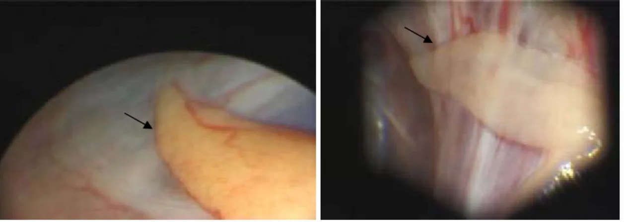
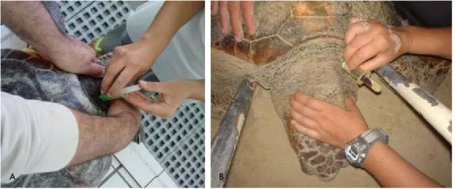
Gonads of living animals were described through laparoscopic inspection. Moreover, a small gonad area was sampled by localised biopsy using the same laparoscopic equipment with an additional biopsy forceps for histological observation and description.
In order to develop a non-invasive sex diagnosing technique and to find the most reliable hormonal indicator of sex in juvenile turtles as well as to understand how hormonal titres vary with gonadal development, hormone titers were measured concurrently with the laparoscopic and histological sexing techniques in juvenile individuals. Thus, a quantitative study of testosterone and estradiol-17β blood plasma levels in loggerhead sea turtles from the north-eastern Atlantic was conducted to:
a) establish sex steroids titres as sexing criteria or thresholds levels, b) estimate the sex ratio for the wild juvenile population.
Summarizing, the objectives mentioned above were attained in three ways:
25 variability assessment;
b) Histological description of gonads and determination of their developmental stage, as well as laparoscopy validation;
c) Determination of sex hormone levels (testosterone and estradiol) and assessment of potential correlations between hormone profiles and individual’s sex, as determined by laparoscopy.
Since only the juvenile life stage is observed in Madeira Island waters the closest nesting population in Boa Vista Island (Cape Verde Archipelago) was chosen to assess the hatchling and the adult life stages.
After sampling, all live turtles were tagged with flipper tags and/or Passive Integrated Transponder (PIT) tags.
This research was granted all the necessary permits by the official authorities to conduct the proposed studies, always having the animal’s welfare in mind.
Materials and Methods
Considering that all sea turtles are listed under Annex I CITES, Annex II Bern Convention and Annex I Bonn Convention, research on these animals must assure that the individuals are kept alive and are released into the wild again. Thus, due to the logistic constraints inherent to working with protected species and the different life stages under assessment, different sampling approaches were adopted for the Madeira and the Cape Verde populations. Sampling in Madeira Island included blood sampling, laparoscopies and gonad biopsies of juvenile individuals. Sampling in Cape Verde included blood sampling in adult females only, and necropsy of dead hatchlings for gonad withdrawal and posterior gonad histology.
26
Sexing sea turtles
Currently 3 methods are available for sex identification of juvenile turtles (laparoscopy, gonad histology and blood sex steroids assessment) (Wibbels et al., 2000). These three methods were used in parallel and are described in the next chapters.
Study Areas
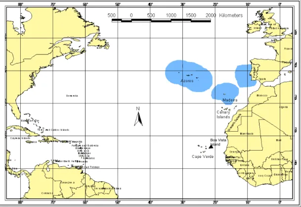
Two study sites were chosen in an effort to assess the whole life cycle: Madeira Island (Portugal) and Boa Vista Island (Cape Verde Archipelago). Both archipelagos are located in the eastern Atlantic Ocean (Fig. 5).
Fig. 5 Madeira and Cape Verde Archipelagos in the north-eastern Atlantic ocean. Blue areas are Portuguese Economic Areas (EEZ’s).
27
Madeira Island (Portugal)
Study area description and population characterization
Madeira Archipelago (Portugal) is located off the north-west coast of Africa (33°N; 17°W), around 1000 km from the European continent and 500 km from the African coastline and is administrated as part of the Portuguese Economic Exclusive Zone (EEZ).
Madeira island is of volcanic origin (6 MY), emerging from the abyssal plain, and thus has no continental shelf. Depths such as 1000-2000 meters can be reached as close as 1-2 nautical miles from the coast, providing a truly pelagic/oceanic nursery environment for loggerheads on these waters.
The archipelago of Madeira is included in the general North Atlantic currents circulation system. The eastern side of the North Atlantic system consists of 4 currents: the Azores current, Portugal Current, the Canary’s current and the North Equatorial Current.
The Canary current is the dominant surface current and the NE trades are the dominant wind regimes. The island mass effect phenomena provides a sheltered leeward area on the south coast, making potential spotting of sea turtles while basking easy during special warm and calm weather. The most probable cause of this phenomenon seems to be the obstruction caused by the island’s interior mountain range to the dominant NE trade winds (Caldeira et al., 2001). Climate is subtropical with small temperature amplitudes, and the weather is highly influenced by the Azores anticyclone.
The Marine Turtle Project at University of Madeira started a tagging and monitoring program in 1994 focused on the pelagic stage ecology of the juvenile loggerheads offshore Madeira Island.
Sea Turtle Capture
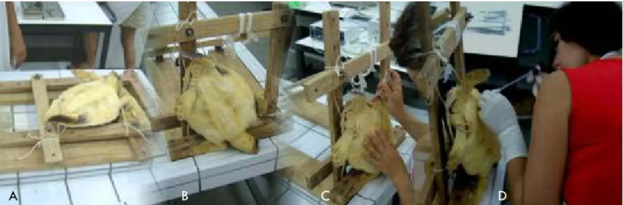
28 belonging to the University of Madeira was used. For safety reasons, the boat crew comprised 2-3 persons, all acting as observers.
Turtles were searched for actively by boat up to 10 nautical miles offshore the Island, and they were captured without regard to their size or location by approaching them at slow speed from behind and picking them with a large dip-net (handle: 2.5m; mouth: 75cm diameter; netting made of thin nylon (gillnet material) (Dellinger et al., 1997). The exact direction of each search depended on sea surface conditions, usually following frontal systems where floating material aggregates.
For every sea turtle captured environmental conditions such as weather and ocean conditions were recorded, as well as behaviour, time, date and GPS position.
Following capture, all turtles were transported to land-based holding facilities at the Marine Biology Station of Funchal where they were maintained in fiberglass tanks filled with sea-water for a minimum of 24 hours to complete sampling procedures. Two large circular tanks (3 m diameter, 1.5m water depth, ~10000l) and 4 smaller squared tanks (~ 1000l) were available to keep the turtles. They were monitored periodically during captivity to assess health and well being. All turtles were returned unharmed to their habitat.
Biometry and Tagging Activities
All turtles were measured, weighted, photographed, and tagged during the holding period. A set of biometrical parameters were taken for posterior correlations with sex (Table 1), as described by Bolten (1999). Straight line measurements were taken using Haglöf forestry callipers (80 and 95cm) for large biometric measurements and Vernier callipers for small biometric measurements, both till the nearest mm. Over the curve measurements were taken using a flexible tailor's tape and weight was determined using an electronic platform balance (Mettler Spider1s60Lst) at 2 g intervals.
29 Loss of monel-style flipper tags has been a problem for most sea turtle species, making it difficult to identify individuals during subsequent observations (Mrosovsky, 1976). Thus, since July 2003 the turtles were tagged with PIT tags (Passive Integrated Transponders, AVID FriendChipTM (AVID
Identification Systems, Inc.)), inserted subcutaneously. Insertion places were the shoulder for small size animals (SCL<30 cm) and the region underneath the scales or between the digits of the dorsal surface of the right front flipper for larger animals. These tagging locations have been used previously by other sea turtle research teams (Balazs, 1999).
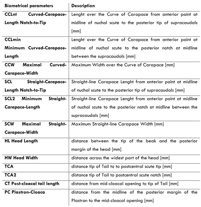
Table 1 - Biometrical parameters taken for hatchling, juvenile and female adult turtles.
Biometrical parameters Description
CCLnt Curved-Carapace-Length Notch-to-Tip
Lenght over the Curve of Carapace from anterior point at midline of nuchal scute to the posterior tip of supracaudals [mm]
CCLmin
Minimum
Curved-Carapace-Length
Lenght over the Curve of Carapace from anterior point at midline of nuchal scute to the posterior notch at midline between the supracaudals [mm]
CCW Maximal
Curved-Carapace-Width
Maximum Width over the Curve of Carapace [mm]
SCL
Straight-Carapace-Length Notch-to-Tip
Straight-line Carapace Lenght from anterior point at midline of nuchal scute to the posterior tip of supracaudals [mm]
SCL2 Minimum
Straight-Carapace-Length
Straight-line Carapace Lenght from anterior point at midline of nuchal scute to the posterior notch at midline between the supracaudals [mm]
SCW Maximal
Straight-Carapace-Width
Maximum Straight-line Carapace Width [mm]
HL Head Length distance between the tip of the beak and the posterior
margin of the head [mm]
HW Head Width distance across the widest part of the head [mm]
TCA distance tip ofTail to to postcentral scute tip [mm]
TCA2 distance tip ofTail to postcentral scute notch [mm]
CT Post-cloacal tail length distance from mid-cloacal opening to tip of Tail [mm]
PC Plastron-Cloaca distance from the midline of the posterior margin of the
30
Biometrical parameters Description
TL TailLength distance from tip of tail to furthest between tail and
capapace [mm]
FFL ForeFlipper-Lenght [mm] R/L State which fore-flipper was measured: R=right, L=left
FFW ForeFlipper-Width [mm] measured just before 1st claw
CLW 1st claw length [mm] measured on Right Foreflipper
WT Weight [g]
Boa Vista Island (Republic of Cape Verde)
Study area description and population characterization
The Cape Verde Islands are an archipelago consisting of 10 volcanic islands and several islets lying in the Atlantic Ocean (14º48’-17º180N, 22º42’-25º18W) about 500 km west of Senegal, West Africa. The climate is dry tropical but ocean conditions are heavily influenced by the cool Canary current that comes from the north, with consistently strong northeast tradewinds.
Five species of marine turtles inhabit the waters of the archipelago, where they feed and/or reproduce, with loggerheads being the most common and with confirmed nesting activity for the islands of Sal, Boa Vista, Maio and São Vicente (López-Jurado et al., 1999b).
Although the existence of marine turtles in these islands was previously known (see López-Jurado et al. (1999a) for a review), in 1977 hawksbill and loggerhead turtles were reported as the species most caught, and an extrapolation from fishermen figures for one island estimated that 1000 adult turtles were caught per year in the whole archipelago (Schleich, 1979). In 1998 three species of marine turtles were recorded for the island of Sal: loggerhead turtle (Caretta caretta), hawksbill turtle (Eretmochelys imbricata) and leatherback turtle (Dermochelys coriacea) although the actual size of their nesting population was unknown (Lazar and Holcer, 1998). Most marine turtles were caught both at sea or during egg-laying and slaughtered both for meat consumption or for the shell’s for tourism trade (Cabrera et al., 1999).