Valproic acid inhibits the invasion of PC3 prostate cancer cells
by upregulating the metastasis suppressor protein NDRG1
Jae Eun Lee and Jung Hwa Kim
Department of Biological Sciences, Inha University, Incheon, South Korea
Abstract
Valproic acid (VPA) is a clinically available histone deacetylase inhibitor with promising anticancer attributes. Recent studies have demonstrated the anticancer effects of VPA on prostate cancer cells. However, little is known about the differential effects of VPA between metastatic and non-metastatic prostate cancer cells and the relationship between the expression of metastasis suppressor proteins and VPA. In the present study, we demonstrate that inhibition of cell viability and invasion by VPA was more effective in the metastatic prostate cancer cell line PC3 than in the tumorigenic but non-metastatic prostate cell line, RWPE2. Further, we identified that the metastasis suppressor NDRG1 is upregulated in PC3 by VPA treatment. In contrast, NDRG1 was not increased in RWPE2 cells. Also, the suppressed invasion of PC3 cells by VPA treatment was relieved by NDRG1 knockdown. Taken together, we sug-gest that the anticancer effect of VPA on prostate cancer cells is, in part, mediated through upregulation of NDRG1. We also conclude that VPA has differential effects on the metastasis suppressor gene and invasion ability between non-metastatic and metastatic prostate cancer cells.
Keywords: VPA, NDRG1, prostate cancer cells.
Received: January 26, 2015; Accepted: July 14, 2015.
Introduction
Valproic acid (VPA, 2-propylvaleric acid) is a branched, short-chain fatty acid that has been widely used as an antiepileptic drug (Chateauvieuxet al., 2010). Re-cently, VPA has attracted a lot of attention due to its anticancer activity, which is thought to be mediated by histone deacetylase (HDACs) inhibition. VPA is a member of a promising new class of anticancer agents that affect proliferation, differentiation, and apoptosis in both solid and hematologic malignancies (Blaheta and Cinatl, 2002). Several studies have demonstrated the anti-tumoral charac-teristics of VPA in prostate cancer cells (Blaheta and Cinatl, 2002; Chateauvieuxet al., 2010). VPA diminishes cell proliferation and migration of prostate cancer cells by modulating multiple pathways, including cell cycle arrest, apoptosis, and angiogenesis (Gaoet al,.2007; Shabbeeret al., 2007; Wedelet al., 2011; Sidanaet al., 2012; Wittet al., 2013). However, the association between the expression of metastasis suppressor genes and VPA has not been studied in detail.
Metastasis suppressor genes inhibit metastasis but do not affect the growth of primary tumors. The expression or function of metastasis suppressor genes is lost primarily in many metastatic cancers. Interestingly, restoration of
me-tastasis suppressor gene expression could inhibit cancer metastasis (Smith and Theodorescu, 2009). Metastasis sup-pressor proteins participate in the regulation of multiple steps in the metastatic process, including cancer cell inva-sion, survival in the bloodstream, and survival at the sec-ondary site (Smith and Theodorescu, 2009). The low ex-pression of metastasis suppressor genes in highly metastatic cancers is dedicated to the epigenetic control and in some cases posttranslational regulation (Ballestar and Esteller, 2008; Elliset al., 2009). N-myc Downstream Reg-ulated Gene-1 (NDRG1) is a metastasis suppressor gene, and its implication in cancer progression and metastasis has been extensively studied. The anti-metastatic function of NDRG1 as a metastasis suppressor protein has been identi-fied in multiple cancers including breast, colon, prostate, and gastric cancers (Bandyopadhyay et al., 2003, 2004; Maruyamaet al., 2006; Jianget al., 2010).
E-cadherin is a tumor suppressor which is highly ex-pressed in epithelial cells and plays crucial roles in cell-cell adhesion. Downregulation of E-cadherin expression is the hallmark of the epithelial-to-mesenchymal transition (EMT) process, the mechanism by which immotile epithe-lial cells convert to the motile mesenchymal phenotype (Yang and Weinberg, 2008). Loss of E-cadherin expression or function is associated with cancer cell invasion and me-tastasis (Frixenet al., 1991).
In this report, we aimed to investigate the differential effects of VPA on metastatic vs. non-metastatic prostate
Send correspondence to Jung Hwa Kim. Department of Biological Sciences, Inha University, 402-751 Incheon, South Korea. E-mail: [email protected].
cancer cells and the relationship between the expression of metastasis suppressor proteins and VPA. We found that the metastatic prostate cancer cell PC3 was more sensitive to VPA treatment, regarding cell viability than the non-metastatic prostate cancer cell RWPE2. Furthermore, VPA induced the metastasis suppressor protein NDRG1 in PC3 cells but not in RWPE2. Finally, we found that induction of E-cadherin expression by VPA treatment was inhibited by NDRG1 knockdown. Moreover, when NDRG1 was knocked down the inhibition of PC3 invasion by VPA was relieved. We, hence, conclude that VPA might function more effectively on metastatic prostate cancer than on non-metastatic prostate cancer and that the anticancer ef-fect of VPA on prostate cells is, in part, mediated by the in-duction of NDRG1.
Methods
Cell lines and Cell culture
PC3 cells were maintained in RPMI1640 medium (Welgene) supplemented with 10% FBS. RWPE2 cells were maintained in Keratinocyte-Serum Free Medium (K-SFM, Invitrogen) supplemented with 12.5 mg/L bovine pi-tuitary extract (BPE) and 1.25mg/L EGF. All cells were supplemented with an antibiotic-antimycotic solution (100 units/mL penicillin, 0.1 mg/mL streptomycin, and 0.25 mg/mL amphotericin B) and grown at 37 °C in stan-dard cell culture conditions (5% CO2, 95% humidity).
Antibodies
Antibodies were purchased from the manufacturers as follows: anti-NDRG1 (ab37897 and ab124689, Abcam), anti-BRMS1 (ab134968, Abcam), anti-NM23H1 (sc-56928, Santa Cruz) anti-E-cadherin (610181, BD Trans-duction Laboratories), anti-vimentin (sc-32322, Santa Cruz), anti-b-actin (A1978, Sigma-Aldrich).
Real time RT-PCR and knockdown
Total RNA was extracted from PC3 treated with VPA (0, 0.75, 1, 3 mM) for 24 h using Trizol reagent (Invi-trogen). Reverse transcription reactions were performed with 2 ug of total RNA using RevertAid M-MuLV reverse transcriptase (Thermo Scientific) and oligo (dT) primers (Fermentas) according to the manufacturer’s protocol. The abundance of mRNA was detected by real-time quantita-tive RT-PCR using the ABI prism 7300 system (Applied Biosystems) and SYBR Green reagent (Molecular Probes). Transcript quantity of the NDRG1 gene was calculated us-ing the DDCt method by normalization to GAPDH. The measurement was performed in three independent biologi-cal experiments and each with three technibiologi-cal replicates. The sequences of the primer pairs were as follows: NDRG1 5’-CGCCAGCACATTGTGAATGAC-3’ and 5’-TTTG AGTTGCACTCCAC CACG-3’ (Changet al., 2013), and GAPDH CCACATCGCTCAGACACCAT-3’ and
5’-TGACAAGCTTCCCGTTCTCA-3’. The target sequence
for small hairpin RNA for NDRG1 was
5’-AGACCACTCTCCTCAAGAT-3’. The primer containing the short hairpin RNA sequence targeting NDRG1 was an-nealed and cloned into a pMSCVpuro vector.
Cell viability assay
PC3 and RWPE2 cells were plated at a density of 4 x 10⁴cells per well in 6-well plates in duplicate, and after 24 h in clutlure they were treated with 0, 0.75, 1, 3 mM VPA. At the indicated time point after VPA treatment, via-ble cells were counted daily by a trypan blue-exclusion as-say.
In vitroinvasion assay
A total of 2.5 x 10⁴PC3 or RWPE2 stable cells were loaded onto the top of a 24-well Matrigel invasion chamber assay plate (BD Biocoat; BD Biosciences). RPMI1640 me-dium containing 15% FBS was added to the bottom cham-ber as a chemoattractant for PC3 cells, and K-SFM medium containing BPE and EGF and supplemented with 15% FBS was used for RWPE2 cells. After incubation for 22 h, the cells that had migrated to the lower surface of the filter were fixed with 100% methanol and stained with 0.5% Giemsa solution. Cells were counted in nine random fields per in-sert.
Results
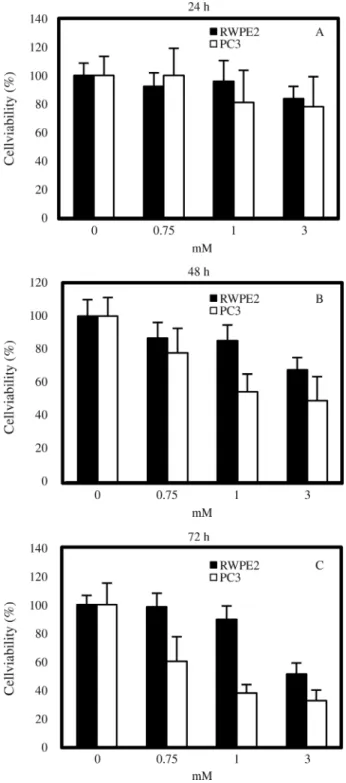
Inhibition of prostate cancer cell cell viability by VPA
In the present study we aimed to determine the differ-ential effects of VPA in relation to the metastatic potdiffer-ential of prostate tumor cells. To do so, the highly metastatic pros-tate cancer cell line PC3 and the tumorigenic but non-metastatic prostate cancer cell line RWPE2 were treated with VPA (0, 0.75, 1, and 3 mM) and the degree of cell via-bility was assessed by counting the viable cells. In both cell types, VPA inhibited cell viability in a dose-dependent fashion, and the duration of VPA treatment also had a posi-tive relationship with cell viability inhibition (Figure 1). The effects of VPA on the viability of PC3 cells were in ac-cordance with those of a previously published study (Xiaet al., 2006). Notably so, PC3 cells were more sensitive than RWPE2 cells to the viability inhibition activity of VPA. At the concentrations of 0.75 and 1 mM VPA, the viability of RWPE2 was not significantly decreased when compared to controls (0 mM VPA). However, after 72 h of treatment with 1 mM VPA, the viability of PC3 cells (38.2%±6.1%) was two-fold lower than that of RWPE2 cells (76.6%±
Suppression of PC3 invasion by VPA treatment
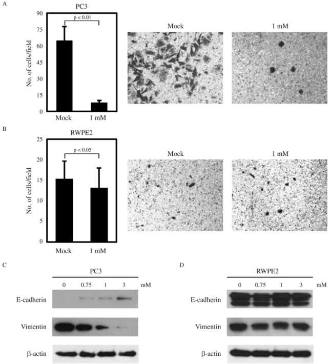
Next, we checked whether the invasive ability of prostate cancer cells is differentially regulated by VPA. PC3 cells have a high invasive potential, and VPA has been reported to significantly inhibit PC3 invasion (Wedelet al.,
2011; Jianget al., 2014) (Figure 2A). As expected, RWPE2 cells showed an extremely low invasive ability, and VPA treatment only slightly reduced their invasiveness. EMT is important for tumor cells to acquire migratory and invasive properties and allows tumor cells to infiltrate surrounding tissues and ultimately metastasize to distant sites (Yang and Weinberg, 2008). The hallmark of EMT is the change in ex-pression of the epithelial marker E-cadherin, as well as of the mesenchymal marker vimentin (Leeet al., 2006; Thiery and Sleeman, 2006). Thus, we examined the effects of VPA treatment on the expression of E-cadherin and vimentin. As shown in Figure 2C, VPA induced the molecular alterations characteristic of the reversion of EMT in PC3 cells, with E-cadherin being increased and vimentin decreased in a dose-dependent manner. In contrast, there was no change in the expression of E-cadherin and vimentin by VPA in RWPE2 cells (Figure 2D). Hence, we conclude that the in-hibitory effect of VPA on cell viability and invasion is more effective in the metastatic prostate cancer cell PC3 than in the non-metastatic prostate cancer cell RWPE2.
Upregulation of NDRG1 by VPA in PC3 cells
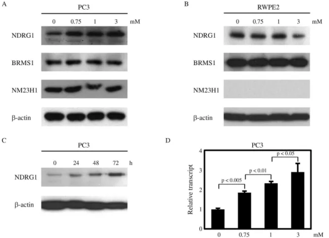
Next, we investigated the effects of VPA on the ex-pression of several metastasis suppressors, including NDRG1, BRMS1, and NM23H1. Interestingly, NDRG1 was upregulated by VPA treatment in a dose-dependent manner in PC3 cells, while the expression of BRMS1 and NM23H1 was not affected (Figure 3A). In contrast, there was no increase in NDRG1 expression in RWPE2 cells treated with VPA (Figure 3B). Furthermore, in PC3 cells the up-regulation of NDRG1 was augmented when the du-ration of VPA treatment was prolonged (Figure 3C). Real-time PCR assays revealed that the NDRG1 transcript level was also increased by VPA in PC3 cells (Figure 3D). From these data, we hypothesize that VPA exerts its anti-tumor activity more specifically in metastatic prostate cells than in non-metastatic prostate cancer cells, in part, by up-regulating NDRG1 expression in metastatic prostate cells.
Implication of NDRG1 in VPA-mediated suppression of invasiveness
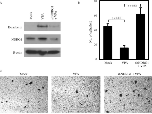
In prostate cancer cells, NDRG1 overexpression has been shown to maintain membrane E-cadherin and inhibit TGF-b-induced EMT (Chen et al., 2012). Also, the in-volvement of NDRG1 in E-cadherin recycling and stabili-zation has been reported (Kachhapet al., 2007). Thus, we investigated whether the increase of E-cadherin by VPA treatment in PC3 may be associated with VPA-mediated NDRG1 induction. We could show that the increase in E-cadherin caused by VPA treatment was inhibited by NDRG1 knockdown even in the presence of VPA (Figu-re 4A). Furthermo(Figu-re, the change in E-cadherin exp(Figu-ression by NDRG1 knockdown was reflected in the invasiveness of PC3, as the suppressed PC3 invasion by VPA treatment was relieved by NDRG1 knockdown (Figure 4B,C). These
results indicate that the anticancer activity of VPA in meta-static prostate cancer cells is in part mediated through the induction of the metastasis suppressor NDRG1.
Discussion
In the present study we showed that VPA treatment has differential effects on metastatic vs. non-metastatic prostate cancer cells, with respect to cell viability and invasiveness, as well as expression of metastasis suppres-sor proteins. We found that highly metastatic PC3 cells
were more sensitive to VPA treatment in the inhibition of cell viability and invasiveness than the non-metastatic RWPE2 cells. The inhibitory effects of VPA on cell viabil-ity and proliferation, as well as invasiveness of PC3 cells had already been investigated by other groups (Xiaet al., 2006; Iacopinoet al., 2008; Wedelet al., 2011; Jianget al., 2014). Furthermore, previous reports had shown that the anti-invasive activity of VPA was valid only in PC3 but not in LNCaP prostate cancer cells with low metastatic poten-tial (Annicotteet al,.2006). This leads us to conclude that
the anticancer activity of VPA is more effective on meta-static prostate cells than non-metameta-static cells.
Epigenetic regulation of NDRG1 expression has been studied in some cancers. In the colon cell line SW620, sev-eral histone markers were related to the transcriptional re-pression of NDRG1, but this was not due to NDRG1 promoter methylation (Li and Chen, 2011). Treatment with 5-Aza-2’-deoxycytidine and Trichostatin A enhanced NDRG1 protein expression in pancreatic cancer cell lines, and again, there was no methylation of CpG island in the NDRG1 promoter (Angstet al., 2010). Recently, it has been shown that promoter methylation of the NDRG1 gene was associated with reduced NDRG1 expression but not with histone modification in gastric cancer cells and tissue samples (Changet al., 2013). Thus, it seems that epigenetic modification of NDRG1 may vary among different cells and tissues. In this report we demonstrated that NDRG1 is upregulated by VPA in highly metastatic prostate cancer cells but not in non-metastatic prostate cancer cells. We also detected an increase in NDRG1 expression in another metastatic prostate cancer cell line DU145, but not in LNCaP (data not shown). This indicates that there are fun-damental epigenetic differences between non-metastatic and metastatic prostate cancer cells that could alter the
transcriptional response to VPA. Thus, we suggest that VPA can have a greater therapeutic benefit in treatments of metastatic prostate cells than non-metastatic cells, partly by up-regulating NDRG1. It has been reported that VPA in-hibits MDA-MB-231 breast cancer cell migration by NM23H1 up-regulation (Liet al., 2012). However, in our study we could not detect an increase in NM23H1 expres-sion, neither in PC3, nor in RWPE2 cells. This means that the effects of VPA on gene expression may be tissue spe-cific.
al., 2007; Songet al., 2010). Furthermore, upregulation of NDRG1 by iron chelators inhibits the TGF-b-induced EMT (Chenet al., 2012). In the present study, we showed that the up-regulation of E-cadherin by VPA is partially mediated through the induction of NDRG1. The invasion inhibitory action of VPA in metastatic cells is, thus, apparently medi-ated through the metastasis suppressor NDRG1, and the restoration of metastasis suppressors by the action of the epigenetic modulator VPA could be a promising strategy for metastatic cancer treatment.
We consider that the differential anticancer action of VPA toward metastatic prostate cancer cells highlights its more beneficial therapeutic effects in the treatment of met-astatic cancers. The involvement of the metastasis suppres-sor NDRG1 in the action of VPA gives certain clues on the mechanism of action of this versatile drug and should con-tribute to the discovery of other powerful epigenetic thera-pies based on the regulation of metastasis suppressors.
Acknowledgments
This research was supported by the Basic Science Re-search Program through the National ReRe-search Foundation
of Korea (NRF) funded by the Ministry of Education, Sci-ence and Technology (NRF-2013R1A1A2005972) and INHA University to J.H.K.
References
Angst E, Dawson DW, Nguyen A, Park J, Go VL, Reber HA, Hines OJ and Eibl G (2010) Epigenetic regulation affects N-myc downstream-regulated gene 1 expression indirectly in pancreatic cancer cells. Pancreas 39:675-679.
Annicotte JS, Iankova I, Miard S, Fritz V, Sarruf D, Abella A, Berthe ML, Noel D, Pillon A, Iborra F,et al.(2006) Pero-xisome proliferator-activated receptor gamma regulates E-cadherin expression and inhibits growth and invasion of prostate cancer. Mol Cell Biol 26:7561-7574.
Ballestar E and Esteller M (2008) Epigenetic gene regulation in cancer. Adv Genet 61:247-267.
Bandyopadhyay S, Pai SK, Gross SC, Hirota S, Hosobe S, Miura K, Saito K, Commes T, Hayashi S, Watabe M,et al.(2003) The Drg-1 gene suppresses tumor metastasis in prostate can-cer. Cancer Res 63:1731-1736.
Bandyopadhyay S, Pai SK, Hirota S, Hosobe S, Takano Y, Saito K, Piquemal D, Commes T, Watabe M, Gross SC, et al., 2004. Role of the putative tumor metastasis suppressor gene Drg-1 in breast cancer progression. Oncogene 23:5675-5681.
Blaheta RA and Cinatl Jr J (2002) Anti-tumor mechanisms of valproate: A novel role for an old drug. Med Res Rev 22:492-511.
Chang X, Zhang S, Ma J, Li Z, Zhi Y, Chen J, Lu Y and Dai D (2013) Association of NDRG1 gene promoter methylation with reduced NDRG1 expression in gastric cancer cells and tissue specimens. Cell Biochem Biophys 66:93-101. Chateauvieux S, Morceau F, Dicato M and Diederich M (2010)
Molecular and therapeutic potential and toxicity of valproic acid. J Biomed Biotechnol 2010:479364.
Chen Z, Zhang D, Yue F, Zheng M, Kovacevic Z and Richardson DR (2012) The iron chelators Dp44mT and DFO inhibit TGF-beta-induced epithelial-mesenchymal transition via up-regulation of N-Myc downstream-regulated gene 1 (NDRG1). J Biol Chem 287:17016-17028.
Ellis L, Atadja PW and Johnstone RW (2009) Epigenetics in can-cer: Targeting chromatin modifications. Mol Cancer Ther 8:1409-1420.
Frixen UH, Behrens J, Sachs M, Eberle G, Voss B, Warda A, Lochner D and Birchmeier W (1991) E-cadherin-mediated cell-cell adhesion prevents invasiveness of human carci-noma cells. J Cell Biol 113:173-185.
Gao D, Xia Q, Lv J and Zhang H (2007) Chronic administration of valproic acid inhibits PC3 cell growth by suppressing tumor angiogenesis in vivo. Int J Urol 14:838-845.
Iacopino F, Urbano R, Graziani G, Muzi A, Navarra P and Sica G (2008) Valproic acid activity in androgen-sensitive and -in-sensitive human prostate cancer cells. Int J Oncol 32:1293-1303.
Jiang K, Shen Z, Ye Y, Yang X and Wang S (2010) A novel mo-lecular marker for early detection and evaluating prognosis of gastric cancer: N-myc downstream regulated gene-1 (NDRG1). Scand J Gastroenterol 45:898-908.
Jiang W, Zheng Y, Huang Z, Wang M, Zhang Y, Wang Z, Jin X and Xia Q (2014) Role of SMAD4 in the mechanism of valproic acid’s inhibitory effect on prostate cancer cell inva-siveness. Int Urol Nephrol 46:941-946.
Kachhap SK, Faith D, Qian DZ, Shabbeer S, Galloway NL, Pili R, Denmeade SR, DeMarzo AM and Carducci MA (2007) The N-Myc down regulated Gene1 (NDRG1) is a Rab4a effector involved in vesicular recycling of E-cadherin. PLoS One 2:e844.
Lee JM, Dedhar S, Kalluri R and Thompson EW (2006) The epi-thelial-mesenchymal transition: New insights in signaling, development, and disease. J Cell Biol 172:973-981. Li GF, Qian TL, Li GS, Yang CX, Qin M, Huang J, Sun M and
Han YQ (2012) Sodium valproate inhibits MDA-MB-231 breast cancer cell migration by upregulating NM23H1 ex-pression. Genet Mol Res 11:77-86.
Li Q and Chen H (2011) Transcriptional silencing of N-Myc downstream-regulated gene 1 (NDRG1) in metastatic colon cancer cell line SW620. Clin Exp Metastasis 28:127-135. Maruyama Y, Ono M, Kawahara A, Yokoyama T, Basaki Y,
Kage M, Aoyagi S, Kinoshita H, and Kuwano M (2006) Tu-mor growth suppression in pancreatic cancer by a putative metastasis suppressor gene Cap43/NDRG1/Drg-1 through modulation of angiogenesis. Cancer Res 66:6233-6242. Shabbeer S, Kortenhorst MS, Kachhap S, Galloway N, Rodriguez
R and Carducci MA (2007) Multiple molecular pathways explain the anti-proliferative effect of valproic acid on pros-tate cancer cellsin vitroandin vivo. Prostate 67:1099-1110. Sidana A, Wang M, Shabbeer S, Chowdhury WH, Netto G,
Lupold SE, Carducci M and Rodriguez R (2012) Mecha-nism of growth inhibition of prostate cancer xenografts by valproic acid. J Biomed biotechnol 2012:e180363. Smith SC and Theodorescu D (2009) Learning therapeutic lessons
from metastasis suppressor proteins. Nat Rev Cancer 9:253-264.
Song Y, Oda Y, Hori M, Kuroiwa K, Ono M, Hosoi F, Basaki Y, Tokunaga S, Kuwano M, Naito S, et al. (2010) N-myc downstream regulated gene-1/Cap43 may play an important role in malignant progression of prostate cancer, in its close association with E-cadherin. Hum Pathol 41:214-222. Thiery JP and Sleeman JP (2006) Complex networks orchestrate
epithelial-mesenchymal transitions. Nat Rev Mol Cell Biol 7:131-142.
Wedel S, Hudak L, Seibel JM, Juengel E, Tsaur I, Wiesner C, Haferkamp A and Blaheta RA (2011) Inhibitory effects of the HDAC inhibitor valproic acid on prostate cancer growth are enhanced by simultaneous application of the mTOR in-hibitor RAD001. Life Sci 88:418-424.
Witt D, Burfeind P, von Hardenberg S, Opitz L, Salinas-Riester G, Bremmer F, Schweyer S, Thelen P, Neesen J and Kaul-fuss S (2013) Valproic acid inhibits the proliferation of can-cer cells by re-expressing cyclin D2. Carcinogenesis 34:1115-1124.
Xia Q, Sung J, Chowdhury W, Chen CL, Hoti N, Shabbeer S, Carducci M and Rodriguez R (2006) Chronic administration of valproic acid inhibits prostate cancer cell growthin vitro
andin vivo. Cancer Res 66:7237-7244.
Yang J and Weinberg RA (2008) Epithelial-mesenchymal transi-tion: At the crossroads of development and tumor metasta-sis. Dev Cell 14:818-829.
Zhang L, Wang G, Wang L, Song C, Wang X and Kang J (2011) Valproic acid inhibits prostate cancer cell migration by up-regulating E-cadherin expression. Pharmazie 66:614-618.
Associate Editor: Emmanuel Dias Neto