Journal of HerbMed Pharmacology
Journal homepage: http://www.herbmedpharmacol.com J HerbMed Pharmacol. 2015; 4(4): 121-124.
The effects of hesperetin on apoptosis induction and
inhibition of cell proliferation in the prostate cancer
PC3 cells
*Corresponding author: Esfandiar Heidarian, Medical Plants Research Center, Shahrekord University of Medical Sciences, Shahrekord, Iran. Email: heidarian46@yahoo.com
Introduction
Prostate cancer is the second major cause of cancer-relat-ed deaths and the most common cancer diagnoscancer-relat-ed in men in the United States and Europe. Prostate cancer incidence increases with age (1,2). It is initially androgen-dependent (AD) but it will progress to become a more invasive and androgen-independent form with resistance to treatment (3). The apoptotic process is an important target in treat-ment of cancer. Apoptosis or programmed cell death is characterized by process of autonomous cellular elimina-tion which avoids eliciting inflammaelimina-tion (4).
Apoptosis can be triggered by 2 main pathways; one is the extrinsic death-receptor pathway and the other is the intrinsic mitochondrial pathway (5). The main treatment
for most cancers is chemotherapy but it has side effects such as decreasing the number of peripheral blood cells or toxic adverse effects (6). Thus, in recent years the medici-nal plant compounds are used to treat cancer. Flavonoids and flavonoid-rich food sources have been reported to be used for the treatment of several diseases with their pow-erful antioxidant activities (7).
Hesperetin (3’,5,7-trihydroxy-4’-methoxyflavanone), a member of the flavonoids, is found in fruits such as or-anges and red fruits (8). Many reports have shown that flavonoids have various anticancer effects such as kinase activity, the inhibition of cell proliferation, and the induc-tion of apoptosis on cancer cells (9). Hesperetin and its metabolites have several biological activities such as an-Moein Shirzad1, Pezhman Beshkar2, Esfandiar Heidarian3*
¹Clinical Biochemistry Research Center, Shahrekord University of Medical Sciences, Shahrekord, Iran ²Cellular and Molecular Research Center, Shahrekord University of Medical Sciences, Shahrekord, Iran ³Medical Plants Research Center, Shahrekord University of Medical Sciences, Shahrekord, Iran
Implication for health policy/practice/research/medical education:
Hesperetin has anti-cancer properties against different cell lines and this effect seems to be through the inhibition of cell proliferation with the induction of cell cycle arrest at the G1 phase. These results can be a step toward targeted combination or alternative chemotherapy in the prostate cancer.
Please cite this paper as:Shirzad M, Beshkar P, Heidarian E. The effects of hesperetin on apoptosis induction and inhibition of cell proliferation in the prostate cancer PC3 cells. J HerbMed Pharmacol. 2015;4(4):121-124.
Introduction: Prostate cancer is the second leading cause of cancer-related deaths and the most common cancer diagnosed in men in the United States and Europe. Hesperetin, a member of the flavonoids with antioxidant property, is found in fruits such as oranges and red fruits. This study was undertaken to evaluate the effects of hesperetin on apoptosis induction and inhibition of cell proliferation in the prostate cancer PC3 cells.
Methods: PC3 cell line was cultured in standard condition. The cells were exposed to different concentrations of hesperetin (0-1000 μM) for 48 hours. Cell viability was measured by MTT assay. Apoptosis induction was assessed by Annexin V-FITC by flow cytometry analysis.
Results: The PC3 cells exposed to hesperetin (0-1000 μM) exhibited an IC50 (inhibitory concentration of 50%) about 450 μM. At different concentrations of hesperetin (400, 450 and 500 µm), the apoptosis increased slightly (not significant) in treated PC3 cells compared to the control group (5.4%, 7.8% and 9.1% respectively vs. 4.2%).
Conclusion: These results clearly show that hesperetin can lead to inhibition of PC3 cells proliferation.
A R T I C L E I N F O
Keywords: Prostate cancer Hesperetin Apoptosis Article History:
Received: 2 August 2015 Accepted: 7 September 2015 Article Type:
Original Article
Shirzad M et al
Journal of HerbMed Pharmacology, Volume 4, Number 4, October 2015 http://www.herbmedpharmacol.com 122
tioxidant, anti-inflammatory, and lipid lowering effects (10). Hesperetin has also been shown to act as a neuro-protective compound (11) and some studies are indicative of hesperetin neuroprotective properties against apoptosis (12,13). Therefore, considering the antioxidant properties of hesperetin, the aim of this study was to examine the ef-fects of hesperetin on apoptosis induction and inhibition of cell proliferation in the prostate cancer PC3 cells.
Materials and Methods
Reagents
The human PC3 prostate cancer cells were purchased from Pasteur Institute of Iran (Tehran, Iran). RPMI1640 medium, PEN/STREP, dimethyl sulfoxid (DMSO), trypan blue, and hesperetin were obtained from Sigma (St. Lou-is, MO). Fetus bovine serum (FBS) was purchased from GIBCO (USA) institute. MTT were acquired from Merck (Darmstadt, Germany).
Cell culture and cell count
The human PC-3 cells were maintained in RPMI 1640 supplemented with 10% fetal bovine serum (FBS), 100 U/mL penicillin, 0.1 mg/mL streptomycin (PS). The cells were maintained at 37°C in a humidified incubator with 5% carbon dioxide. Total and viable cell counts (survival rate) were determined by standard hemocytometer proce-dures with the trypan blue exclusion test. Live viable cells were seen as colorless (impermeable to the dye due to in-tact membrane) and dead cells were seen as blue (perme-able to dye due to disruption of cell membrane).
Determination of cell viability (MTT assay)
The cytotoxic effects of the hesperetin on the PC3 cells were determined by the MTT assay (14). The principle behind this technique depends on the capacity of living cells to reduce tetrazolium salt [3-(4,5-dimethylthiazol-2-yl)-2,5-diphenyltetrazolium bromide] to a formazan crystal in their metabolizing mitochondria. Briefly, PC3 cells were seeded at an amount of 5 × 10³ cells per well
in 96-well plates and allowed to grow overnight. The cells were treated with different concentrations (0-1000 µM) of hesperetin (solution in DMSO with 0.1% final concentra-tion). After incubation for 48 hours, supernatant was dis-carded and was added 100 µL colorless RPMI. Then, add-ing 10 µL MTT (5 mg/mL in phosphate buffered saline) to each well and incubated for 4 hours. After the removal of the medium, 150 µL DMSO was added to each well and shaken gently and carefully. The absorbance was read at a wavelength of 490-620 nm using an ELISA microplate reader (State Fax® 2100, Awareness, USA).
Analysis of apoptosis and necrosis
The analysis of apoptosis was conducted by flow cytom-etry. Apoptosis was measured by kits specifically (BD Biosciences) according to the manufacturer’s instructions. Briefly, PC3 cells were cultured in 6-well plates and al-lowed to attach to plate overnight. The cells were treated
with 450 μM of hesperetin for 48 hours and they were har-vested by trypsinization and, after washing with cold PBS, were diluted with 500 µL of binding buffer, and incubated in 10 µL of Annexin-binding buffer with 2 µL of Annexin V-FITC for 20 minutes in the dark. Then, the cells were acquired and analyzed by flow cytometry (15).
Statistical analysis
The data were expressed as mean ± S.D. They were ana-lyzed by SPSS software (version 20) and Graph Pad Prism (version 5.01, USA). Nonparametric data were analyzed with Kruskal-Wallis test. A P ≤ 0.05 was considered sig-nificant. All experiments were repeated at least 3 times.
Results
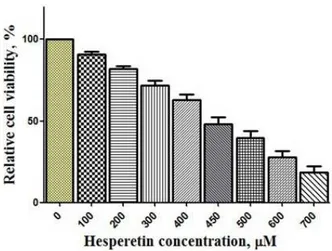
Figure 1 shows that hesperetin has decreased significantly cell proliferation in a dose-dependent manner (P < 0.05) after 48 hours. The cells that exposed to hesperetin (0-1000 μM) exhibited an IC50 (inhibitory concentration of 50%) about 450 μM. In this concentration, hesperetin eliminated 50% of the cells.
Effect of hesperetin on apoptosis in PC3 cells
Figures 2 and 3 show flow cytometric analysis using An-nexin V staining for apoptosis in PC3 cells. Apoptosis in PC3 cells was slightly induced (not significantly) 5.4%, 7.8% and 9.1% at 400, 450, and 500 µM of hesperetin, re-spectively.
Discussion
Chemotherapy is considered as the main treatment for most types of cancers but because of side effects, major ef-forts have been made to recognize natural compound that can prevent the development of cancers. Many natural compounds have been found to induce apoptosis in vari-ous cancer cells and there are obvivari-ous evidences indicating that these compounds are potent inhibitors of cancer cell
Figure 1. Inhibition of cell proliferation by hesperetin for 48 h. The cells were cultured at the density of 5 × 10³ cells per well in
Efects of hesperetin on apoptosis
Journal of HerbMed Pharmacology, Volume 4, Number 4, October 2015
http://www.herbmedpharmacol.com 123
proliferation (16,17). This beneficial effect has often been attributed to the high content of naturally-occurring an-tioxidants, such as vitamins and flavonoids. In particular, studies have indicated a specific link between citrus intake and the reduced risk of diseases such as cancer (18,19). In the present study, we analyzed the effects of hesperetin (a flavonoid) on PC3 cells. The effects of this antioxidant were assessed on growth and proliferation of PC3 cells in vitro using an MTT assay or cytotoxicity assay test (Figure 1). Hesperetin in 450 μM led to the reduction of cell vi-ability as reported by other investigators (20,21). Our re-sults of flow cytometry indicate that apoptosis increased slightly (not significantly) in hesperetin-treated PC3 cells compared to the control group (Figures 2 and 3). Previous studies have also shown that hesperetin treatment leads to the inhibition of cell proliferation with the induction of cell cycle arrest at the G1 phase (22). Nevertheless, Palit et al (23) showed that hesperetin would induce apoptosis in breast carcinoma by triggering aggregation of reactive oxygen species (ROS) and activation of ASK1/JNK sig-naling pathway (23) which runs counter to our findings. On the other hand, some studies have demonstrated that hesperetin has antioxidant, anti-proliferation, and apop-tosis effects (24). Also, it was found that hesperetin at 100 nM was effective in preventing neuronal apoptosis via a mechanism involving the activation/phosphorylation of both ERK1/2 and Akt/PKB signaling pathways (11). Choi et al showed that hesperetin and epigallocatechin gallate exerted antiapoptosis effects in endothelial cells exposed to oxidized LDL. They showed that hesperetin was medi-ated by interrupting both JNK and p38 MAPK-responsive death pathways (25). Also, in another study it was found
Figure 2. Effect of hesperetin on apoptosis in PC3 cells. The cells were cultured for 24 hours. Then, the cells were treated with different concentrations of hesperetin (0, 400, 450, 500 µM) for 48 hours. The appearance of apoptosis cells was detected by
low cytometry using Annexin V-FITC staining. A; Control without treatment, B, C and D the cells were treated with 400, 450, and 500 μM of hesperetin, respectively.
Figure 3. The effect of different concentrations of hesperetin
on PC3 cells apoptosis after 48 hours incubation compared to control group. No signiicant change (P > 0.05) was observed in
apoptosis and necrosis between hesperetin-treated PC3 cells and control group cells different concentrations of hesperetin.
that protective effects of hesperetin and naringenin against apoptosis in ischemia/reperfusion induced retinal injury in rats. As a result hesperetin and naringenin can prevent harmful effects induced by I/R injury in the rat retina by inhibiting apoptosis of retinal cells, which proposes that those flavanones have a therapeutic potential for the pro-tection of ocular ischemic diseases (10). Therefore, in this study the prevention of PC3 apoptosis probably resulted in the changes of cellular signaling proteins.
In this study, we did not evaluate the effect of hesperetin on cellular signaling proteins such as the nuclear translo-cation level of p65, ERK1/2, Akt/PKB, and regulatory ef-fectors such as Bcl2 levels, and Bax levels. These factors can influence cell apoptosis and survival. Therefore, we suggest that future studies focus on the effects of EA on other cell signaling and effector proteins.
In conclusion, our data confirmed that hesperetin led to the reduction of PC3 cells without considerable increasing apoptosis considerably. Therefore, the study of the mecha-nism of apoptosis induction could be a step of progress toward target therapy which might be considered in the future studies.
Acknowledgements
We would like to express our gratitude to those who have helped us in Clinical Biochemistry Research Center of Shahrekord University of Medical Sciences.
Authors’ contributions
MS performed the experimental work and helped the writing, EH contributed in the conception of the work, design, writing, analysis, revising the draft, approval of the final version of the manuscript, and agreed for all aspects of the work, PB led the flowcytometry analysis.
Conflict of interests
Shirzad M et al
Journal of HerbMed Pharmacology, Volume 4, Number 4, October 2015 http://www.herbmedpharmacol.com 124
Ethical considerations
Ethical issues (including plagiarism, misconduct, data fabrication, falsification, double publication or submis-sion, redundancy) have been completely observed by the authors.
Funding/Support
This study was funded by Shahrekord University of Medi-cal Sciences (grant No. 1145), Shahrekord, Iran.
References
1. Gong Y, Chippada-Venkata UD, Oh WK. Roles of matrix metalloproteinases and their natural inhibitors in prostate cancer progression. Cancers (Basel). 2014;6(3):1298-327.
2. Culig Z, Santer FR. Androgen receptor signaling in prostate cancer. Cancer Metastasis Rev. 2014;33(2-3):413-427.
3. Goldberg AA, Titorenko VI, Beach A, Sanderson JT. Bile acids induce apoptosis selectively in androgen-dependent and-inandrogen-dependent prostate cancer cells. Peer J. 2013;1:e122.
4. Fink SL, Cookson BT. Apoptosis, pyroptosis, and necrosis: mechanistic description of dead and dying eukaryotic cells. Infect Immun. 2005;73(4):1907-1916.
5. Cerella C, Grandjenette C, Dicato M, Diederich M. Roles of apoptosis and cellular senescence in cancer and aging. Curr Drug Targets. 2015.
6. Islambulchilar M, Asvadi I, Sanaat Z, Esfahani A, Sattari M. Taurine attenuates chemotherapy-induced nausea and vomiting in acute lymphoblastic leukemia. Amino Acids. 2015;47(1):101-109.
7. Heidarian E, Saffari J, Jafari-Dehkordi E. Hepatoprotective action of Echinophora platyloba DC leaves against acute toxicity of acetaminophen in rats. J Diet Suppl. 2014;11(1):53-63.
8. Kim GD. Hesperetin inhibits vascular formation by suppressing of the PI3K/AKT, ERK, and p38 MAPK signaling pathways. Prev Nutr Food Sci. 2014;19(4):299.
9. Kim SY, Lee JY, Park YD, Kang KL, Lee JC, Heo JS. Hesperetin alleviates the inhibitory effects of high glucose on the osteoblastic differentiation of periodontal ligament stem cells. PLoS One. 2013;8(6):e67504.
10. Kara S, Gencer B, Karaca T, et al. Protective effect of hesperetin and naringenin against apoptosis in ischemia/reperfusion-induced retinal injury in rats. Sci World J. 2014;2014:797824.
11. Mansuri ML, Parihar P, Solanki I, Parihar MS. Flavonoids in modulation of cell survival signalling pathways. Genes Nutr. 2014;9(3):1-9.
12. Roohbakhsh A, Parhiz H, Soltani F, Rezaee R, Iranshahi M. Molecular mechanisms behind the
biological effects of hesperidin and hesperetin for the prevention of cancer and cardiovascular diseases. Life Sci. 2015;124:64-74.
13. Cho J. Antioxidant and neuroprotective effects of hesperidin and its aglycone hesperetin. Arch Pharm Res. 2006;29(8):699-706.
14. Delle Monache S, Sanità P, Trapasso E, et al. Mechanisms underlying the anti-tumoral effects of Citrus bergamia juice. PLoS One. 2013;16(4):e61484. 15. Barrasa JI, Olmo N, Pérez-Ramos P, et al. Deoxycholic
and chenodeoxycholic bile acids induce apoptosis via oxidative stress in human colon adenocarcinoma cells. Apoptosis. 2011;16(10):1054-67.
16. Chinembiri TN, du Plessis LH, Gerber M, Hamman JH, du Plessis J. Review of natural compounds for potential skin cancer treatment. Molecules. 2014;19(8):11679-721.
17. Korkina L. Phenylpropanoids as naturally occurring antioxidants: from plant defense to human health. Cell Mol Biol. 2007;53(1):15-25.
18. Le Marchand L, Murphy SP, Hankin JH, Wilkens LR, Kolonel LN. Intake of flavonoids and lung cancer. J Natl Cancer Inst. 2000;92(2):154-60.
19. Kris-Etherton PM, Hecker KD, Bonanome A, Coval SM, Binkoski AE, Hilpert KF, et al. Bioactive compounds in foods: their role in the prevention of cardiovascular disease and cancer. Am J Med. 2002;113(9):71-88.
20. Alshatwi AA, Ramesh E, Periasamy V, Subash‐Babu P. The apoptotic effect of hesperetin on human cervical cancer cells is mediated through cell cycle arrest, death receptor, and mitochondrial pathways. Fundam Clin Pharmacol. 2013;27(6):581-92. 21. Pollard SE, Whiteman M, Spencer JP. Modulation of
peroxynitrite-induced fibroblast injury by hesperetin: a role for intracellular scavenging and modulation of ERK signalling. Biochem Biophys Res Commun. 2006;347(4):916-23.
22. Choi EJ. Hesperetin induced G1-phase cell cycle arrest in human breast cancer MCF-7 cells: involvement of CDK4 and p21. Nutr Cancer. 2007;59(1):115-9. 23. Palit S, Kar S, Sharma G, Das PK. Hesperetin
induces apoptosis in breast carcinoma by triggering accumulation of ROS and activation of ASK1/JNK pathway. J Cell Physiol. 2015;230(8):1729-39. 24. Rainey-Smith S, Schroetke LW, Bahia P, Fahmi
A, Skilton R, Spencer JP, et al. Neuroprotective effects of hesperetin in mouse primary neurones are independent of CREB activation. Neurosci Lett. 2008;438(1):29-33.