R E S E A R C H
Open Access
The effects of the novel A53E
alpha-synuclein mutation on its oligomerization
and aggregation
Diana F. Lázaro
1†, Mariana Castro Dias
1†, Anita Carija
2, Susanna Navarro
2, Carolina Silva Madaleno
3,
Sandra Tenreiro
3, Salvador Ventura
2and Tiago F. Outeiro
1,3,4*Abstract
α-synuclein (aSyn) is associated with both sporadic and familial forms of Parkinson’s disease (PD), the second most common neurodegenerative disorder after Alzheimer’s disease. In particular, multiplications and point mutations in the gene encoding for aSyn cause familial forms of PD. Moreover, the accumulation of aSyn in Lewy Bodies and Lewy neurites in disorders such as PD, dementia with Lewy bodies, or multiple system atrophy, suggests aSyn misfolding and aggregation plays an important role in these disorders, collectively known as synucleinopathies. The exact function of aSyn remains unclear, but it is known to be associated with vesicles and membranes, and to have an impact on important cellular functions such as intracellular trafficking and protein degradation systems, leading to cellular pathologies that can be readily studied in cell-based models. Thus, understanding the molecular effects of aSyn point mutations may provide important insight into the molecular mechanisms underlying disease onset. We investigated the effect of the recently identified A53E aSyn mutation. Combining in vitro studies with studies in cell models, we found that this mutation reduces aSyn aggregation and increases proteasome activity, altering normal proteostasis.
We observed that, in our experimental paradigms, the A53E mutation affects specific steps of the aggregation process of aSyn and different cellular processes, providing novel ideas about the molecular mechanisms involved in synucleinopathies.
Keywords:Alpha-synuclein, Parkinson’s disease, Oligomerization, Aggregation, Neurodegeneration
Introduction
Parkinson’s disease (PD) is a highly debilitating and pro-gressive neurodegenerative disorder affecting around seven million people worldwide. PD is typically known as a movement disorder, due to the characteristic motor manifestations associated with the loss of dopaminergic neurons from thesubstantia nigra, although it also affects other areas of the brain. PD and other neurodegenera-tive disorders, such as demential with Lewy bodies, and multiple system atrophy, are also characterized by the accumulation of aggregated alpha-synuclein (aSyn) in
proteinaceous inclusions known as Lewy bodies (LBs) or Lewy neurites [54]. Together, these diseases are known as synucleinopathies [17, 55]. However, it is still unclear whether LBs are themselves toxic or protective [7, 43], with smaller oligomeric species of aSyn being the culprits as recent studies suggest [31, 45, 58, 63]. aSyn is a disordered and abundant neuronal protein whose normal function is still elusive. Familial forms of PD associated with duplication and triplication of the SNCAgene [53], along with studies of aSyn overexpres-sion, in cellular and animal models, suggest the protein may acquire a toxic function. The cellular pathologies associated with increased levels and accumulation of aSyn include disruption of vesicular transport [6, 42], mitochondrial dysfunction, impairment of autophagy and proteasome, and oxidative stress [2, 21], suggesting aSyn plays a multitude of roles in the cell, perhaps due * Correspondence:[email protected]
†
Equal contributors
1Department of Neurodegeneration and Restorative Research, University Medical Center Göttingen, Waldweg 33, 37073 Göttingen, Germany 3Chronic Disease Research Center (CEDOC), NOVA Medical School, Campo dos Mártires da Pátria, 130, 1169-056 Lisbon, Portugal
Full list of author information is available at the end of the article
to its intrinsically disordered nature. Under physiological conditions, aSyn is considered to be a pre-synaptic protein [37] that associates with vesicles and membranes [11].
According to the “Braak hypothesis”, PD pathology is thought to start from the periphery (gut or nose), and progress until it reaches the brain [4, 5, 49], spreading in a prion-like manner [20, 29, 36]. However, this hypothesis is still controversial, and the molecular mechanisms underlying this phenomenon are not fully understood [25].
The vast majority of PD cases are sporadic but single point mutations in the gene encoding for aSyn (SNCA) cause familial forms of the disease [10]. The most recently identified aSyn mutation causes the substitution of alanine at position 53 by a glutamate residue (A53E), identified in a 36 year-old Finnish patient with atypical PD. The patient displayed a dense accumulation ofSNCAinclusions in the striatum and a severe cortical pathology, affecting both the superficial and deep laminae [3]. In vitro, the A53E mutation was shown to reduce aSyn aggregation and fi-bril formation without changing the secondary structure content of the protein, when compared to WT aSyn [15]. These data suggest that the negatively charged glutamate residue may affect the folding and, consequently, the ag-gregation process of the protein.
In our study, we conducted a detailed study of the ef-fects of the A53E mutation on aSyn using a combination of in vitro and cellular models of aSyn oligomerization and aggregation [40, 45]. Our results showed that the A53E mutation modulates aSyn aggregation in vitro and in vivo and impacts on distinct cellular pathways.
Altogether, the study of specific aSyn mutants provides novel insight into the spectrum of functions and cellular pathologies associated, opening novel avenues for the design of therapeutic strategies for PD and other synucleinopathies.
Materials and methods
Protein expression and purification
pET21a vectors (Novagen) encoding for WT aSyn and the A53E mutant were transformed into E. coli BL21 (DE3) cells. For protein expression, 10 ml overnight culture of transformed cells was used to inoculate 1 L of LB medium with 100 μg/mL ampicillin, which was further incubated at 37 °C and 250 rpm. At an OD600 0.6, protein expression was induced with 1 mM of isopropyl-1-thio-β-D-galactopyranoside (IPTG) for 4 h at 37 °C. Afterwards, the cultures were centrifuged and the cell pellet frozen at−80 °C.
For cell lysis, pellets were resuspended in 15 mL of lysis buffer (50 mM Tris · HCl, 150 mM NaCl, 1 μg/mL Pepstatin A, 20 μg/ml Aprotinin, 1 mM Benzamidine, 1 mM PMSF and 1 mM EDTA) and sonicated on ice. The lysate was boiled for 10 min at 95 °C and soluble
and insoluble fractions were separated by centrifugation at 48,384 × g, for 15 min. 136 μL/mL of 10% strepto-mycin sulfate and 228 μL/mL of glacial acetic acid was added to the supernatant. The resulting solution was centrifuged for 5 min at 48,384 ×g, and the supernatant was collected and precipitated with saturated ammo-nium sulphate at 4 °C in a ratio 1:1 (v/v) with the super-natant. The pellets were washed with a 1:1 (v/v) solution of ammonium sulphate and water. The pellets were re-suspended in 900μL of ammonium acetate 100 mM and the same volume of 100% ethanol was added to precipi-tated aSyn.
Purification protocol was as adapted from [59]. Briefly, ethanol precipitated aSyn was resuspended in starting buffer (25 mM Tris · HCl at pH 8.0) and filtered through a Millex-HP filter syringe-driven filter unit (0.45 μm, Millipore). Anion exchange high-performance liquid-chromatography was carried out on an AKTA-FPLC (GE Healthcare). The sample was loaded, bounded to Hi-Trap column (GE Healthcare) and eluted with a NaCl linear gradient of elution buffer (25 mM Tris · HCl at pH 8.0, 1 M NaCl). The fractions containing aSyn were collected and buffer was exchanged for 20 mM ammo-nium acetate. The identity, and purity of the recombin-ant proteins was assessed by Mass Spectrometry and SDS-PAGE being higher than 99%.
Aggregation assays
The protein stocks were prepared by resuspending ly-ophilized protein in native buffer (10 mM sodium phos-phate at pH 7.0) to a concentration of approximately 100 μM. Then, the protein stocks were filtered through 0.22 μm filter unit. The integrity of the protein and the absence of soluble oligomers at the beginning of the re-action was confirmed by gel filtration chromatography in a Superdex 75 10/300 column (GE Healthcare Life Sciences). Three aliquots of 300 μL of aSyn WT and A53E mutant were prepared from the protein stocks, in native buffer to a final concentration of 60μM. Samples were incubated in an Eppendorf Thermomixer Comfort (Eppendorf, USA) with 0.02% sodium azide at 600 rpm and 37 °C.
Light scattering spectroscopy
Thioflavin T binding assay
Th-T binding to amyloid fibrils was recorded using a JASCO FP-8200 spectrofluorometer (Jasco Inc, MD, USA) with an excitation wavelength of 445 nm and emission range from 460 to 600 nm at 25 °C, using a slit width of 5 nm for excitation and emission. The final concentration of Th-T was 25μM and final protein con-centration was 10μM in native buffer. Solutions without protein were used as negative controls. All experiments were carried out in triplicates.
Congo red binding assay
CR binding to amyloid fibrils was tested using a Cary-400 Varian spectrophotometer (Varian Inc., Palo Alto, CA, USA) by recording the absorbance spectra from 375 to 675 nm, at 25 °C. A final concentration of 10μM CR was added to 10 μM protein samples in native buffer. Protein solutions in the presence and absence of CR were used to calculate the differential CR spectra. All ex-periments were carried out in triplicates.
Transmission electron microscopy (TEM) assays
For negative staining, incubated samples were diluted in Mili-Q water to 10μM and 10 μL were then placed on carbon-coated copper grids, and left to stand for 5 min. The grids were washed with distilled water and stained with 2% (w/v) uranyl acetate for 1 min. The morphology of aggregates of WT and A53E aSyn was observed using a JEOL JEM 1400 transmission electron microscope (JEOL, USA) at an accelerating voltage of 120 kV. The width of fibrils for WT and necklace-like structures for A53E was measured using ImageJ (250 measurements). Results are provided as Mean ± SE.
ATR-FTIR spectroscopy
Attenuated total reflectance Fourier transform infrared spectroscopy (ATR FT-IR) analysis of amyloid fibrils was performed using a Bruker Tensor 27 FTIR Spectrometer (Bruker Optics Inc.) with a Golden Gate MKII ATR accessory. Incubated samples were centrifuged and the insoluble fraction was resuspended in water. Each spectrum consists of 16 independent scans, measured at a spectral resolution of 4 cm−1 within the 1800
–1500 cm−1 range.
Second derivatives of the spectra were used to determine the frequencies at which the different spectral components were located. FTIR spectra were fitted to overlapping Gaussian curves using PeakFit package software (Systat Software).
Aggregation kinetics
Aggregation of aSyn, departing from soluble monomeric form, was monitored by measuring the transition from non-aggregated to aggregated state according to the Th-T fluorescence at 486 nm on a 96-wells microplate reader
for 72 h at 37 °C (Victor Microplate reader, Perkin Elmer, USA). The reactions were carried out with 70μM soluble purified WT and A53E aSyn in native buffer. Experiments were carried out in triplicates.
Sedimentation assay
aSyn aggregation was measured by sedimentation using centrifugation. The incubated samples of WT and A53E mutant were centrifuged at 48,384 × g for 30 min, and the supernatants were carefully removed. The amount of soluble aSyn was measured by absorbance at 280 nm using aSyn extinction coefficient Ɛ280= 5960 M−1/cm−1,
before and after centrifugation. All measurements were carried out in triplicates.
Primer design
The primers were designed according with the manufac-turer’s instructions (Table 1).
Generation of A53E aSyn constructs for expression in mammalian cells
A53E was inserted in the Venus-BiFC system [45] or SynT [40] by site-directed mutagenesis (QuickChange II Site-Directed Mutagenesis Kit, Agilent Technologies, SC, USA) following the manufacturer’s instructions. All con-structions were confirmed by sequencing.
Cell culture
Human Embryonic Kidney 293 (HEK) cells were grown in Dulbecco’s Modified Eagle Medium (DMEM, Life Technologies- Invitrogen, Carlsbad, CA, USA), and hu-man neuroglioma cells (H4) in Opti-MEM I with Gluta-max (Life Technologies- Gibco, Carlsbad, CA, USA), both supplemented with 10% Fetal Bovine Serum Gold (PAA, Cölbe, Germany) and 1% Penicillin-Streptomycin (PAN, Aidenbach, Germany). Cells were grown at 37 °C, with 5% of CO2.
Cell transfection HEK cells
The day before transfection, 100 000 cells were plated in 12-well plates (Costar, Corning, New York, USA). The cells were transfected with equimolar amounts of the plas-mids using Metafectene (Biotex, Munich, Germany) as specified by the manufacturer. After twenty-four hours, the cells were collected or stained for further analysis.
Table 1Primers used to perform site-directed mutagenesis and generate the A53E mutant
A53E forward 5′GAGTGGTGCATGGTGTGGAAACAGTGGCTGAGAAGAC 3′
H4 cells
Eighty thousand cells were plated in 12-well plates (Costar, Corning, New York, USA). After 24 h, equal amount of SynT and Synphilin-1 were transfected using FuGENE6 Transfection Reagent (Promega, Madison, USA) in a ratio of 1:3 according to the manufacturer’s recommendation. Forty-eight hours after transfection, the cells were proc-essed for different assays.
Immunocytochemistry
After transfection, cells were fixed with 4% paraformal-dehyde at room temperature (RT), followed by a permeabilization with 0.5% Triton X-100 (SigmaAldrich, St. Louis, MO, USA). The cells were blocked in 1.5% normal goat serum (PAA, Cölbe, Germany)/1xPBS (1.37 M NaCl, 27 mM KCl, 101.4 mM Na2HPO47.H2O, 16.7 mM KH2PO4), and then incubated with primary antibody. Primary antibodies used in this study were: mouse Syn1 (1:1000, BD Transduction Laboratory, New Jersey, USA) or rabbit anti-aSyn (1:1000, Abcam, Boston, USA), anti-Giantin (1:1000, Abcam, Boston, USA), aSyn-S129 1:1000 (Wako Chemicals USA, Inc., Richmond, USA) overnight, and secondary antibody (Alexa Fluor 488 donkey anti-mouse IgG and/or Alexa Fluor 555 goat anti rabbit IgG, (Life Technologies- Invitrogen, Carlsbad, CA, USA)) for 2 h at RT. Cells were finally stained with Hoechst 33258 (Life Technologies- Invitrogen, Carlsbad, CA, USA) (1:5000 in DPBS) for 5 min, and maintained in 1xPBS for imaging.
Yeast transformation and plasmids
The p426GAL-aSyn-GFP plasmid carries the human gene of aSyn with a C-terminal fusion to GFP, under the regulation of GAL1 inducible promoter [44]. This plas-mid was used to generate aSyn A53E by site directed mutagenesis. The yeast cells W303-1A (MATa; can1-100; his3-11,15; leu2-3,112; trp1-1;ura3-1; ade2-1) were transformed with the indicated plasmids using lithium acetate standard method [16].
Yeast growth
For all experiments, an inoculum was prepared to obtain cells inlog growth phase, using synthetic complete (SC) medium [0.67% (w/v) yeast nitrogen base without amino acids (Difco), 1% (w/v) raffinose and 0.79 g.L−1complete
supplement mixture (CSM) (QBiogene)], 200 rpm, 30 °C, as we described [57]. To induce aSyn expression, in liquid medium, cells were grown (OD600 nm0.2) in SC selective medium 1% (w/v) galactose (aSyn ON) for 7 h at 30 °C, 200 rpm. aSyn cytotoxicity was evaluated by spotting as-says. OD600 nmwas set to 0.1 ± 0.005 and 1:10 serially dilu-tions of each sample were prepared [57]. Then, 4 μL of each dilution was spotted in solid SC selective medium containing 2% glucose (aSyn OFF) or 1% (w/v) galactose
(aSyn ON) and incubated at 30 °C for 36–42 h. Images were acquired using ChemiDoc Touch (Bio-Rad).
Fluorescence microscopy
HEK cells expressing the aSyn Venus-BiFC assay were visualized using the Olympus IX81-ZDC microscope system, with a 20× objective. One hundred images were randomly taken out of four independent experiments. Total intensity was measured using the Olympus Scan^R Image Analysis Software.
In order to determine the percentage of yeast cells with aSyn inclusions, cells were grown as described above and GFP fluorescence was visualized with a Zeiss Z2 Widefield Fluorescence microscope. The percentage of cells presenting aSyn inclusions was then determined by counting at least 300 cells for each treatment using ImageJ software.
Quantification of aSyn inclusions
Cells expressing aSyn were scored according with the presence or absence of inclusion on transfected cells. Three independent experiments were performed, and the results were expressed as the percentage of the total number of transfected cells.
Thioflavin S staining
Freshly prepared, 0.5% of Thio-S (Sigma-Aldrich, St. Louis, MO, USA) was incubated within the cells for 5 min. The cells were washed three times with 80% ethanol, and main-tained in 1xPBS for fluorescence microscopy.
Quantification of Golgi fragmentation
Golgi morphology was assessed in transfected HEK and H4, and scored into three groups, was we previously published [32] (normal, diffused and fragmented). Three independent experiments were performed.
Western blot analysis
HEK and H4 cells were lysed with Radio-Immunopre-cipitation Assay (RIPA) lysis buffer (50 mM Tris pH 8.0,
0.15 M NaCl, 0.1% SDS, 1% NP40, 0.5%
Sigma-Aldrich, St. Louis, MO, USA), and incubated with Syn1 (1:1000, BD Biosciences, San Jose, CA, USA), and 1:2000 anti-b-actin (Sigma-Aldrich, St. Louis, MO, USA) overnight at 4 °C. After washing, the membranes were incubated for 1 h with secondary antibody, anti-mouse IgG, or anti-rabbit IgG, horseradish peroxidase labeled secondary antibody (GE Healthcare, Bucks, UK) at 1:10,000. Proteins were detected by ECL chemiluminescent detection system (Millipore, Billerica, MA, USA) in Fusion FX (Vilber Lourmat). The band intensity was estimated using the ImageJ software (NIH, Bethesda, MD, USA) and normalized against b-actin.
For aSyn quantification total yeast protein extraction and western blot was performed following standard pro-cedures as described before [57]. Antibodies used: aSyn (BD Transduction Laboratories, San Jose, CA, USA), pS129-aSyn (Wako Chemicals USA, Inc., Richmond VA, USA) PGK (Life Technologies, PaisleyUK). Triton soluble and insoluble fractions were processed and analyzed as de-scribed before [56].
Native PAGE
For native PAGE, HEK cells were lysed in 1xPBS pH 7.4 with Protease Inhibitor Cocktail tablet and separated in 4–16% gradient Native pre-cast gel (SERVA Electrophor-esis GmbH, Heidelberg, Germany). Gels were run accord-ing to the manufacturer’s instructions, and transferred as previously described.
Proteinase K digestion
H4 cells samples were digested with Proteinase K (2.5 μg/mL) (Roth, Carlsbad, Germany) for 1, 3, and 5 min at 37 °C. The enzyme reaction was stopped with protein sample buffer, and the samples were separated in a SDS-page gel, as described above.
Flow cytometry (FCM)
FCM was performed in a BD FACSCanto II. To analyze cell viability yeast cells transformed with the indicated plasmids were incubated with 5 μg.mL−1PI, for 15 min
at 30 °C, 200 rpm and protected from light. Cells were then washed with PBS and used to FCM. A minimum of 10,000 events were collected for each experiment. Re-sults were expressed as median fluorescence intensity (MFI) of a molecule.
Measurement of 26S Proteasome Catalytic Activity The chymotrypsin-like activity of the 26S proteasome was determined has previously described [27]. Briefly, after 48 h transfected cells were collected in lysis buffer (50 mM Tris, pH 7.5, 250 mM Sucrose, 5 mM MgCl2, 1 mM DTT, 0.5 mM EDTA, 0.025% Digitonin, 2 mM ATP). The reaction was initiated with 15μg from the total protein lysates, together with the addition of reaction buffer
(50 mM Tris (pH 7.5), 40 mM KCl2, 5 mM MgCl2, 1 mM DTT, 0.5 mM ATP, 100μM Suc-LLVYAMC) were mix in 100μl final volume. The fluorescence of AMC (380 nm ex-Citation and 460 nm emission) was monitored in a micro-plate fluorometer (Infinite M1000, Tecan) at 37 °C. As control, proteasome was inhibited with 20 μM MG132 (Sigma, Hamburg, Germany) prior to the measurements.
Statistical analyses
Data were analyzed using GraphPad Prism 6 (San Diego California, USA) software and were expressed as the mean +−SD. Statistical differences from WT aSyn were calculated using unpaired Student t-test and one-way ANOVA with post-hoc Tukey’s test. Significance was assessed for, where * corresponds to p< 0.05, ** corre-sponds top< 0.01 and *** corresponds top< 0.001.
Results
A53E mutant forms protofibrils by reducing aSyn fibrilization
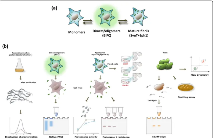
To understand the effect of the A53E substitution in the aggregation process of aSyn (Fig. 1a), we assembled a pipeline of both in in vitro and cellular studies, in hu-man and yeast cells (Fig. 1b), and conducted a battery of experiments to characterize the behavior of this recently identified aSyn mutant form.
We started to compare the in vitro aggregation prop-erties of WT and A53E aSyn using synchronous light scattering, sedimentation and binding to amyloid-binding dyes. For these assays, the soluble forms of both proteins were incubated at 60μM under agitation at 37 °C for two weeks. The light scattering signal was 6-fold higher for WT aSyn than for the A53E mutant, suggesting differ-ences in aggregation process (Fig. 2a). Using spectropho-tometry, we quantified the levels of aggregated protein in both solutions after separating aSyn in two fractions (sol-uble and insol(sol-uble), by centrifugation. The amount of pro-tein in the insoluble fraction in the WT aSyn preparation was two times higher than with A53E (*p< 0.05, Fig. 2b).
transmission electron microscopy (TEM). Although we detected the presence of higher order complexes in both preparations, their size and morphology was different. For WT aSyn, we observed the typical long and unbranched amyloid fibrils (Fig. 2e), 11.6 ± 0.4 nm with a width of 11.6 ± 0.4 nm (Fig. 2e). In contrast, the structures formed by A53E aSyn exhibited a protofibrillar appearance, with small round oligomeric structures that seemed to be linked in a necklace fashion, with a width of 28.5 ± 0.7 nm (Fig. 2f-g).
To assess the secondary structure content of the as-semblies formed by WT and A53E aSyn, we analyzed the amide I region of the FTIR spectrum (1700–1600 cm−1).
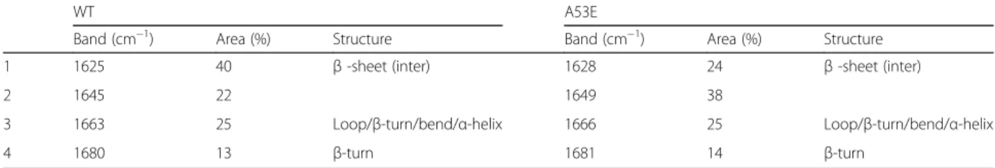
This region of the spectrum corresponds to the absorp-tion of the carbonyl peptide bond of the main amino acid chain of the protein, and is a sensitive marker of the protein secondary structure. After deconvolution of the FTIR spectra of the aSyn solutions, we were able to assign the individual secondary structure elements and their relative contribution to the main absorbance signal at the end of the aggregation reaction (Fig. 2g and h and Table 2). The absorbance spectra were radically different for WT and A53E aSyn. While the spectrum of WT aSyn was dominated by a peak at 1625 cm−1, attributable to
the presence of amyloid-like inter-molecular β–sheet structure (Fig. 2g), the spectrum of the A53E mutant was dominated by a peak at 1649 cm−1corresponding to
disordered/random coil conformation (Fig. 2h).
Next, we monitored how the mutation impacted on the aggregation kinetics of aSyn by continuously moni-toring the changes in Th-T binding over time for WT and A53E variants. The kinetics of amyloid fibril forma-tion usually follows a sigmoidal curve that reflects a nucleation-dependent growth mechanism. The aggrega-tion of both proteins followed this pattern, with an ap-parent lag phase of 8 h (Fig. 2i). After this lag phase, the two aggregation reactions diverged significantly, with an exponential increase for WT aSyn that plateaued at around 55 h, and a steady and much slower increase for A53E aSyn, reaching a 3.5-times lower fluorescence in-tensity. Altogether, our data demonstrates that the A53E mutation reduces aSyn amyloid formation in vitro.
The A53E mutation decreases aSyn oligomerization in cellular models
We next investigated the effects of the A53E mutation on the behavior of aSyn in the context of living human cell models. First, we used the Bimolecular Fluorescence
Fig. 1Aggregation process and study design.aaSyn aggregation process in cell models.bExperimental design used in the study. In vitro
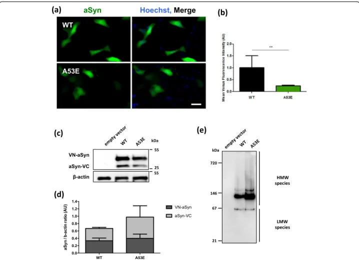
Complementation (BiFC) assay to monitor aSyn oligomerization, as we previously described [45]. Briefly, non-fluorescence Venus fragments are fused to either the N- or C-terminus of aSyn and, upon dimerization/ oligomerization of the protein, the fluorophore is reconsti-tuted resulting in fluorescence signal. While this assay in-volves the tagging of aSyn with fragments of fluorescent proteins, it constitutes a powerful paradigm to assess aSyn oligomerization. We observed that A53E aSyn, similarly to WT, formed dimers/oligomers (Fig. 3a). However, the fluorescence signal was lower than that observed with WT aSyn, suggesting differences in the dimerization/ oligomerization process (**p< 0.01) (Fig. 3b), since the levels of expression of WT and A53E aSyn were identical (Fig. 3c and d, Additional file 1: Figure S2.1 and Additional file 1: Figure S2.2). We also found that the A53E mutant produced high molecular weight species, similar to WT aSyn (Fig. 3e). Thus, we refer to the species formed as oligomers, for simplicity.
The A53E mutation alters the biochemical properties of aSyn inclusions
Next, we investigated if the later stages of the aSyn ag-gregation process were altered by the A53E mutation. For that, we used a well-established cell-based aggrega-tion model that consists in the co-expression of SynT (C-terminally modified aSyn) and Synphilin-1 [40], since expression of aSyn alone does not result in inclusion for-mation. In this aSyn aggregation model, aSyn inclusions are readily detected by immunocytochemistry using anti-bodies against aSyn, allowing the characterization of differ-ent types of inclusions [32], and screening modulators of aSyn aggregation [41]. 48 h after transfection, we analyzed
inclusion formation in the cells (Fig. 4a), and observed that the A53E mutation did not alter inclusion formation when compared to WT aSyn (Fig. 4b). Again, no differences in the levels of aSyn or Synphilin-1 were detected between WT and A53E mutant aSyn (Fig. 4c-e).
aSyn is phosphorylated on Serine 129 (pS129) in LBs found in the brains of PD patients, linking this post-translational modification with disease [14, 52]. We pre-viously showed that specific aSyn mutations, such as the E46K, can alter pS129 on aSyn [39]. Thus we investigated the effect of the A53E mutation on S129 phosphorylation. We found that both WT and A53E aSyn inclusions were positive for pS129 (Fig. 4f).
To further investigate the biochemical nature of the inclusions formed by the A53E aSyn mutant, we stained the cells with Thioflavin S (Th-S), a dye that binds to β-sheet rich amyloid structures [33]. We observed that the larger inclusions formed by WT and A53E aSyn stained positive for Th-S (Fig. 4g). In addition, we also used pro-teinase K (PK) resistance as a marker of aggregate for-mation, as protein inclusions tend to be more resistance to PK digestion. Interestingly, we found that the A53E mutant was less resistant when compared to WT aSyn (Fig. 4h and i), suggesting the inclusions formed by the A53E mutant have a less-compact nature than those formed by the WT protein.
The ubiquitin-proteasome system (UPS) is the major non-lysosomal pathway for selective protein degradation. In cell models, it has been shown that aSyn accumula-tion can affect the activity of the UPS system [61]. In our experimental conditions, we observed that cells ex-pressing A53E aSyn mutant display increased proteolytic activity of the proteasome (Fig. 4j and k).
(See figure on previous page.)
Fig. 2Aggregation properties of WT and A53E aSyn variants. aSyn WT and A53E mutant, prepared at 60μM in 10 mM sodium phosphate,
pH 7.0, were incubated for 2 weeks under agitation at 37 °C.aStatic light scattering of 10μM aSyn in 10 mM sodium phosphate, WT (solid line) and A53E mutant (dashed line).bDistribution of aSyn between the soluble and insoluble fractions.cFluorescence emission spectra of Th-T upon incubation with 10μM aSyn WT (solid line) and A53E mutant (dashed line). Free Th-T emission spectrum is represented in grey.dCR absorbance spectra in the presence of 10μM aSyn WT (solid line) and A53E mutant (dashed line). Free CR absorbance spectrum is represented in grey.
f-gMorphology of WT and A53E aSyn aggregates TEM micrographs. Negatively stained aggregates formed by aSyn WT (left panel) and A53E mutant (right panel) incubated for two weeks.h-iSecondary structure of WT and A53E aSyn aggregates. Secondary structure content of the aSyn WT and A53E mutant after two weeks incubation. ATR-FTIR absorbance spectra in the amide I region was acquired (thick line) and the fitted individual bands after Gaussian deconvolution are shown (thin lines).iAggregation kinetics of WT and A53E aSyn. Aggregation kinetics of aSyn were monitored by following the changes in relative ThioT fluorescence emission. Concentration of protein was 70μM WT aSyn (crosses) and A53E mutant (dots) in a final volume of 150μL. The evolution of Th-T fluorescence in the absence of protein is represented ingrey,n= 3
Table 2Assignment of secondary structure components of aSyn variants in the amide I region of the FTIR spectra
WT A53E
Band (cm−1) Area (%) Structure Band (cm−1) Area (%) Structure
1 1625 40 β-sheet (inter) 1628 24 β-sheet (inter)
2 1645 22 1649 38
3 1663 25 Loop/β-turn/bend/α-helix 1666 25 Loop/β-turn/bend/α-helix
aSyn aggregation leads to Golgi fragmentation
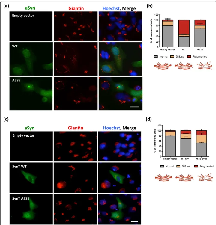
aSyn induces several cellular pathologies that have been documented over the years and are routinely used to as-sess the effect of specific mutations or genetic interactors [60]. One particular type of cellular pathology associated with aSyn toxicity is the fragmentation of the Golgi appar-atus [13]. This is also evident in other neurodegenerative diseases [18], suggesting it might be a more general re-sponse to the proteotoxicity associated with protein mis-folding and aggregation. To assess whether expression of A53E mutant aSyn affected the integrity of the Golgi, we analyzed the morphology of this organelle in both the aSyn oligomerization and aggregation models (Fig. 5a and c). We classified the morphology of the Golgi as normal, diffuse and fragmented, as we previously described [32].
In the oligomerization model, we observed that WT aSyn reduced the percentage of cells exhibiting normal Golgi morphology (~40%, ***p< 0.001 Fig. 5a and b and Additional file 1: Figure S3.1–3.3), as we previously
reported [32]. However, in cells expressing the A53E mutant the effects were not as pronounced as with WT, and the phenotype was more similar to that of cells carrying an empty vector (~70% and 80% of the transfected cells displayed normal Golgi morphology for A53E, and empty vector, respectively) (*p< 0.05) (Fig. 5b and Additional file 1: Figure S3.1).
In the aSyn aggregation model, we observed the op-posite effect. Around 50% of the cells expressing the A53E SynT displayed normal Golgi morphology whereas around 70% of the cells expressing WT SynT displayed normal Golgi (Fig. 5c and d and Additional file 1: Figure S2.4–2.6; ***p< 0.001 and *p< 0.01 for empty vector, and WT, respectively). This suggests that the aggregation of A53E aSyn induces Golgi alterations.
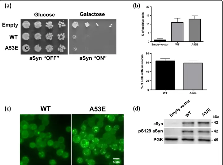
A53E aSyn behaves identically to WT aSyn in yeast cells Yeast cells have been extremely useful to assess cellular pathologies associated with the expression of aSyn. Thus,
Fig. 3A53E reduces aSyn oligomerization.aFluorescent cells, expressing VN-aSyn and aSyn-VC constructs, as a result of the aSyn interaction.
in order to assess if the A53E mutation alters the cytotox-icity and aggregation of aSyn, we expressed this mutant in S. cerevisiaeand monitored phenotypes previously estab-lished [44]. We expressed WT or mutant A53E aSyn fused to eGFP using multi-copy (2 μ) plasmids and under the regulation of a galactose-inducible promoter (GAL1). aSyn cytotoxicity was first evaluated by a spotting assay. The growth of the cells expressing A53E aSyn was compared to that of cells expressing WT aSyn (Fig. 6a). As described before, expression of WT aSyn is toxic and results in re-duced cell growth (Fig. 6a). We found that cells expressing A53E aSyn display a similar phenotype, suggesting that this familial mutation does not significantly affect aSyn toxicity in yeast (Fig. 6a). To further dissect the cytotox-icity of the A53E mutant, we performed propidium iodide (PI) staining and flow cytometry analysis, a readout of plasma membrane integrity (Fig. 6b). We observed that, 7 h after induction of aSyn expression, no significant dif-ferences were observed between cells expressing either WT or the A53E aSyn (Fig. 6b).
Next, we evaluated the subcellular distribution of the A53E mutant aSyn, using fluorescence microscopy. 7 h after induction of aSyn expression we assessed inclusion formation in the cells (Fig. 6c). The expression of A53E aSyn resulted in the formation of cytoplasmic inclusions that looked similar to those formed in cells expressing aSyn WT (Fig. 6c). In addition, no significant differences were observed in the percentage of cells with inclusions between WT and the A53E (Fig. 6c). We also evaluated the levels of aSyn expression by immunoblot analyses and found that both proteins were expressed at similar levels (Fig. 6d). The levels of phosphorylation on serine 129 were also indistinguishable between WT and A53E mutant aSyn (Fig. 6d).
Discussion
aSyn plays a major role in the pathological processes in-volved in neurodegenerative diseases, like PD or Dementia with Lewy Bodies [26]. When overexpressed in cells, to mimic familial forms of PD associated with multiplications of the aSyn gene, aSyn can promote cytotoxicity and impair vital processes, thereby contributing to cell death [9, 38, 44]. However, the precise molecular mechanisms underlying aSyn toxicity are still unclear, compromising
our ability to intervene therapeutically. Both mutations and multiplications of theSNCAgene cause familial forms of PD [8, 22, 64]. Currently, six missense mutations in aSyn have been associated with autosomal dominant forms of parkinsonism (A30P, E46K, H50Q, G51D, A53E, A53T) [1, 30, 34, 46–48, 65]. Of these, the A53E was the last one to be identified and, therefore, has been less investigated.
In this study, we aimed to investigate the effect of the substitution of the alanine at position 53 by a glutamic acid residue that introduces an additional negative charge in the protein. For this purpose, we used in vitro tech-niques to characterize the biophysical effects of the muta-tion, and exploited cell-based models to assess the effects of the expression of the A53E mutant on the distribution, aggregation, and toxicity of the protein.
In vitro,we observed that A53E attenuates aSyn aggre-gation and reduces amyloid fibril formation when com-pared to WT aSyn. In fact, the formation of amyloid structures by the A53E mutant is marginal, since A53E is unable to bind CR. Overall, these observations are in line with a previous report showing that the presence of a negatively-charged residue can reduce the intrinsic ag-gregation propensity of aSyn [15]. Nevertheless, because the change in net charge in aSyn is small, −9 and −10 for the WT and A53E proteins, respectively, the effect this mutation has on aggregation has more likely a local origin. We used the Amylpred2 consensus aggregation predictor to analyze if the A53E mutation might have an impact in the intrinsic aggregation propensity of the aSyn sequence. Amylpred2 identifies a hot spot of aggre-gation corresponding to the 49–55 sequence stretch (VHGVATV), including Ala53. The A53E mutation shortens the aggregation region now including only resi-dues 49–53 (VHGVE). The AGGRESCAN algorithm en-ables the comparison of the aggregation propensity of the two regions, showing that it is 2.1 lower for A53E than for WT aSyn.
In the context of a cell, the behavior of a protein is subjected not only to the crowded environment but also to the action of various protein quality control mecha-nisms. In human cells, we observed that the A53E muta-tion reduces aSyn oligomerizamuta-tion without changing the aggregation pattern. Interestingly, the inclusions formed
(See figure on previous page.)
Fig. 4A53E does not change the inclusion pattern.a-bAt least 50 cellspercondition were classified according to the pattern formed. We
by A53E aSyn are more sensitive to PK digestion than those formed by WT aSyn, suggesting that the inclusions formed are less compact or, possibly, more immature. The negative charge introduced by the glutamate residue can perhaps alter the intermolecular interactions between aSyn molecules, and prevent the formation of tighter inclusions.
This may also correlate with the increase in proteasome ac-tivity that we observed, since this is an important degrad-ation system to eliminate soluble proteins and smaller assemblies that are not degraded by autophagy.
In our previous studies, we did not observe major differ-ences when comparing another PD-associated aSyn mutant
Fig. 5Golgi morphology in the oligomerization and aggregation models.a-bRepresentative pictures of transfected cells with the BiFC system.
at position 53 (A53T) with WT protein. We found that the aggregation of A53T aSyn was identical to that of WT aSyn [32], suggesting again that the change in charge at position 53 may influence the initial steps of the aggregation process (dimerization/oligomerization) of aSyn, and not the later steps that culminate with the formation of the mature in-clusions. Also, the differences might result by difference spatial sequestration of aSyn. Misfolded proteins can be tar-getted to specific compartments, like aggresomes, to facili-tate their degradation [23, 24, 62]. These compartments rely on filament network proteins like vimentin, actin, and tubulin, or proteins that can assist in the transportation of misfolded proteins through the microtubule network, such as p62 (24843142, 12093283, 14623963, 24086678). For ex-ample, Tubulin Polymerization Promoting Protein (TPPP/ p25), belongs to the microtubule network, and associates
with aSyn in pathological conditions, possibly affecting aSyn aggregation (17123092).
The Golgi apparatus plays a determinant role in the intracellular flow of several endogenous proteins and ex-ogenous macromolecules, regulating the trafficking to their final destination inside or outside cells [12]. Frag-mentation of the Golgi apparatus is a characteristic fea-ture in several neurodegenerative diseases, including PD [13, 18, 19]. Interestingly, in our cell-based aggregation models, we observed a striking loss of the typical Golgi morphology in cells expressing A53E aSyn. Previous studies about the effect of this recently identified PD mutation in aSyn showed that A53E is as toxic as WT aSyn [15, 50]. While this was also the trend we observed in our cellular models, our findings suggest that the A53E mutant may cause specific alterations in cell physiology
Fig. 6Phenotypic characterization of yeast cells expressing the A53E aSyn mutant.aCytotoxicity of WT and A53E aSyn in yeast cells compared
that are still not understood and demand additional investigation.
Conclusions
Overall, our study demonstrates that the A53E aSyn mu-tation influences the ability of aSyn to aggregate in vitro and in vivo, and that it may induce different cellular pathologies that should be further investigated in vivo. Ultimately, a deeper understanding of the effect of aSyn mutations on the behavior of the protein will enable the design of novel models to assess the potential value of future therapeutic strategies for PD and other synucleinopathies.
Additional file
Additional file 1: Figure S1.1.Biochemical characterization of WT and
A53E recombinant aSyn. Analysis of purified WT and A53E aSyn variants by SDS-PAGE (18%) stained with Coomassie Brilliant Blue.S1.2.MALDI-TOF mass spectrometry analysis of purified WT (up) and A53E (down) aSyn variants. A MW of 14462.98 Da was obtained for WT (theoretical of 14460.16 da) and of 14519.47 Da for A53E (theoretical of 14518.19 Da) aSyn variants. The peaks of 7231.82 and 7260.97 Da correspond to the M+2ions of WT and A53E aSyn, respectively.Figure S2.1 and S2.2.
Immunoblot quantifications. Levels of VN-aSyn (S2.1) and aSyn-VC (S2.1).n=3.Figure S3.1-S3.3.Morphological analysis of Golgi apparatus in the aSyn BiFC system. The morphology of the Golgi was analyzed as normal (S3.1), diffuse (S3.2) and fragmented (S3.3). One-way ANOVA with post-hoc Tukey’s test (*p<0.05, **p<0.01, ***p<0.001).n=3.Figure S3.4-3.6Morphological analysis of Golgi apparatus in the aSyn aggregation model. Transfected cells were analyzed according to the morphology of the Golgi: normal (S3.4), diffuse (S3.5) and fragmented (S3.6) One-way ANOVA with post-hoc Tukey’s test (*p<0.05, **p<0.01, ***p<0.001).n=3. (PDF 389 kb)
Abbreviations
A53E:Substitution of alanine at position 53 by a glutamate residue; aSyn: alpha-synuclein; ATR FT-IR: Attenuated total reflectance Fourier transform infrared spectroscopy; BiFC: Bimolecular fluorescence complementation assay; CR: Congo red; H4: Human Neuroglioma cells; HEK: Human embryonic kidney cells; IPTG: isopropyl-1-thio-β-D-galactopyranoside; LBs: Lewy bodies; PD: Parkinson’s disease; PI: Propidium iodide; PK: Proteinase K; RT: Room temperature; SC: synthetic complete medium; TEM: Transmission electron microscopy; Th-S: Thioflavin S; Th-T: Thioflavin T; UPS: Ubiquitin-proteasome system
Acknowledgements
TFO is supported by the DFG Center for Nanoscale Microscopy and Molecular Physiology of the Brain, by the German-Israeli Foundation for Scientific Research and Development (GIF), and by a grant from the Niedersachsisches Ministerium fur Wissenschaft und Kultur (MWK). TFO and SV are supported by a grant from Fundación La Marato de TV3 (Ref. 20144330). The funders had no role in study design, data collection and analysis, decision to publish, or preparation of the manuscript.
Authors’contributions
Conceived and designed the experiments: DFL, MD, SV, TFO. Performed the experiments: DFL, MD, AC, SN, CSM, ST. Analyzed the data: DFL, MD, SV, ST, TFO. Wrote the paper: DFL, MD, SV, TFO. All authors read and approved the final manuscript.
Competing interests
The authors declare that they have no competing interests.
Author details
1Department of Neurodegeneration and Restorative Research, University Medical Center Göttingen, Waldweg 33, 37073 Göttingen, Germany.2Institut de Biotecnologia i Biomedicina and Departament de Bioquímica i Biologia Molecular, Universitat Autònoma de Barcelona, 08193, Bellaterra, Barcelona, Spain.3Chronic Disease Research Center (CEDOC), NOVA Medical School, Campo dos Mártires da Pátria, 130, 1169-056 Lisbon, Portugal.4Max Planck Institute for Experimental Medicine, Göttingen, Germany.
Received: 29 October 2016 Accepted: 3 December 2016
References
1. Appel-Cresswell S, Vilarino-Guell C, Encarnacion M, Sherman H, Yu I, Shah B, Weir D, Thompson C, Szu-Tu C, Trinh J et al (2013) Alpha-synuclein p.H50Q, a novel pathogenic mutation for Parkinson’s disease. Mov Disord 28:811–813 2. Auluck PK, Caraveo G, Lindquist S (2010) alpha-Synuclein: membrane
interactions and toxicity in Parkinson’s disease. Annu Rev Cell Dev Biol 26:211–233
3. Barona-Lleo L, Zulueta-Santos C, Murie-Fernandez M, Perez-Fernandez N (2014) Recent onset disequilibrium mimicking acute vestibulopathy in early multiple sclerosis. Am J Otolaryngol 35:529–534
4. Braak H, Del Tredici K, Rub U, de Vos RA, Jansen Steur EN, Braak E (2003) Staging of brain pathology related to sporadic Parkinson’s disease. Neurobiol Aging 24:197–211
5. Braak H, Ghebremedhin E, Rub U, Bratzke H, Del Tredici K (2004) Stages in the development of Parkinson’s disease-related pathology. Cell Tissue Res 318:121–134
6. Cai H, Reinisch K, Ferro-Novick S (2007) Coats, tethers, Rabs, and SNAREs work together to mediate the intracellular destination of a transport vesicle. Dev Cell 12:671–682
7. Chandra S, Gallardo G, Fernandez-Chacon R, Schluter OM, Sudhof TC (2005) Alpha-synuclein cooperates with CSPalpha in preventing
neurodegeneration. Cell 123:383–396
8. Chartier-Harlin MC, Dachsel JC, Vilarino-Guell C, Lincoln SJ, Lepretre F, Hulihan MM, Kachergus J, Milnerwood AJ, Tapia L, Song MS et al (2011) Translation initiator EIF4G1 mutations in familial Parkinson disease. Am J Hum Genet 89:398–406
9. Cooper AA, Gitler AD, Cashikar A, Haynes CM, Hill KJ, Bhullar B, Liu K, Xu K, Strathearn KE, Liu F et al (2006) Alpha-synuclein blocks ER-Golgi traffic and Rab1 rescues neuron loss in Parkinson’s models. Science 313:324–328 10. Coppede F (2012) Genetics and epigenetics of Parkinson’s disease. Sci
World J 2012:489830
11. Dikiy I, Eliezer D (2014) N-terminal acetylation stabilizes N-terminal helicity in lipid- and micelle-bound alpha-synuclein and increases its affinity for physiological membranes. J Biol Chem 289:3652–3665
12. Fan J, Hu Z, Zeng L, Lu W, Tang X, Zhang J, Li T (2008) Golgi apparatus and neurodegenerative diseases. Int J Dev Neurosci 26:523–534
13. Fujita Y, Ohama E, Takatama M, Al-Sarraj S, Okamoto K (2006) Fragmentation of Golgi apparatus of nigral neurons with alpha-synuclein-positive inclusions in patients with Parkinson’s disease. Acta Neuropathol 112:261–265
14. Fujiwara H, Hasegawa M, Dohmae N, Kawashima A, Masliah E, Goldberg MS, Shen J, Takio K, Iwatsubo T (2002) alpha-Synuclein is phosphorylated in synucleinopathy lesions. Nat Cell Biol 4:160–164
15. Ghosh D, Sahay S, Ranjan P, Salot S, Mohite GM, Singh PK, Dwivedi S, Carvalho E, Banerjee R, Kumar A, Maji SK (2014) The newly discovered Parkinson’s disease associated Finnish mutation (A53E) attenuates alpha-synuclein aggregation and membrane binding. Biochemistry 53:6419–6421 16. Gietz D, St Jean A, Woods RA, Schiestl RH (1992) Improved method for high
efficiency transformation of intact yeast cells. Nucleic Acids Res 20:1425 17. Goedert M, Spillantini MG, Del Tredici K, Braak H (2013) 100 years of Lewy
pathology. Nat Rev Neurol 9:13–24
18. Gonatas NK, Stieber A, Gonatas JO (2006) Fragmentation of the Golgi apparatus in neurodegenerative diseases and cell death. J Neurol Sci 246:21–30 19. Gosavi N, Lee HJ, Lee JS, Patel S, Lee SJ (2002) Golgi fragmentation occurs
in the cells with prefibrillar alpha-synuclein aggregates and precedes the formation of fibrillar inclusion. J Biol Chem 277:48984–48992
21. Hernandez-Vargas R, Fonseca-Ornelas L, Lopez-Gonzalez I, Riesgo-Escovar J, Zurita M, Reynaud E (2011) Synphilin suppresses alpha-synuclein neurotoxicity in a Parkinson’s disease Drosophila model. Genesis 49:392–402 22. Ibanez P, Bonnet AM, Debarges B, Lohmann E, Tison F, Pollak P, Agid Y,
Durr A, Brice A (2004) Causal relation between alpha-synuclein gene duplication and familial Parkinson’s disease. Lancet 364:1169–1171 23. Johnston JA, Ward CL, Kopito RR (1998) Aggresomes: a cellular response to
misfolded proteins. J Cell Biol 143:1883–1898
24. Kaganovich D, Kopito R, Frydman J (2008) Misfolded proteins partition between two distinct quality control compartments. Nature 454:1088–1095 25. Kannarkat GT, Boss JM, Tansey MG (2013) The role of innate and adaptive
immunity in Parkinson’s disease. J Park Dis 3:493–514
26. Kim WS, Kagedal K, Halliday GM (2014) Alpha-synuclein biology in Lewy body diseases. Alzheimers Res Ther 6:73
27. Kisselev AF, Goldberg AL (2005) Monitoring activity and inhibition of 26S proteasomes with fluorogenic peptide substrates. Methods Enzymol 398:364–378
28. Klunk WE, Pettegrew JW, Abraham DJ (1989) Quantitative evaluation of congo red binding to amyloid-like proteins with a beta-pleated sheet conformation. J Histochem Cytochem 37:1273–1281
29. Kordower JH, Brundin P (2009) Propagation of host disease to grafted neurons: accumulating evidence. Exp Neurol 220:224–225
30. Kruger R, Kuhn W, Muller T, Woitalla D, Graeber M, Kosel S, Przuntek H, Epplen JT, Schols L, Riess O (1998) Ala30Pro mutation in the gene encoding alpha-synuclein in Parkinson’s disease. Nat Genet 18:106–108
31. Lashuel HA, Overk CR, Oueslati A, Masliah E (2013) The many faces of alpha-synuclein: from structure and toxicity to therapeutic target. Nat Rev Neurosci 14:38–48
32. Lazaro DF, Rodrigues EF, Langohr R, Shahpasandzadeh H, Ribeiro T, Guerreiro P, Gerhardt E, Krohnert K, Klucken J, Pereira MD et al (2014) Systematic comparison of the effects of alpha-synuclein mutations on its oligomerization and aggregation. PLoS Genet 10:e1004741
33. Lee HJ, Lee SJ (2002) Characterization of cytoplasmic alpha-synuclein aggregates. Fibril formation is tightly linked to the inclusion-forming process in cells. J Biol Chem 277:48976–48983
34. Lesage S, Anheim M, Letournel F, Bousset L, Honore A, Rozas N, Pieri L, Madiona K, Durr A, Melki R et al (2013) G51D alpha-synuclein mutation causes a novel parkinsonian-pyramidal syndrome. Ann Neurol 73:459–471 35. LeVine H 3rd (1993) Thioflavine T interaction with synthetic Alzheimer’s
disease beta-amyloid peptides: detection of amyloid aggregation in solution. Protein Sci 2:404–410
36. Li JY, Englund E, Holton JL, Soulet D, Hagell P, Lees AJ, Lashley T, Quinn NP, Rehncrona S, Bjorklund A et al (2008) Lewy bodies in grafted neurons in subjects with Parkinson’s disease suggest host-to-graft disease propagation. Nat Med 14:501–503
37. Maroteaux L, Campanelli JT, Scheller RH (1988) Synuclein: a neuron-specific protein localized to the nucleus and presynaptic nerve terminal. J Neurosci 8:2804–2815
38. Martin LJ, Pan Y, Price AC, Sterling W, Copeland NG, Jenkins NA, Price DL, Lee MK (2006) Parkinson’s disease alpha-synuclein transgenic mice develop neuronal mitochondrial degeneration and cell death. J Neurosci 26:41–50 39. Mbefo MK, Fares MB, Paleologou K, Oueslati A, Yin G, Tenreiro S, Pinto M, Outeiro T, Zweckstetter M, Masliah E, Lashuel HA (2015) Parkinson disease mutant E46K enhances alpha-synuclein phosphorylation in mammalian cell lines, in yeast, and in vivo. J Biol Chem 290:9412–9427
40. McLean PJ, Kawamata H, Hyman BT (2001) Alpha-synuclein-enhanced green fluorescent protein fusion proteins form proteasome sensitive inclusions in primary neurons. Neuroscience 104:901–912
41. Moree B, Yin G, Lazaro DF, Munari F, Strohaker T, Giller K, Becker S, Outeiro TF, Zweckstetter M, Salafsky J (2015) Small molecules detected by second-harmonic generation modulate the conformation of monomeric alpha-synuclein and reduce its aggregation in cells. J Biol Chem 290: 27582–27593
42. Murphy DD, Rueter SM, Trojanowski JQ, Lee VM (2000) Synucleins are developmentally expressed, and alpha-synuclein regulates the size of the presynaptic vesicular pool in primary hippocampal neurons. J Neurosci 20:3214–3220
43. Olanow CW, Perl DP, DeMartino GN, McNaught KS (2004) Lewy-body formation is an aggresome-related process: a hypothesis. Lancet Neurol 3:496–503 44. Outeiro TF, Lindquist S (2003) Yeast cells provide insight into alpha-synuclein
biology and pathobiology. Science 302:1772–1775
45. Outeiro TF, Putcha P, Tetzlaff JE, Spoelgen R, Koker M, Carvalho F, Hyman BT, McLean PJ (2008) Formation of toxic oligomeric alpha-synuclein species in living cells. PLoS One 3:e1867
46. Pasanen P, Myllykangas L, Siitonen M, Raunio A, Kaakkola S, Lyytinen J, Tienari PJ, Poyhonen M, Paetau A (2014) Novel alpha-synuclein mutation A53E associated with atypical multiple system atrophy and Parkinson’s disease-type pathology. Neurobiol Aging 35:2180.e2181–2185
47. Polymeropoulos MH, Lavedan C, Leroy E, Ide SE, Dehejia A, Dutra A, Pike B, Root H, Rubenstein J, Boyer R et al (1997) Mutation in the alpha-synuclein gene identified in families with Parkinson’s disease. Science 276:2045–2047 48. Proukakis C, Dudzik CG, Brier T, MacKay DS, Cooper JM, Millhauser GL,
Houlden H, Schapira AH (2013) A novel alpha-synuclein missense mutation in Parkinson disease. Neurology 80:1062–1064
49. Rey NL, Steiner JA, Maroof N, Luk KC, Madaj Z, Trojanowski JQ, Lee VM, Brundin P (2016) Widespread transneuronal propagation of alpha-synucleinopathy triggered in olfactory bulb mimics prodromal Parkinson’s disease. J Exp Med 213:1759–1778
50. Rutherford NJ, Giasson BI (2015) The A53E alpha-synuclein pathological mutation demonstrates reduced aggregation propensity in vitro and in cell culture. Neurosci Lett 597:43–48
51. Sabate R, Rodriguez-Santiago L, Sodupe M, Saupe SJ, Ventura S (2013) Thioflavin-T excimer formation upon interaction with amyloid fibers. Chem Commun 49:5745–5747
52. Saito Y, Kawashima A, Ruberu NN, Fujiwara H, Koyama S, Sawabe M, Arai T, Nagura H, Yamanouchi H, Hasegawa M et al (2003) Accumulation of phosphorylated alpha-synuclein in aging human brain. J Neuropathol Exp Neurol 62:644–654
53. Singleton AB, Farrer M, Johnson J, Singleton A, Hague S, Kachergus J, Hulihan M, Peuralinna T, Dutra A, Nussbaum R et al (2003) alpha-Synuclein locus triplication causes Parkinson’s disease. Science 302:841
54. Spillantini MG, Schmidt ML, Lee VM, Trojanowski JQ, Jakes R, Goedert M (1997) Alpha-synuclein in Lewy bodies. Nature 388:839–840
55. Spillantini MG, Crowther RA, Jakes R, Hasegawa M, Goedert M (1998) alpha-Synuclein in filamentous inclusions of Lewy bodies from Parkinson’s disease and dementia with lewy bodies. Proc Natl Acad Sci U S A 95:6469–6473 56. Tenreiro S, Reimao-Pinto MM, Antas P, Rino J, Wawrzycka D, Macedo D,
Rosado-Ramos R, Amen T, Waiss M, Magalhaes F et al (2014) Phosphorylation modulates clearance of alpha-synuclein inclusions in a yeast model of Parkinson’s disease. PLoS Genet 10:e1004302
57. Tenreiro S, Rosado-Ramos R, Gerhardt E, Favretto F, Magalhaes F, Popova B, Becker S, Zweckstetter M, Braus GH, Outeiro TF (2016) Yeast reveals similar molecular mechanisms underlying alpha- and beta-synuclein toxicity. Hum Mol Genet 25:275–290
58. Tsigelny IF, Sharikov Y, Wrasidlo W, Gonzalez T, Desplats PA, Crews L, Spencer B, Masliah E (2012) Role of alpha-synuclein penetration into the membrane in the mechanisms of oligomer pore formation. FEBS J 279:1000–1013
59. Volles MJ, Lansbury PT Jr (2007) Relationships between the sequence of alpha-synuclein and its membrane affinity, fibrillization propensity, and yeast toxicity. J Mol Biol 366:1510–1522
60. Wales P, Pinho R, Lazaro DF, Outeiro TF (2013) Limelight on alpha-synuclein: pathological and mechanistic implications in neurodegeneration. J Park Dis 3:415–459
61. Webb JL, Ravikumar B, Atkins J, Skepper JN, Rubinsztein DC (2003) Alpha-Synuclein is degraded by both autophagy and the proteasome. J Biol Chem 278:25009–25013
62. Weisberg SJ, Lyakhovetsky R, Werdiger AC, Gitler AD, Soen Y, Kaganovich D (2012) Compartmentalization of superoxide dismutase 1 (SOD1G93A) aggregates determines their toxicity. Proc Natl Acad Sci U S A 109:15811–15816
63. Winner B, Jappelli R, Maji SK, Desplats PA, Boyer L, Aigner S, Hetzer C, Loher T, Vilar M, Campioni S et al (2011) In vivo demonstration that alpha-synuclein oligomers are toxic. Proc Natl Acad Sci U S A 108:4194–4199 64. Xu W, Tan L, Yu JT (2015) Link between the SNCA gene and parkinsonism.
Neurobiol Aging 36:1505–1518