Histone deacetylase inhibitors as potential treatment
for spinal muscular atrophy
Jafar Mohseni
1, Z.A.M.H. Zabidi-Hussin
2and Teguh Haryo Sasongko
11
Human Genome Centre, School of Medical Sciences, Universiti Sains Malaysia, Health Campus, Kubang
Kerian, Kelantan, Malaysia.
2
Department of Pediatrics, School of Medical Sciences, Universiti Sains Malaysia, Health Campus, Kubang
Kerian, Kelantan, Malaysia.
Abstract
Histone acetylation plays an important role in regulation of transcription in eukaryotic cells by promoting a more re-laxed chromatin structure necessary for transcriptional activation. Histone deacetylases (HDACs) remove acetyl groups and suppress gene expression. HDAC inhibitors (HDACIs) are a group of small molecules that promote gene transcription by chromatin remodeling and have been extensively studied as potential drugs for treating of spinal muscular atrophy. Various drugs in this class have been studied with regard to their efficacy in increasing the expres-sion of survival of motor neuron (SMN) protein. In this review, we discuss the current literature on this topic and sum-marize the findings of the main studies in this field.
Keywords: HDACi, molecular therapy, spinal muscular atrophy.
Received: January 1, 2013; Accepted: June 20, 2013.
Introduction
Proximal spinal muscular atrophy (SMA) is a fatal, autosomal recessive pediatric neuromuscular disorder that is characterized by the destruction ofa-motor neurons in the anterior horn of the spinal cord. SMA has an estimated incidence of 1/6,000 to 1/10,000 live births, with a carrier frequency of ~1/50 individuals (Burletet al.,1996; Feldkot-teret al., 2002; Kernochan et al., 2005). The criteria for classifying SMA include age of onset and disease progres-sion, based on which SMA patients can be classified into one of four types. Entire gene deletion as well as a variety of intragenic deletions, point mutations and other truncat-ing mutations of survival of motor neuron1 (SMN1) on chromosome 5q13that lead to loss of gene function are the cause of SMA (Clermontet al., 1994; Lefebvreet al., 1995; Burglenet al., 1996; Burletet al., 1996). A highly related homolog of the gene,SMN2 or centromeric SMN, is re-tained (with a variable copy number) in all SMA patients. The substitution of a C by T at position+6 disrupts a exon splice-enhancing region in exon 7. This change results in mostSMN2transcripts lacking exon 7 and encodes a trun-cated protein (Feldkotter et al., 2002; Kernochan et al., 2005).
SMN2has, for many years, provided a promising op-portunity for correcting SMN deficiency. The fact that
SMN2produces SMN protein, although at an insufficiently low amount, led investigators to search for ways of increas-ing the full-length expression of this gene in order to ensure a sufficient level of the protein. Studies in transgenic mice have shown that the insertion of eight copies of human
SMN2into the mouse genome completely rescued
Smn-/-mice (Smn-/-; hSMN2+/+) from the SMA phenotype (Mo-naniet al., 2003). In humans, a high copy number ofSMN2
may preventSMN1-deficient individuals from manifesting the SMA phenotype (Prioret al., 2004). An increase in full-length SMN protein production through enhanced
SMN2expression may be achieved through promoter acti-vation, modulation of exon 7 splicing (inclusion of exon 7 in theSMN2transcript) or both. Another therapeutic target includesSMN1subtle mutations. A subset of SMA patients carrying SMN1 subtle mutations is susceptible to non-sense-mediated mRNA decay (NMD) (Brichta et al., 2008). In this regard, studies aimed at identifying sub-stances that can stabilizeSMNmRNA, especially those that express the full-length protein, are of interest.
Various approaches have been proposed as potential means of treating and/or preventing SMA, including: (1) the use of compounds that enhanceSMN2promoter activity, (2) the use of compounds that modulateSMN2splicing, (3) the use of drugs that stabilizeSMN2mRNA or SMN protein, (4) gene therapy and (5) stem cell therapy (Simic, 2008).
Send correspondence to Teguh Haryo Sasongko. Human Genome Center, School of Medical Sciences, Universiti Sains Malaysia, USM Health Campus, 16150 Kubang Kerian, Kelantan, Malaysia. E-mail: [email protected], [email protected].
One group of drugs in particular, namely, histone deacetylase(HDAC)inhibitors, has been found to increase
SMN2promoter activity. Histone acetylation is an impor-tant epigenetic mechanism that regulates gene expression. When the N-terminus of core histones is acetylated the cor-responding chromatin region is more actively transcribed because of increased accessibility to the DNA. Several drugs in this group have shown promising results in in-creasingSMNpromoter activity as will be summarized be-low.
This article focuses on HDAC inhibitors that target classic HDACs and provides a comprehensive overview of current research on SMA therapy using these inhibitors. Specifically, we will discuss the characteristics and thera-peutic potential of valproic acid, phenylbutyrate, benza-mide M344, suberoylanilidehydroxamic acid, LBH589, trichostatin A, MS-275, romidepsin, resveratrol, curcumin and epigallocathecin gallate.
HDACs and HDAC inhibitors
Histone remodeling by acetylation and/or deacetyl-ation plays an important role in the transcriptional regula-tion of eukaryotic cells. Histone acetylaregula-tion produces a more relaxed chromatin structure that allows trans-criptional activation (Kernochanet al., 2005; Riesteret al., 2007). This is achieved through the acetylation of lysine residues that imparts a negative charge to the affected amino acid which in turn relaxes the chromatin. In this re-gard, HDACs are actually “lysinedeacetylases” (Grayson
et al., 2010; Xuet al., 2007). HDACs therefore repress tran-scription through histone deacetylation.
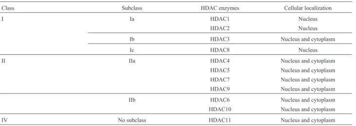
HDACs form a large family of enzymes and have been classified into two groups based on their co-enzyme requirements and sequence similarity to yeast HDACs. These two groups, known as classic HDACs and Sir2-related HDACs (Sirtuins or Class III HDACs), are acti-vated by Zn2+and NAD+, respectively. Classic HDACs are subdivided into three smaller classes that include HDAC-I
(Ia, Ib and Ic), HDAC-II (IIa and IIb) and HDAC-IV. Each of these smaller classes consists of functional HDAC en-zymes (HDAC1 to HDAC11) that are targeted by different HDAC inhibitors (Table 1A,B).Overall, there are 11 classic HDAC enzymes while the Sirtuins contain seven members (Sirt1-Sirt7) (Xu et al., 2007; Nakagawa and Guarente,
2011).
HDAC inhibitors selectively alter gene transcription through chromatin remodeling and by changing the protein structure of transcription factor complexes (Kernochanet al., 2005; Riesteret al., 2007). HDAC inhibitors generally consist of three domains: a linker region, a capping group and a metal moeity (Dayangac-Erdenet al., 2011).
Valproic acid
Valproic acid (VPA) or Depakene is a Federal Drug Administration (FDA)-approved drug with a terminal half-life (t1/2) of 8-10 h in human serum and is frequently used to treat epilepsy, mood disorders and migraine (Brichtaet al., 2003). Although VPA is associated with few neurological side effects, hematological and hepatic side effects are well known (Cotariu and Zaidman, 1988; Lackmann, 2004; Tong
et al., 2005). VPA increases SMN protein levels through transcriptional activation but also increases the expression of additional serine/arginine (SR)- rich proteins that may have important implications for disorders (including SMA) caused by mutations that result in alternative splicing. While promising results have been obtainedin-vitro, clinical trials have yielded variable results (Table 2).
Chemical characteristics: VPA is a simple eight-carbon branched fatty acid (carboxylic acid;C8H14O2) des-ignated as 2-propylpentanoic acid but is also known as dipropylacetic acid.
Phenylbutyrate
Phenyl butyric acid (PBA) or buphenyl is a short-chain fatty acid that has been clinically tested as an anti-cancer drug. In normal tissues, PBA shows little toxicity
Table 1A- Classification of classic histone deacetylases (HDAC).
Class Subclass HDAC enzymes Cellular localization
I Ia HDAC1 Nucleus
HDAC2 Nucleus
Ib HDAC3 Nucleus and cytoplasm
Ic HDAC8 Nucleus
II IIa HDAC4 Nucleus and cytoplasm
HDAC5 Nucleus and cytoplasm
HDAC7 Nucleus and cytoplasm
HDAC9 Nucleus and cytoplasm
IIb HDAC6 Nucleus and cytoplasm
HDAC10 Nucleus and cytoplasm
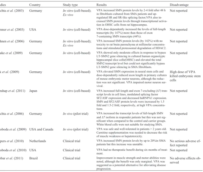
Table 1B- Histone deacetylase (HDAC) inhibitors and their target enzymes.
Inhibitor Target HDAC IC50 Fold increase of full-length SMN2
transcript or SMN protein
VPA HDAC1, HDAC2, HDAC3 0.7-20 mM 2-4
PBA HDAC1, HDAC2 16 nM 0.4-2.4
M344 HDAC6 423 nM 3-7
LBH589 Pan HDACs 5-20 nM 10
SAHA HDAC1, HDAC2, HDAC3, HDAC8, HDAC9 10 nM 5
TSA HDAC5 1.8 nM 2
MS-275 HDAC1, HDAC2, HDAC3, HDAC9 0.5mM Unknown
Romidepsin HDAC1 HDAC2 36 & 47 nM 5
Resveratrol HDAC8 650mM 1.3
Curcumin HDAC8 25mM 1.7
EGCG Unknown Unknown 1.4
EGCG – epigallocathecin gallate; M344 – benzamide 344; MS-275 – entinostat; PBA – phenylbutyrate; SAHA – suberoylanilidehydroxamic acid;TSA – trichostatin A;VPA – valproic acid.
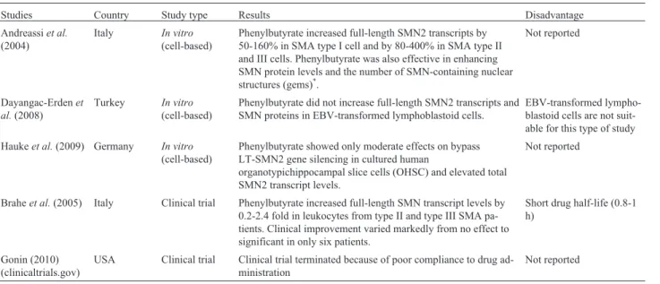
Table 2- Summary of studies on valproic acid (VPA) for the treatment of spinal muscular atrophy.
Studies Country Study type Results Disadvantage
Brichtaet al.(2003) Germany In vitro(cell-based);
Ex vivo
VPA increased SMN protein levels by 2-4 fold after 48 h in fibroblasts cultured from SMA patients and up-regulated SR and SR-like splicing factor;VPA also in-creased SMN protein levels through transcriptional activa-tion in OHSC cells from rat hippocampus.
Not reported
Sumneret al.(2003) USA In vitro(cell-based) VPA dose-dependently increased the levels of full-length transcripts (by 147%) more than those of exon 7-containing SMN transcripts (44%).
Not reported
Hahnenet al.(2006) Germany In vitro(cell-based);
Ex vivo
VPA increased SMN protein levels (by 142%) with no toxicity to rat brain parenchyma at millimolar concentra-tions and stimulated proteosomal degradation of HDAC2.
Not reported
Haukeet al.(2009) Germany In vitro(cell-based) VPA showed only moderate effects in response to bypass LT-SMN2 gene silencing in cultured human organotypic hippocampal slice cells(OHSC) and elevated the total SMN2 transcript level but could not significantly bypass LT-SMN2 gene silencing in SMA fibroblasts.
Not reported
Raket al.(2009) Germany In vitro(cell-based) VPA elevated SMN expression in neural stem cells and dose-dependently reduced axon length in primary cultures of mouse embryonic motor neurons, although the reduc-tion was not significant. VPA impaired motor neuron sur-vival.
High dose of VPA killed embryonic stem cells
Harahapet al.(2011) Japan In vitro(cell-based) VPA increased full-length and exon 7-excluding (D7) tran-script levels in cell lines, modulated splicing factor SF2/ASF expression and decreased hnRNPA1 expression. SMN and SF2/ASF protein levels were increased by 1.5 fold and 1.5-2 fold, respectively, at high VPA concentra-tions.
Not reported
Brichtaet al.(2006) Germany In vivo(pilot trial) VPA increased the transcript levels of full-length SMN andD7 isoform in responder patients but this was not sig-nificant when compared to the control and carrier groups. White blood cells were not suitable for studying SMA.
Not reported
Swobodaet al.(2009) USA and Canada In vivo(pilot trial) VPA was safe and well-tolerated in patients > 2 years old. Carnitine supplementation was needed to decrease the risk of muscle weakness or hepatotoxicity.
Not reported
Pieperset al.(2010) Netherlands Clinical trial VPA increased SMN protein levels by up to 20%in SMA patients but this increase was unstable.
No serious adverse ef-fect reported Swobodaet al.(2010) USA Clinical trial VPA had no therapeutic benefit during six months of
treat-ment.
Not reported
Darbaret al.(2011) Brazil Clinical trial Improvement in muscle strength and motor abilities were noted, although the benefit was only marginal. VPA was suggested as a potential alternative for alleviating disease progression.
and provides protection against various stimuli. Sodium PBA is a pro-drug that is rapidly metabolized to phenyl-acetate, a metabolically-active derivative. Phenylacetate conjugates with glutamine via acetylation to form phenyl-acetylglutamine that is excreted by the kidneys. PBA shows anticancer activity that is generally attributed to its activity as an HDAC inhibitor. Table 3 summarizes studies that have investigated PBA in SMA.
Chemical characteristics: PBA (molecular weight: 186; C10H11O2Na) is known chemically as 4-phenylbutyric acid and is usually supplied as a sodium salt.
Benzamide M344
M344 is a HDAC inhibitor that increases the level of hyperacetylated histone H4 and significantly increases
SMN2mRNA/protein levels in SMA cells by inducing
ter-minal cell differentiation. M344 shows a three-fold selectivity for inhibition of HDAC6 over HDAC1.Table 4 summarizes studies that have investigated benzamide M344 in SMA.
Chemical characteristics: M344 (N-hydroxyl-7-aminoheptanamide) is a benzamide with the molecular for-mula C16H25N3O3.
LBH589
LBH589 (Panobinostat) is a potent putative anti-cancer drug in numerous anti-cancer cell lines and was given or-phan drug status for the treatment of cutaneous T-cell lym-phoma (CTCL) by the FDA in 2007. LBH589 is also a novel hydroxamic-acid-derived HDAC inhibitor that is ac-tive against all classes of HDACs at low nanomolar
con-Table 4- Summary of studies on benzamide M344 for the treatment of spinal muscular atrophy.
Study Country Study type Results Disadvantage
Riesslandet al.(2006) Germany In vitro
(cell-based)
M344 increased FL-SMN2 mRNA levels by restoring the splicing pattern and transcriptional activation of SMN2; there was also an increase in the level of SR and SR-like splicing factors and in the number of nuclear gems. M344 increased the SMN protein levels by 3-7 folds at concen-trations of 30-50mM after 64 h of treatment.
Cytotoxic at > 50mM (MTT assay)
Hahnenet al.(2006) Germany In vitro
(cell-based)Ex vivo
M344 increased the SMN protein levels in human SMA-affected fibroblasts by up to 168% at 10mM. In rat OHSC the SMN transcript levels increased by 149% after a 48 h exposure to M344.
Cytotoxic for rat OHSC at > 20mM (propidium iodide staining)
Haukeet al.(2009) Germany In vitro
(cell-based)
M344 increased the total SMN2 transcript levels in human OHSC by up to 188% at 16mM by bypassing gene silenc-ing.
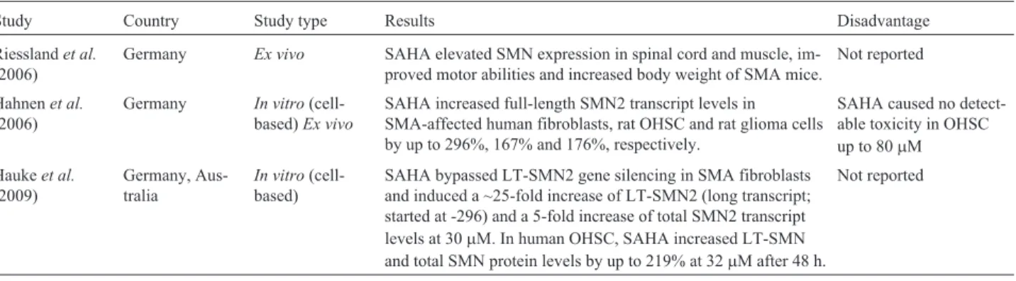
Not reported Table 3- Summary of studies on phenylbutyrate for the treatment of spinal muscular atrophy.
Studies Country Study type Results Disadvantage
Andreassiet al.
(2004)
Italy In vitro
(cell-based)
Phenylbutyrate increased full-length SMN2 transcripts by 50-160% in SMA type I cell and by 80-400% in SMA type II and III cells. Phenylbutyrate was also effective in enhancing SMN protein levels and the number of SMN-containing nuclear structures (gems)*.
Not reported
Dayangac-Erdenet al.(2008)
Turkey In vitro
(cell-based)
Phenylbutyrate did not increase full-length SMN2 transcripts and SMN proteins in EBV-transformed lymphoblastoid cells.
EBV-transformed lympho-blastoid cells are not suit-able for this type of study
Haukeet al.(2009) Germany In vitro
(cell-based)
Phenylbutyrate showed only moderate effects on bypass LT-SMN2 gene silencing in cultured human
organotypichippocampal slice cells (OHSC) and elevated total SMN2 transcript levels.
Not reported
Braheet al.(2005) Italy Clinical trial Phenylbutyrate increased full-length SMN transcript levels by 0.2-2.4 fold in leukocytes from type II and type III SMA pa-tients. Clinical improvement varied markedly from no effect to significant in only six patients.
Short drug half-life (0.8-1 h)
Gonin (2010) (clinicaltrials.gov)
USA Clinical trial Clinical trial terminated because of poor compliance to drug ad-ministration
Not reported
centrations. Table 5 summarizes a study that investigated LBH589 in SMA.
Chemical characteristics: LBH589 (Panobinostat, NVP-LBH589) belongs to the hydroxamate class of inhibi-tors. The molecular formula is C21H23N3O2.
Suberoylanilidehydroxamic acid (SAHA)
Suberoylanilidehydroxamic acid (SAHA;zolinza or vorinostat) was initially approved for the treatment of cuta-neous T-cell lymphoma (CTCL). Vorinostat, an FDA-approved pan-histone deacetylase inhibitor, is a potentially useful drug for clinical trials in SMA patients. Some of this drugs side-effect includes gastrointestinal symptoms, con-stitutional symptoms (thrombocytopenia, anemia), taste disorders, pulmonary embolism and anemia. Severe thrombocytopenia and gastrointestinal bleeding have been reported with the concomitant use of zolinza and other HDAC inhibitors,e.g.,valproic acid. Table 6 summarizes studies that have investigated SAHA in SMA.
Chemical characteristics: SAHA (N-hydroxy-N’-phenyloctanediamide; C14H20N2O3) is poorly soluble in
water, slightly soluble in ethanol, isopropanol and acetone, freely soluble in dimethyl sulfoxide and insoluble in meth-ylene chloride.
Trichostatin A (TSA)
Trichostatin A (TSA), originally developed as an antifungal drug, is a member of a large class of HDAC in-hibitors that has a broad spectrum of epigenetic activities. TSA selectively inhibits class I and II mammalian HDAC. TSA alters gene expression by interfering with the removal of acetyl groups from histones by HDAC and therefore al-ters the ability of DNA transcription factors to access the DNA within chromatin. TSA is harmful by inhalation and is irritating to the eyes, respiratory system and skin. Table 7 summarizes the studies on TSA in SMA.
Chemical characteristics:TSA (7-[4-(dimethylami-no)phenyl]-N-hydroxy-4,6R-dimethyl-7-oxo-2E,4E-hepta dienamide; C17H22N2O3) is extracted from Streptomyces platensisand is soluble in ethanol and dimethylsulfoxide (DMSO).
Table 5- Summary of a study on LBH589for the treatment of spinal muscular atrophy.
Study Country Study type Results Disadvantage
Garbeset al.(2009) Germany In vitro(cell-based) The SMN protein level increased by up to 10 fold at 400 nM LBH589 after a 64-h exposure. A number of gems and a stable increase in SMN protein were also observed.
No cytotoxic effects at up to 500 nM
Table 6- Summary of studies on SAHA for the treatment of spinal muscular atrophy.
Study Country Study type Results Disadvantage
Riesslandet al.
(2006)
Germany Ex vivo SAHA elevated SMN expression in spinal cord and muscle, im-proved motor abilities and increased body weight of SMA mice.
Not reported
Hahnenet al.
(2006)
Germany In vitro (cell-based)Ex vivo
SAHA increased full-length SMN2 transcript levels in SMA-affected human fibroblasts, rat OHSC and rat glioma cells by up to 296%, 167% and 176%, respectively.
SAHA caused no detect-able toxicity in OHSC up to 80mM
Haukeet al.
(2009)
Germany, Aus-tralia
In vitro (cell-based)
SAHA bypassed LT-SMN2 gene silencing in SMA fibroblasts and induced a ~25-fold increase of LT-SMN2 (long transcript; started at -296) and a 5-fold increase of total SMN2 transcript levels at 30mM. In human OHSC, SAHA increased LT-SMN and total SMN protein levels by up to 219% at 32mM after 48 h.
Not reported
Table 7- Summary of studies on TSA for the treatment of spinal muscular atrophy.
Study Country Study type Results Disadvantage
Avilaet al.(2007) USA, Italy In vitro (cell--based)Ex vivo
TSA induced SMN2 promoter activation by approximately two fold after 2-4 h of exposure. TSA markedly improved motor per-formance, attenuated weight loss, increased survival and im-proved the pathology of the motor unit in SMA mice
One-quarter of SMA mice showed no response to TSA treatment
Narveret al.
(2008)
USA Ex vivo TSA improved short-term function and produced long-lasting stabilization of the SMA motor unit. In affected mice treated with TSA and a dietary supplementation the median survival time increased by up to 38 days (170%) as compared to non-treated mice.
Entinostat (MS-275)
Entinostat(MS-275;n-2-aminophenyl-4-n-pyridine-3 -ylmethoxycarbonylaminomethyl-benzamide), is a cell-permeable benzamide analog that inhibits HDAC and in-duces differentiation and transcription of growth factorbII receptor (TbRII), in addition to inhibiting the proliferation of human breast cancer cells. Table 8 summarizes studies that have investigated Entinostat in SMA.
Chemical characteristics:The molecular formula of Entinostat is C21H20N4O3.
Romidepsin
Romidepsin (Istodex or FK228), an HDAC inhibitor from Chromobacterium violaceum, is a bicyclic depsi-peptide. Romidepsin is indicated for the treatment of CTCL in patients who have received at least one prior systemic therapy. Romidepsin shows hematologic and non-hema-tologic toxicity at high doses. Table 9 summarizes a study that investigated the usefulness of romidepsinin SMA.
Chemical characteristics: Romidepsin is described chemically as (1S,4S,7Z,10S,16E,21R)-7-ethylidene-4,21-bis(1 methylethyl)-2-oxa-12,13-dithia-5,8,20,23-tetra azabicyclo[8.7.6]tricos-16ene-3,6,9,19,22-pentone with the molecular formula C24H36N4O6S2.
Resveratrol
Resveratrol (Kojo-Kon, Phytoalexin, Phytoestrogen and SRT-501) is a chemical found in red wine, red grape skins, purple grape juice, mulberries and in smaller amounts in peanuts. Resveratrol is used against hardening of the arteries (atherosclerosis), high cholesterol and for the prevention of cancer. Resveratrol may increase the risk of bleeding. Table 10 summarizes studies that have investi-gated resveratrol in SMA.
Chemical characteristics: Resveratrol, a poly-phenolic compound ((E)-resveratrol (3,5,4’ -trihydroxy-trans-stilbene)), belongs to the stilbene class of molecules
and is classified as anti-cancer, antioxidant and enzyme inhibitor. The molecular formula is C14H12O3.
Curcumin
Curcumin is a mixture of compounds derived from the curry spice turmeric and is used as an herbal supple-ment. Curcumin (diferuloylmethane) is a new HDAC in-hibitor that inhibits the expression of class I HDACs (HDAC1, HDAC3and HDAC8). Curcumin possesses a spectrum of pharmacological properties that have been at-tributed primarily to its inhibition of metabolic enzymes. Curcumin has been alleged to have antioxidant, antiviral, anti-inflammatory and anticancer activities, as well as cho-lesterol-lowering effects.
Chemical characteristics:Curcumin, a natural poly-phenol and the major component of turmeric has the molec-ular formula C21H20O6.
Epigallocatechin gallate
Epigallocatechin gallate (EGCG; Sinecatechins or Veregen), a partially purified fraction obtained from a wa-ter extract of green tea (Camellia sinensis) leaves, is used topically and is a potent antioxidant.Table 11 summarizes studies that have tested curcumin and EGCG in SMA.
Chemical characteristics:The molecular formula for epigallocatechin gallate is C15H14O7.
Discussion
Eight of the 11 known HDACs were inhibited by the compounds reviewed here; HDAC4, HDAC7 and HDAC10 were not inhibited by any of the compounds. As shown in Table 1B, the fold increase of full-lengthSMN2
transcripts or SMN protein varied considerably (from 0.4 to 10).
Five compounds (VPA, M344, resveratrol, EGCG and curcumin) acted by two mechanisms, namely, (1) by increasing the overallSMN2expression through inhibition
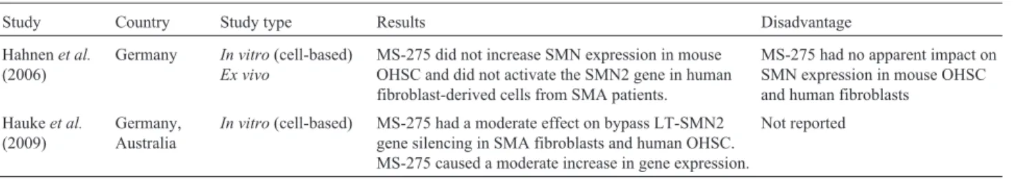
Table 8- Summary of studies on MS-275 for the treatment of spinal muscular atrophy.
Study Country Study type Results Disadvantage
Hahnenet al.
(2006)
Germany In vitro(cell-based)
Ex vivo
MS-275 did not increase SMN expression in mouse OHSC and did not activate the SMN2 gene in human fibroblast-derived cells from SMA patients.
MS-275 had no apparent impact on SMN expression in mouse OHSC and human fibroblasts
Haukeet al.
(2009)
Germany, Australia
In vitro(cell-based) MS-275 had a moderate effect on bypass LT-SMN2 gene silencing in SMA fibroblasts and human OHSC. MS-275 caused a moderate increase in gene expression.
Not reported
Table 9- Summary of a study on romidepsin for the treatment of spinal muscular atrophy.
Study Country Study type Results Disadvantage
Haukeet al.(2009) Germany, Australia In vitro(cell-based) Romidepsin bypassed LT-SMN2 gene silencing and re-sulted in a five-fold increase in the total SMN2 tran-script level in human fibroblasts.
of targeted HDACs and (2) by increasing the incorporation of exon 7 into theSMN2transcripts through the activation of splicing factors. However, the latter three compounds in-duced only a minimal increase in the totalSMN2transcript level. Nevertheless, these compounds may still have useful chemical properties because they are derived from natural products and show few or no adverse effects. In this regard,
insilicoanalyses may be helpful in optimizing the design of molecules with greater effect on SMN2 while retaining their safety.
In addition to HDAC inhibition, an increase in the overall SMN2 transcript level can also be achieved by de-methylation of theSMN2gene. An increase inSMN2 ex-pression through de-methylation, i.e., bypassing SMN2
gene silencing, was recently suggested for SAHA, MS275 and Romidepsin (Haukeet al., 2009), and indicated that these three drugs to have a double mechanism of action in addition to inhibiting targeted HDACs. However, de-methylation contributed to only 5% of the total increase in full-length transcripts.
In contrast, inhibition of HDAC6 by LBH-589 and M344 resulted in the highest fold increase of full-length transcripts, even when compared to inhibition of multiple HDACs. Liet al.(2013) indicated that, unlike other dea-cetylases, HDAC6 has a unique substrate specificity for non-histone proteins. This diversity of functions for HDAC6 suggests that this enzyme could be a potential ther-apeutic target for the treatment of a wide range of diseases. In this regard, finding an inhibitor of HDAC6 may help in the search for a potentSMN2expression activator. It would also be worthwhile to study the effects of currently known HDAC6 inhibitors in SMA cell lines. Once the structure of HDAC6 is known molecular docking strategies may be
used to identify natural or synthetic inhibitors of this en-zyme.
Only two of the HDAC inhibitors discussed here (PBA and VPA) have entered clinical trials for human use. The results of these clinical trials have varied considerably and a systematic review of potential drugs for treating SMA found that none of them, including HDAC inhibitors, were efficacious in treating this condition (Wadman et al., 2012a,b).
Conclusion
We have summarized various studies that have exam-ined the usefulness of HDAC inhibitors for treating SMA. Naturally-derived HDAC inhibitors (also summarized here) are less toxic but also show less therapeutic promise. Given the therapeutic potential of HDAC inhibitors and their theoretical mechanism of action, a search for further inhibitors is warranted in an effort to identify molecules with suitable properties (high blood-brain barrier penetra-tion and minimal/tolerable adverse effects) that can be used to correct the molecular pathology of SMA.
Acknowledgments
This work was supported by Universiti Sains Malay-sia Research University grants 1001/PPSP/812072 and 1001/PPSP/812048 to THS. JM is the recipient of a Uni-versiti Sains Malaysia graduate assistant scholarship.
References
Andreassi C, Angelozzi C, Tiziano FD, Vitali T, De Vincenzi E, Boninsegna A, Villanova M, Bertini E, Pini A, Neri G,et al.
(2004) Phenylbutyrate increases SMN expression in vitro: Table 11- Summary of studies on curcumin and EGCG for the treatment of spinal muscular atrophy.
Study Country Study type Results Disadvantage
Sakla and Lorson (2008)
USA In vitro(cell-based) Polyphenolic compounds (curcumin and EGCG) increased the efficiency ofSMN2exon 7 inclusions. There was in-crease in SMN protein levels and number of activated gems after exposure to these compounds. Total SMN protein ele-vation was 1.4 fold after exposure to EGCG.
Not reported
Dayangac-Erden
et al.(2011)
Turkey In vitro(cell-based) Curcumin increased FL-SMN mRNA level significantly by up to 1.7 fold and caused a concentration-dependent in-crease in exon 7 inclusions.
No Reported Table 10- Summary of studies on resveratrol for the treatment of spinal muscular atrophy.
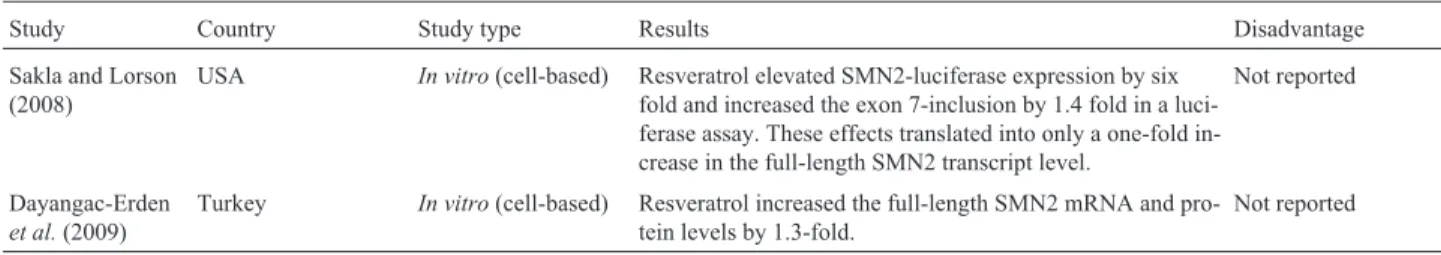
Study Country Study type Results Disadvantage
Sakla and Lorson (2008)
USA In vitro(cell-based) Resveratrol elevated SMN2-luciferase expression by six fold and increased the exon 7-inclusion by 1.4 fold in a luci-ferase assay. These effects translated into only a one-fold in-crease in the full-length SMN2 transcript level.
Not reported
Dayangac-Erden
et al.(2009)
Turkey In vitro(cell-based) Resveratrol increased the full-length SMN2 mRNA and pro-tein levels by 1.3-fold.
Relevance for treatment of spinal muscular atrophy. Eur J Hum Genet 12:59-65.
Avila AM, Burnett BG, Taye AA, Gabanella F, Knight MA, Hartenstein P, Cizman Z, Di Prospero NA, Pellizzoni L, Fischbeck KH,et al.(2007) Trichostatin A increases SMN expression and survival in a mouse model of spinal muscular atrophy. J Clin Invest 117:659-671.
Brahe C, Vitali T, Tiziano FD, Angelozzi C, Pinto AM, Borgo F, Moscato U, Bertini E, Mercuri E and Neri G (2005) Phenyl-butyrate increases SMN gene expression in spinal muscular atrophy patients. Eur J Hum Genet 13:256-259.
Brichta L, Hofmann Y, Hahnen E, Siebzehnrubl FA, Raschke H, Blumcke I, Eyupoglu IY and Wirth B (2003) Valproic acid increases the SMN2 protein level: A well-known drug as a potential therapy for spinal muscular atrophy. Hum Mol Genet 12:2481-2489.
Brichta L, Holker I, Haug K, Klockgether T and Wirth B (2006)In vivoactivation of SMN in spinal muscular atrophy carriers and patients treated with valproate. Ann Neurol 59:970-975. Brichta L, Garbes L, Jedrzejowska M, Grellscheid SN, Holker I,
Zimmermann K and Wirth B (2008) Nonsense-mediated messenger RNA decay of survival motor neuron 1 causes spinal muscular atrophy. Hum Genet 123:141-153. Burglen L, Lefebvre S, Clermont O, Burlet P, Viollet L, Cruaud C,
Munnich A and Melki J (1996) Structure and organization of the human survival motor neurone (SMN) gene. Genomics 32:479-482.
Burlet P, Burglen L, Clermont O, Lefebvre S, Viollet L, Munnich A and Melki J (1996) Large scale deletions of the 5q13 re-gion are specific to Werdnig-Hoffmann disease. J Med Genet 33:281-283.
Clermont O, Burlet P, Burglen L, Lefebvre S, Pascal F, McPher-son J, Wasmuth JJ, Cohen D, Le Paslier D, Weissenbach J,
et al.(1994) Use of genetic and physical mapping to locate the spinal muscular atrophy locus between two new highly polymorphic DNA markers. Am J Hum Genet 54:687-694. Darbar IA, Plaggert PG, Resende MBD, Zanoteli E and Reed UC
(2011) Evaluation ofmuscle strength and motor abilities in children with type II and III spinal muscle atrophy treated with valproic acid. BMC Neurology11:36.
Dayangac-Erden D, Topaloglu and Erdem-Yurter H (2008) A pre-liminary report on spinal muscular atrophy lymphoblastoid cell lines: Are they an appropriate tool for drug screening? Adv Ther 25:274-279.
Dayangac-Erden D, Bora-Tara G, Ayhan P, Kocaefe C, Dalkara S, Yelekc IK, Erdem-Yurter H and Demir AS (2009) Histone deacetylase inhibition activity and molecular dock-ing of (E)-resveratrol: Its therapeutic potential in spinal muscular atrophy. Chem Biol Drug Des73:355-364. Dayangac-Erden D, Bora-Tatar G, Dalkara S, Demir AS and
Erdem-Yurter H (2011) Carboxylic acid derivatives of his-tone deacetylase inhibitors induce full length SMN2 tran-scripts: A promising target for spinal muscular atrophy ther-apeutics. Arch Med Sci 7:230-234.
Feldkotter M, Schwarzer V, Wirth R, Wienker TF and Wirth B (2002) Quantitative analyses of SMN1 and SMN2 based on real-time lightCycler PCR: Fast and highly reliable carrier testing and prediction of severity of spinal muscular atrophy. Am J Hum Genet 70:358-368.
Garbes L, Riessland M, Holker I, Heller R, Hauke J, Trankle C, Coras R, Blumcke I, Hahnen E and Wirth B (2009) LBH589
induces up to 10-fold SMN protein levels by several inde-pendent mechanisms and is effective even in cells from SMA patients non-responsive to valproate. Hum Mol Genet 18:3645-3658.
Grayson DR, Kundakovic M and Sharma RP (2010) Is there a fu-ture for histone deacetylase inhibitors in the pharmacothe-rapy of psychiatric disorders? Mol Pharmacol 77:126-135. Hahnen E, Eyupoglu IY, Brichta L, Haastert K, Trankle C,
Sieb-zehnrubl FA, Riessland M, Holker I, Claus P, Romstock J,et al. (2006) In vitro and ex vivo evaluation of second-generation histone deacetylase inhibitors for the treatment of spinal muscular atrophy. J Neurochem 98:193-202. Harahap ISK, Saito T, San LP, Sasaki N, Gunadi, Nurputra DKP,
Yusoff S, Yamamoto T, Morikawa S, Nishimura N,et al.
(2011) Valproic acid increases SMN2 expression and modu-lates SF2/ASF and hnRNPA1 expression in SMA fibroblast cell lines. Brain Dev 34:213-222.
Hauke J, Riessland M, Lunke S, Eyupoglu IY, Blumcke I, El-Osta A, Wirth B and Hahnen E (2009) Survival motor neuron gene 2 silencing by DNA methylation correlates with spinal muscular atrophy disease severity and can be bypassed by histone deacetylase inhibition. Hum Mol Genet 18:304-317. Kernochan LE, Russo ML, Woodling NS, Huynh TN, Avila AM,
Fischbeck KH and Sumner CJ (2005) The role of histone acetylation in SMN gene expression. Hum Mol Genet 14:1171-1182.
Kissel JT, Scott CB, Reyna SP, Crawford TO, Simard LR, Kros-schell KJ, Acsadi G, Elsheik B, Schroth MK, D’Anjou G,et al.(2011) SMA CARNIVAL Trial Part II: A prospective, single-armed trial of L-carnitine and valproic acid in ambu-latory children with spinal muscular atrophy. PLoS One 6:e21296.
Lackmann GM (2004) Valproic-acid-induced thrombocytopenia and hepatotoxicity: Discontinuation of treatment? Pharma-cology 70:57-58.
Lefebvre S, Burglen L, Reboullet S, Clermont O, Burlet P, Viollet L, Benichou B, Cruaud C, Millasseau P, Zeviani M,et al.
(1995) Identification and characterization of a spinal muscu-lar atrophy-determining gene. Cell 80:155-165.
Li Y, Shin D and Kwon SH (2013) Histonedeacetylase6 plays a role as a distinctregulator of diversecellular processes. FEBS J 280:775-793.
Monani UR, Pastore MT, Gavrilina TO, Jablonka S, Le TT, Andreassi C, DiCocco JM, Lorson C, Androphy EJ, Send-tner M,et al.(2003) A transgene carrying an A2G missense mutation in the SMN gene modulates phenotypic severity in mice with severe (type I) spinal muscular atrophy. J Cell Biol 160:41-52.
Nakagawa T and Guarente L (2011) Sirtuins at a glance. J Cell Sci 124:833-838.
Narver HL, Kong L, Burnett BG, Choe DW, Bosch-Marce M, Taye AA, Eckhaus MA and Sumner CJ (2008) Sustained improvement of spinal muscular atrophy mice treated with trichostatin A plus nutrition. Ann Neurol 64:465-470. Piepers S, Cobben JM, Sodaar P, Jansen MD, Wadman RI,
Prior TW, Swoboda KJ, Scott HD and Hejmanowski AQ (2004) Homozygous SMN1 deletions in unaffected family mem-bers and modification of the phenotype by SMN2. Am J Med Genet 130A:307-10.
Rak K, Lechner BD, Schneider C, Drexl H, Sendtner M and Jablonka S(2009) Valproic acid blocks excitability in SMA type I mouse motor neurons. Neurobiol Dis 36:477-487. Riessland M, Brichta L, Hahnen E and Wirth B(2006) The
benza-mide M344, a novel histone deacetylase inhibitor, signifi-cantly increases SMN2 RNA/protein levels in spinal muscu-lar atrophy cells. Hum Genet 120:101-110.
Riessland M, Ackermann B, Forster A, Jakubik M, Hauke J, Garbes L, Fritzsche I, Mende Y, Blumcke I, Hahnen E,et al.
(2010) SAHA ameliorates the SMA phenotype in two mou-se models for spinal muscular atrophy. Hum Mol Genet 19:1492-1506.
Riester D, Hildmann C and Schwienhorst A (2007) Histone dea-cetylase inhibitors - Turning epigenic mechanisms of gene regulation into tools of therapeutic intervention in malignant and other diseases. Appl Microbiol Biotechnol 75:499-514. Sakla M, Sand Lorson CL (2008) Induction of full-length survival motor neuron by polyphenol botanical compounds. Hum Genet 122:635-643.
Simic G (2008) Pathogenesis of proximal autosomal recessive spinal muscular atrophy. Acta Neuropathol 116:223-234. Sumner CJ, Huynh TN, Markowitz JA, Perhac JS, Hill B, Coovert
DD, Schussler K, Chen X, Jarecki J, Burghes AH,et al.
(2003) Valproic acid increases SMN levels in spinal muscu-lar atrophy patient cells. Ann Neurol 54:647-654.
Swoboda KJ, Scott CB, Reyna SP, Prior TW, LaSalle B, Sorenson SL, Wood J, Acsadi G, Crawford TO, Kissel JT,et al.(2009) Phase II open label study of valproic acid in spinal muscular atrophy. PLoS One 4:e5268.
Swoboda KJ, Scott CB, Crawford TO, Simard LR, Reyna SP, Krosschell KJ, Acsadi G, Elsheik B, Schroth MK, D’Anjou
G,et al.(2010) SMA CARNI-VAL Trial Part I: Double-blind, randomized, placebo-controlled trial of L-carnitine and valproic acid in spinal muscular atrophy. PLoS One 5:e12140.
Tong V, Teng XW, Chang TK and Abbott FS (2005) Valproic acid II: Effects on oxidative stress, mitochondrial membrane potential, and cytotoxicity in glutathione-depleted rat hepa-tocytes. Toxicol Sci 86:436-443.
Wadman RI, Bosboom WM, van den Berg LH, Wokke JH, Ian-naccone ST and Vrancken AF (2012a) Drug treatment for spinal muscular atrophy type I. Cochrane Database Syst Rev CD006281.
Wadman RI, Bosboom WM, van den Berg LH, Wokke JH, Ian-naccone ST and Vrancken AF (2012b) Drug treatment for spinal muscular atrophy types II and III. Cochrane Database Syst Rev CD006282.
Xu WS, Parmigiani RB and Marks PA (2007) Histone deacetylase inhibitors: Molecular mechanisms of action. Oncogene 26:5541-5552.
Internet Resources
ClinicalTrials.gov, http://www.clinicaltrials.gov/ (22 December, 2012).
PubChem Substance database,
http://www.ncbi.nlm.nih.gov/pcsubstance (22 December, 2012).
Selleck Chemicals website, http://www.selleckchem.com (24 April, 2013).
Associate Editor: Maria Rita Passos-Bueno