Vol.54, n. 6: pp. 1159-1165, November-December 2011
ISSN 1516-8913 Printed in Brazil BRAZILIAN ARCHIVES OF
BIOLOGY AND TECHNOLOGY
A N I N T E R N A T I O N A L J O U R N A L
The Cytogenetic Effects of Black Tea and Green Tea on
Cultured Human Lymphocytes
Halil Erhan Ero
ğ
lu
*Department of Biology; Faculty of Science and Art; Bozok University; 66200; Yozgat - Turkey
ABSTRACT
In this study, the cytogenetic effects of black tea and green tea were determined in cultured peripheral blood lymphocytes. Results showed that black tea and green tea induced the mitotic and replication indexes and decreased micronuclei. But these data were not statistically significant for green tea. The effects of black tea on the micronucleus formation and mitotic index were statistically significant. The decrease in micronucleus counts indicated that black tea and green tea had considerable anticlastogenic and antigenotoxic effects as observed in vitro in human lymphocytes. Thus, it could be concluded that tea polyphenols protected the normal cells from genotoxic or carcinogenic agents, which indicated the therapeutic and antioxidative role of catechins, flavonoids or other tea compounds.
Key words: Tea; catechins, micronucleus, mitotic index, replication index
*Author for correspondence: [email protected]
INTRODUCTION
Tea (Camellia sinensis L.) is an evergreen shrub of the Theaceae family. Tea plant is native to South East Asia but is currently cultivated in more than 30 countries. According to its processing, tea can be classified in to the three major forms. These forms are black tea (fully aerated or fermented), green tea (unaerated or unfermented) and oolong tea (partially aerated or semi-fermented) (Ratnasooriya and Fernando, 2008). Of the tea produced worldwide, 78% is black tea, which is usually consumed in the Western countries, 20% is green tea, which is commonly consumed in Asian countries, and 2% is oolong tea which is produced mainly in southern China (Ju et al., 2007; Khan and Mukhtar, 2007). Green tea is produced by steaming or pan-frying the fresh tea leaves, which
inactivate the enzymes and prevents the oxidation of tea constituents. Oolong tea is a partially fermented tea and has the flavor and health characteristics of both the green and black teas (Sharangi, 2009).
(ECG), and epigallocatechin gallate (EGCG) (Alexis et al., 1999). The usual concentration of total polyphenols in dried green tea leaves is about 8 to 12% (Min and Peigen, 1991; Graham, 1992). The approximate mean percentages of components of solid extracts in the black tea are catechins (10-12%), theaflavins (3-6%), thearubigins (12-18%), flavonols (6-8%), phenolic acids (10-12%), amino acids (13-15%), methylxanthines (8-11%), carbohydrates (15%), proteins (1%), mineral matter (10%), and volatiles (<0.1%).
There have been descriptions of various biological properties of tea, including antibacterial (Nance and Shearer, 2003; Bandyopadhyay et al., 2005), antiviral (Nakayama et al., 1993; Yamamoto et al.,
1997), antioxidative (Matsuzaki and Hara, 1985; Mukhtar and Ahmad, 2000; Erba et al., 2005; Frei and Higdon, 2003), anti-inflammatory (Alexis et al., 1999) antitumor (Katiyar et al., 1993), antimutagenic (Constable et al., 1996; Kuroda, 1996; Yen and Chen, 1996; Kennedy et al., 1998) and anticarcinogenic (Lambert et al., 2005; Khan et al., 2006; Lu et al., 2006; Siddiqui et al., 2006) activities. Health benefits of tea consumption on human have been determined. Tea reduces the risk of cardiovascular diseases, treats respiratory diseases, corrects skin disorder, aids in indigestion, prevents diabetes, improves oral health, keeps away from liver disease, and gives a boost to immunity, treats arthritis, burns fat (Sharangi, 2009).
The micronucleus (MN), mitotic index (MI), and replication index (RI) analysis methods are cytogenetic tests that are used both in vivo and in vitro. Micronuclei may originate from the acentric fragments (chromosome fragments lacking a centromere), or whole chromosomes that are unable to migrate with the rest of the chromosomes during anaphase in cell division. The MI, or the percentage of metaphases among the harvested, fixed lymphocytes requires the addition of colchicine, or colcemid to arrest the progression of the cells from the metaphase to anaphase, ensuring a sufficient number of metaphases for the cytogenetic analysis. The RI measures the cell division kinetics by counting the percentage of the cells in the first, second, third or more metaphase (Ozkul et al., 2005).
In this study, the in vitro cytogenetic effects of the black tea and green tea were investigated in the cultured human lymphocytes.
MATERIAL AND METHODS
Preparation of aqueous extracts
The black tea and green tea samples (50, 100, 200, and 400 mg) were added to 100 mL of deionised water and boiled for 5 min. The extracts were allowed to cool. Both the preparations were sterilized through a 0.22 m filter and stored +4ºC until tested. The concentrations (0.5, 1, 2, and 4 mg/mL) of the black tea and green tea used in this study were a result of trial and error.
Chemicals
Peripheral blood (PB) karyotyping medium (Biological Industries, Israel), colcemid (Sigma, Germany) and giemsa stain (Merck, Germany) were used in peripheral blood cultures. PB karyotyping medium was composed of RPMI-1640 basal medium supplemented with L -glutamine, fetal bovine serum, antibiotics (gentamycin) and phytohemagglutinin (PHA).
In vitro mitotic index assay
slide. The slides were air dried and stained with 5% Giemsa.
The MI was calculated as the proportion of metaphase for 1000 cells in each donor and concentration (total counted cells = 4000).
In vitro micronucleus assay
For the MN analysis, the peripheral lymphocytes were incubated at 37ºC for 72 h. The cells were treated with the black tea and green tea extracts at concentrations of 0.5, 1, 2, and 4 mg/mL. For RI, 5-bromo-2-deoxyuridine (BrdU, 10 g/mL, Sigma) was added at the initiation of the cultures. For MN, Cytochalasin B (Sigma) was added at 44 h of the incubation at a final concentration of 5 µg/mL to block the cytokinesis. At the end of the incubation at 37ºC, the cells were harvested by centrifugation. The MN staining was performed according to Ozkul et al., (2005).
The slides were scored by a single observer. Five hundred cells were examined at 600x magnification from each slide and when micronucleated binucleate cells were located, they were examined at 1000x magnification.
The RI was calculated for 500 cells per culture according to the following formula: RI = (1 x M1 + 2 x M2 + 3 x M3)/500, where M1, M2, and M3 stood for the number of cells in the first
metaphase, second metaphase and third or more metaphases, respectively (Tuylu et al., 2007).
Statistical analysis
The computer software program SPSS 10.0 was used to analyze the data. The statistical significance of the effects of the extracts of the black tea and green tea on the MN formation, mitotic division, and nuclear division was assessed using the repeated measures of the analysis of variance (ANOVA) and the differences between the groups were determined by the least significant differences (LSD) test with p < 0.05 and p < 0.01 was considered significant.
RESULTS
Micronucleus
The results of the MN test are given in Figure 1. The MN rates were decreased by the extracts of both black tea and green tea. When the MN formation was analyzed after the treatment of the black tea with different extract concentrations, significant changes were detected in the percentage of MN for all the concentrations (p < 0.05). The extracts of green tea did not afford any protection against MN formation (p = 0.05).
Mitotic index
The results of the MI test are given in Table 1. Although both the black tea and green tea extracts increased the MI rates, these increases were not significant for the green tea. When the extracts in the lymphocyte cultures were analyzed through the
MI evaluation, a significant increase was found at 2 and 4 mg/mL concentrations for the black tea (p
< 0.01). The mean values of the MI rates were between 2.92 ± 0.27 – 3.27 ± 0.35 for the black tea and 1.95 ± 0.43 – 2.12 ± 0.30 for thegreen tea.
Table 1 - Mitotic index (%) (mean ± SDs) in human lymphocyte cultures exposed to extracts of black tea and green tea.
Species Concentrations (mg/mL) Total number: dividing cells Mean ± SDs (%)
Control 90 2.25 ± 0.55
0.5 117 2.92 ± 0.27
1 114 2.85 ± 0.45
2 131 3.27 ± 0.35 *
Black Tea
4 128 3.20 ± 0.45 *
Control 64 1.60 ± 0.24
0.5 78 1.95 ± 0.43
1 82 2.05 ± 0.35
2 79 1.97 ± 0.50
Green Tea
4 85 2.12 ± 0.30
ANOVA: * p < 0.01 (significantly different from control)
Replication index
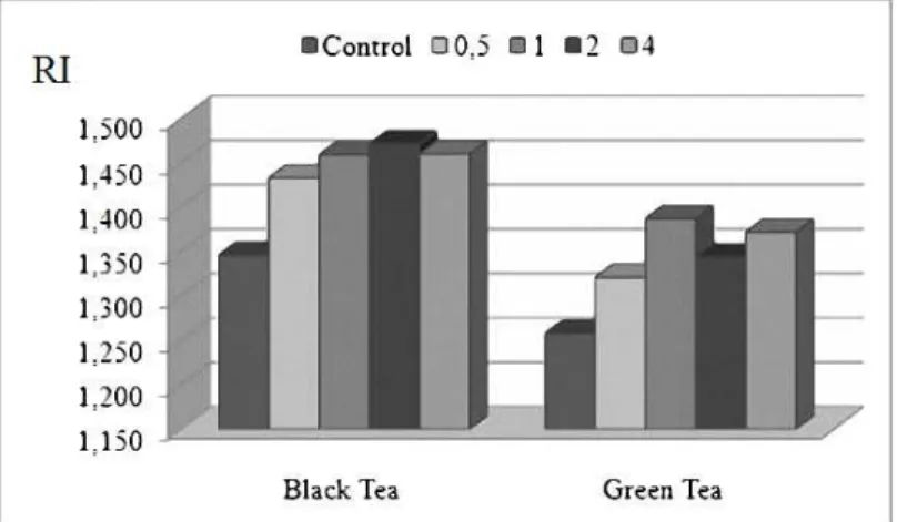
All the tested concentrations of black and green tea did not lead to a marked decrease in the RI in comparison with the control group. As shown in
Figure 2, increasing extract concentrations of the teas increased the RI. But these increases were not statistically significant (p = 0.05).
Figure 2 - Replication index changes in the lymphocyte, according to the control and treatment with different concentrations of the green tea and black tea. The RI rates were increased by the extracts of both the black tea and green tea. But these increases were not statistically significant. The RI rates of control and black tea extracts (0.5, 1, 2, and 4 mg/mL) were 1.346, 1.433, 1.459, 1.471, and 1.460, respectively. The RI rates of the control and green tea extracts (0.5, 1, 2, and 4 mg/mL) were 1.258, 1.321, 1.387, 1.344, and 1.372, respectively. The most RI values were 2 mg/mL concentration for the black tea and 1 mg/mL concentration for the green tea.
DISCUSSION
Tea is a pleasant, popular, socially accepted, economical and safe drink that is enjoyed every
to human health. In the present study, genotoxic and anticlastogenic effects of the black tea and green tea were investigated in cultured human lymphocytes.
The peripheral lymphocytes are the best materials for the determination of cytogenetic effects. The MN technique has been proposed as a useful tool for the measurement of genotoxicity in vivo and in vitro cultures. The MN arises during the cell division either from the chromosomes that lag in anaphase, or from the chromosome fragments (Fenech and Morley, 1985). In the living creatures, which are exposed to a mutagen factor, the probability of formation of mitotic and meiotic defects is increased and the rate of MN could increase due to this increase (Ramalho 1988). Alcohol consumption, smoking and viral infections increase the MN rates in peripheral blood lymphocytes (Seitz 1998). The donors chosen for this study did not smoke or consume alcohol. They had not been exposed to X-ray and gamma-ray and they did not have any viral infections. According to the results, the extracts of both the black tea and green tea decreased MN rates (Fig. 1). The decrease in the MN indicated that the black tea and green tea were not genotoxic and clastogenic agents. The antigenotoxic and anticlastogenic properties of the teas might be due to the catechins (polyphenols) present in the tea. Many studies have demonstrated that tea catechins could suppress the genotoxic activity of various carcinogens with both in vitro and in vivo systems (Kuroda, 1996; Sinha et al., 2005; Isbrucker et al.,
2006). Chang et al., (2003) have shown that there is minimal genotoxic concern with a decaffeinated green tea catechin mixture.
The antigenotoxic and anticlastogenic activities of the tea are mostly due to its antioxidant activity that inactivates the direct carcinogens. The antioxidant property has been highly attributed to the polyphenolic compounds in the tea. Catechins and flavonoids from the polyphenols are primarily responsible for the beneficial healthful properties of the tea. The flavonoids have antioxidant, anti-inflammatory, antiallergic and antimicrobial effects (Venditti et al., 2010; Nie and Xie, 2011). Tea catechins have been found to be better antioxidants than the vitamins C and E, tocopherol and carotene (Sharangi, 2009). Tea contains six primary catechin compounds, namely catechin, gallocatechin (GC), EC, EGC, ECG, and EGCG, the latter being the most active component. Many studies have confirmed the free radicals
scavenging activity of catechins in vitro and in vivo (Matsuzaki and Hara, 1985; Mukhtar and Ahmad, 2000; Frei and Higdon, 2003). It has been reported that the green tea consumed within a balanced controlled diet improves the overall antioxidative status and protects against the oxidative damage in the humans (Erba et al.,
2005). A significant rise in the plasma antioxidant capacity was detected after brewed green tea, or black tea solids were consumed (Leenen et al.,
2000). Besides, the most active tea component (EGCG) possesses significant cancer chemopreventive activity (Katiyar and Mukhtar, 1996). It elicits a variety of cellular and molecular responses (Roy et al., 2001) which include antimutagenic activity (Kuroda and Hara, 1999), suppression of oxidative DNA damage (Chung, 1999) and induction of the apoptosis in the tumor cells (Yokoyama et al., 2001).
The MI and RI are used as indicators of adequate cell proliferation biomarkers. The MI measures the proportion of the cells in the M-phase of the cell cycle and its inhibition could be considered as cellular death, or delay in the cell proliferation kinetics (Rojas, 1993). The present results also showed that the MI and RI values of the extracts were higher than in the controls. This could be the reason that many cells survived the first cell cycle in the culture to inhibit the MN, as they would have not entered a process of necrosis, or apoptosis before this event. A negative correlation was observed between the MN induction and cell proliferation; namely the lower the MN frequencies were detected in exposed cells, the higher the values of nuclear division progression were expressed as RI. This could mean that the cells, not exposed to chromosomal damage, might live.
REFERENCES
Alexis, A. F., Jones. V. A. and Stiller, M. J. (1999), Potential therapeutic applications of tea in dermatology. Int. J. Dermatol., 38, 735–743.
Bandyopadhyay, D., Chatterjee, T. K., Dasgupta, A., Lourduraja, J. and Dastidar, S. G. (2005), In vitro and in vivo antimicrobial action of tea: the commonest beverage of Asia. Biol. Pharm. Bull., 28, 2125–2127. Chang, P. Y., Mirsalis, J., Riccio, E. S., Bakke, J. P.,
Lee, P. S., Shimon, J., Phillips, S., Fairchild, D., Hara, Y. and Crowell, J. A. (2003), Genotoxicity and toxicity of the potential cancer-preventive agent polyphenon E. Environ. Mol. Mutagen., 41, 43–54. Chung, F. L. (1999), The prevention of lung cancer
induced by a tobacco-specific carcinogen in rodents by green and black tea. Proc. Soc. Exp. Biol. Med., 4, 244–248.
Constable, A., Varga, N., Richoz, J. and Stadler, R. H. (1996), Antimutagenicity and catechin content of soluble instant teas. Mutagenesis, 11, 189–194. Erba, D., Riso, P., Bordoni, A., Foti, P., Biagim, P. L.
and Testolin, G. (2005), Effectiveness of moderate green tea consumption on antioxidative status and plasma lipid profile in humans. J. Nutr. Biochem., 16, 144–149.
Fenech, M. and Morley, A. (1985), Measurement of micronuclei in lymphocytes. Mutat. Res., 147, 29–36. Frei, B. and Higdon, J. V. (2003), Antioxidant activity
of tea polyphenols in vivo: Evidence from animal studies. J. Nutr., 133, 3275–3284.
Graham, H. N. (1992), Green tea composition, consumption, and polyphenol chemistry. Prev. Med., 21, 334–350.
Isbrucker, R. A., Bausch, J., Edwards, J. A. and Wolz, E. (2006), Safety studies on epigallocatechin gallate (EGCG) preparations Part 1: Genotoxicity. Food Chem. Toxicol., 44, 626–635.
Ju, J., Lu, G., Lambert, J. D. and Yang, C. S. (2007), Inhibition of carcinogenesis by tea constituents. Semin Cancer Biol., 17, 395–402.
Katiyar, S. K., Agarwal, R., Zaim, M. T. and Mukhtar, H. (1993), Protection against N-nitrosodiethylamine and benzo[a]pyrene-induced forestomach and lung tumorigenesis in A/J mice by green tea. Carcinogenesis, 14, 849–855.
Katiyar, S. K. and Mukhtar, H. (1996), Tea in chemoprevention of cancer: epidemiologic and experimental studies. Int. J. Oncol., 8, 221–238. Kennedy, D. O., Nishimura, S., Hasuma, T., Yano, Y.,
Otani, S. and Matsui-Yuasa, I. (1998), Involvement of protein tyrosine phosphorylation in the effect of green tea polyphenols on Ehrlich ascites tumor cells in vitro. Chem-Biol. Interact., 110, 59–172.
Khan, N., Afaq, F., Saleem, M., Ahmad, N. and Mukhtar, H. (2006), Targeting multiple signaling pathways by green tea polyphenol (−)-epigallocatechin-3-gallate. Cancer Res., 66, 2500– 2505.
Khan, N. and Mukhtar, H. (2007), Tea polyphenols for health promotion. Life Sci., 81, 519–533.
Kuroda, Y. (1996), Bio-antimutagenic activity of green tea catechins in cultured Chinese hamster V79 cells. Mutat. Res., 36, 1179–1186.
Kuroda, Y. and Hara, Y. (1999), Antimutagenic and anticarcinogenic activity of tea polyphenols. Mutat. Res., 436, 69–97.
Lambert, J. D., Hong, J., Yang, G. Y., Liao, J. and Yang, C. S. (2005), Inhibition of carcinogenesis by polyphenols: evidence from laboratory investigations. Am. J. Clin. Nutr., 81, 284–291.
Leenen, R., Roodenburg, A., Tijburg, L. and Wiseman, S. A. (2000), A single dose of tea with or without milk increases plasma antioxidant activity in humans. Eur. J. Clin. Nutr., 54, 87–92.
Lu, G., Liao, J., Yang, G., Reuhl, K. R., Hao, X. and Yang, C. S. (2006), Inhibition of adenoma progression to adenocarcinoma in a 4-(methylnitrosamino)-1-(3-pyridyl)-1-butanone-induced lung tumorigenesis model in A/J mice by tea polyphenols and caffeine. Cancer Res., 66, 11494–11501.
Matsuzaki, T. and Hara, Y. (1985), Antioxidative activity of tea leaf catechins. Nippon Kog. Kaish., 59, 129–134.
Min, Z. and Peigen, X. (1991), Quantitative analysis of the active constituents in green tea. Phytother. Res., 5, 239–240.
Mukhtar, H. and Ahmad, N. (2000), Tea polyphenols: prevention of cancer and optimizing health. Am. J. Clin. Nutr., 71, 1698–1702.
Nakayama, M., Suzuki, K., Toda, M., Okubo, S., Hara, Y. and Shimamura, T. (1993), Inhibition of the infectivity of influenza virus by tea polyphenols. Antivir. Res., 21, 289–299.
Nance, C. L. and Shearer, W. T. (2003), Is green tea good for HIV-1 infection? J. Allergy Clin. Immun., 112, 851–853.
Nie, S. P. and Xie, M. Y. (2011), A review on the isolation and structure of tea polysaccharides and their bioactivities. Food Hydrocolloid., 25,144–149 Ozkul, Y., Silici, S. and Eroglu, E. (2005), The
anticarcinogenic effect of propolis in human lymphocytes culture. Phytomedicine12, 742–747. Ramalho, A. (1988), Use of the frequencies of
Ratnasooriva, W. D. and Fernando, T. S. P. (2008), Effect of black tea brew of Camellia sinensis on sexual competence of male rats. J. Ethnopharmacol., 118, 373–377.
Rojas, E. (1993), Mitotic index and cell proliferation kinetics for the identification of antineoplastic activity. Anti-Cancer Drug., 4, 637–640.
Roy, M., Siddiqi, M. and Bhattacharya, R. K. (2001), Cancer chemoprevention: tea polyphenol induced cellular and molecular responses. Asian Pac. J. Cancer Prev., 2, 109–116.
Seitz, H. K. (1998), Alcohol and cancer. Recent Dev. Alcohol., 14, 67–95.
Sharangi, A. B. (2009), Medicinal and therapeutic potentialities of tea (Camellia sinensis L.). Food Res. Int., 42, 529–535.
Siddiqui, I. A., Adhami, V. M., Saleem, M. and Mukhtar, H. (2006), Beneficial effects of tea and its polyphenols against prostate cancer. Mol. Nutr. Food Res., 50, 130–143.
Sinha, D., Bhattacharya, R. K., Siddiqi, M. and Roy, M. (2005), Amelioration of sodium arsenite-induced clastogenicity by tea extracts in Chinese hamster v79 cells. J. Environ. Pathol. Tox., 24, 129–140.
Tuylu, B. A., Zeytinoglu, H. S. and Isikdag, I. (2007), Synthesis and mutagenicity of 2-aryl-substitute (o-hydroxy-, m-bromo-, o-methoxy-, o-nitro-phenyl or 4–pyridyl) benzothiazole derivatives on Salmonella typhimurium and human lymphocytes exposed in vitro. Biologia, 62, 626–632.
Venditti, E., Bacchetti, T., Tiano, L., Carloni, P., Greci, L. and Damiani, E. (2010), Hot vs. cold water steeping of different teas: Do they affect antioxidant activity? Food Chem., 119, 1597–1604
Yamamoto, T., Juneja, L. R., Chu, D. C. and Kim, M. (1997), Chemistry and applications of green tea. CRC Press, LLC, Florida.
Yen, G. C. and Chen, H. Y. (1996), Relationship between antimutagenic activity and major components of various teas. Mutagenesis, 11, 37–41.
Yokoyama, S., Hirano, H., Wakimaru, N., Sarker, K. P. and Kuratsu, J. (2001). Inhibitory effect of epigallocatechin gallate on brain tumor cell lines in vitro. Neuro-Oncology., 3, 22–28.