www.cbpv.com.br/rbpv
Immune response of calves inoculated with proteins of
Anaplasma marginale bound to an immunostimulant complex
Resposta imune de bezerros inoculados com proteínas de
Anaplasma marginale
ligadas a um complexo imunoestimulante
Marcela Ribeiro Gasparini1; Rafael Felipe da Costa Vieira2; Denise Amaral Gomes do Nascimento1; João Luis Garcia1; Odilon Vidotto1; Marilda Carlos Vidotto1*
1Department of Preventive Veterinary Medicine, Londrina State University – UEL, Londrina, PR, Brazil
2Department of Veterinary Sciences, Federal University of Paraiba – UFPB, Campus II, Areia, PB, Brazil
Received October 1, 2012 Accepted May 16, 2013
Abstract
Despite our current knowledge of the immunology, pathology, and genetics of Anaplasma marginale, prevention in
cattle is currently based on old standbys, including live attenuated vaccines, antibiotic treatment, and maintaining enzootic stability in cattle herds. In the present study, we evaluated the use of an immunostimulant complex (ISCOMATRIX) adjuvant, associated with a pool of recombinant major surface proteins (rMSP1a, rMSP1b, rMSP4 and rMSP5) to improve the humoral immune response triggered in calves mainly by IgG2. Ten calves were divided in three groups: 4 calves were inoculated with the ISCOMATRIX/rMSPs (G1); 2 calves were inoculated with ISCOMATRIX adjuvant (G2); and 4 calves received saline (G3). Three inoculations were administered at 21-day intervals. In G1, the calves showed significant increases in total IgG, IgG1 and IgG2 levels 21 days after the second inoculation, compared to the control group (p < 0.05), and G1 calves remained above the cut-off value 28 days after the third inoculation (p < 0.05). The post-immunized sera from calves in G1 reacted specifically for each of the rMSPs used. In conclusion, the ISCOMATRIX/rMSPs induced antigen-specific seroconversion in calves. Therefore, additional testing to explore the protection induced by rMSPs, both alone and in conjunction with proteins previously identified as subdominant epitopes, is warranted.
Keywords: Bovine anaplasmosis, vaccine, MSP1a, MSP1b, MSP4, MSP5.
Resumo
Apesar dos avanços da imunologia, patologia e genética de Anaplasma marginale, a prevenção em bovinos ainda é baseada nas vacinas vivas atenuadas, na terapia com antibiótico e estabilidade enzoótica dos rebanhos bovinos. No presente estudo, avaliou-se o uso de um complexo imunoestimulante (ISCOMATRIX), associado às proteínas recombinantes de superfície (rMSP1a, rMSP1b, rMSP4 e rMSP5) para melhorar a resposta imune humoral desencadeada em bezerros, principalmente por IgG2. Dez animais foram divididos em três grupos: 4 bezerros foram inoculados com o ISCOMATRIX/rMSPs (G1), 2 bezerros foram inoculados com ISCOMATRIX adjuvante (G2) e 4 bezerros receberam salina (G3). Três doses vacinais foram administradas em intervalos de 21 dias. No G1, os bezerros apresentaram aumentos significativos nos níveis de IgG total, IgG1 e IgG2 21 dias após a segunda inoculação, em comparação com o grupo de controle (p <0,05). Nos bezerros do G1 esses níveis de anticoprpos permaneceram acima do ponto de corte 28 dias após a terceira inoculação (p < 0,05). Os soros pós-imunização de bezerros do G1 reagiram especificamente com cada uma das rMSPs utilizadas. Em conclusão, o ISCOMATRIX/rMSPs induziu soroconversão antígeno-específica em bezerros. Portanto, se justifica a realização de ensaios adicionais para explorar a proteção induzida pela rMSPs, tanto sozinhas como em conjunto com novas proteínas identificadas com epitopos subdominantes.
Palavras-chave: Anaplasmose bovina, vacina, MSP1a, MSP1b, MSP4, MSP5.
*Corresponding author: Marilda Carlos Vidotto
Departamento de Medicina Veterinária Preventiva, Universidade Estadual de Londrina – UEL, Pr 445, Km 380, CEP 86051-990, Londrina, PR, Brasil
e-mail: [email protected]
Introduction
Anaplasma marginale, of the family Anaplasmataceae, is an obligate intraerythrocytic bacterium that causes bovine anaplasmosis (DUMLER et al., 2001). Transmission of A. marginale to cattle occurs biologically by ticks and mechanically by biting flies and by blood-contaminated fomites. Both male ticks and cattle hosts can become persistently infected with A. marginale, serving as reservoirs of infection (KOCAN et al., 2010). This disease is responsible for serious economic losses, mainly in tropical and subtropical regions, due to fever and hemolysis with associated severe anemia, which results in high rates of morbidity and mortality in susceptible animals (RICHEY; PALMER, 1990). The use of vaccines have been suggested as an alternative to control the disease; however, the commercially available vaccines consist of live or dead organisms, and the present limitations consist of the need for cryopreservation, insufficient efficacy, and the possibility of induction of iso-antibodies against erythrocytes that are transferred by colostrum, resulting in isoerythrolysis at birth (KUTTLER, 1984).
Six major surface proteins (MSPs) of A. marginale have been well characterized as MSP1a, MSP1b, MSP2, MSP3, MSP4, and MSP5 (PALMER et al., 1989; TEBELE et al., 1991; OBERLE et al., 1993; ALLEMAN; BARBET, 1996; VISSER et al., 1992). These proteins are responsible for the interaction of A. marginale with host cells, and they include adhesion proteins and MSPs from multigene families (PALMER et al., 1999, KOCAN et al., 2010). MSP1a, MSP4, and MSP5 are present and have been conserved in many Brazilian isolates of A. marginale (KANO et al., 2002),
suggesting their potential use as components for a recombinant protein vaccine for anaplasmosis.
Recently, in addition to the well-characterized major surface proteins MSP1 through MSP5, other proteins, classified as immunologically ‘subdominant’ antigens, have been identified as protective antigens ofthe type IV secretion system (TFSS) (LOPEZ et al., 2005, 2007). Sera from cattle experimentally and naturally infected with A. marginale have recognized recombinant VirB9, VirB10, and other sub-dominant antigens, which stimulate the production of IgG2, proliferation of T-lymphocytes and secretion gamma interferon (LOPEZ et al., 2007; ARAÚJO et al., 2008), suggesting the inclusion of these proteins in the development of vaccines against A. marginale.
The use of new generation vaccines, particularly those based on recombinant proteins and DNA, has demonstrated less adverse reactions compared to conventional vaccines; however, their immunogenic activity is reduced (SINGH; O’HAGAN, 2002). Preliminary studies, using recombinant plasmids of the PR1 strain of A. marginale (pcDNA-msp1a, pcDNA-msp1b,
pcDNA-msp4, and pcDNA-msp5), have demonstrated good immune
humoral and cellular responses in BALB/c mice (KANO et al., 2008). The association of these recombinant plasmids during the immunization of mice resulted in elevated antibody titers when analyzed by ELISA, reacted with the recombined proteins (rMSP1a, rMSP1b, and rMSP5) of A. marginale by Western blot, and induced strong proliferation of T lymphocytes (KANO et al., 2008). Additionally, the humoral immune response of BALB/c
mice to recombinant MSPs (rMSPs) (MSP1a, MSP1b, MSP4, and MSP5), incorporated into immunostimulant complex (ISCOM), induced the production of specific antibodies to each rMSP (KAWASAKI et al., 2007b). Thus, the aim of this study was to evaluate the humoral immune response in calves using adjuvant ISCOMATRIX and rMSPs (rMSP1a, rMSP1b, rMSP4 and rMSP5) of Anaplasma marginale (PR1 strain).
Materials and Methods
The study was approved by the Ethics Committee for Animal Experimentation and Animal Welfare at the Londrina State University, Paraná State, Brazil (CEEA number 47/05).
Expression of MSP genes and purification of
recombi-nant MSPs proteins
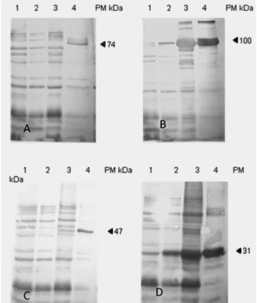
Escherichia coli BL21 Star (DE3) One Shot cells (Invitrogen Life Technologies, Sao Paulo, BRA) were transformed with the recombinant plasmids MSP1a, MSP1b, pET102-MSP4 and pET102-MSP5 to produce recombinant MSP1a and MSP1b (TAMEKUNI et al., 2009), MSP4 (KAWASAKI et al., 2007a) and MSP5 (MARANA et al., 2009) as a fusion product with 6His. The transformants were grown in Luria Bertani (LB) medium to an optical density (OD) of OD500nm 0.7, and Isopropil-thio-D-Galactoside (IPTG) was added to a final concentration of 1 mM. Bacteria were harvested at 5 h post-induction by centrifugation; pellets were re-suspended in lysis buffer, and expression was analyzed from soluble and insoluble fractions on 12% SDS-PAGE gels. The culture of induced bacteria was centrifuged and lysed by sonication. The suspension was then centrifuged and the supernatant utilized for protein purification using the Ni-NTA resin columns method (Qiagen, Valencia, CA, USA); protein concentrations were determined as previously described (BRADFORD, 1976). The induced rMSP1a and MSP1b proteins (74 kDa and 100 kDa) were purified under native conditions (TAMEKUNI et al., 2009), and MSP4 and MSP5 (47 kDa and 31 kDa) were purified as described by Kawasaki et al. (2007a).
ISCOMATRIX/MSP preparation
The ISCOMATRIX adjuvant was prepared and examined, using negative staining transmission electron microscopy, to confirm the formation of characteristic vesicles, as previously described (KAWASAKI et al., 2007b). ISCOMATRIX/rMSPs were prepared by a previously described technique (KAWASAKI et al., 2007b). The ISCOMATRIX/rMSPs were produced by the addition of 200 µg of each rMSP (rMSP1a, rMSP1b, rMSP4 and rMSP5)
(1:1) to the adjuvant.
Inoculation
The animals were kept in three separated cattle pens and were fed silage and concentrate twice per day and water ad libidum. The
to keep them free from ticks and biting flies. At the start of the experiment, all of the calves were approximately 240 days old and were determined to be free of A. marginale infection by PCR (LEW et al., 2002).
Ten calves were randomly divided into three groups: Four calves in group G1 were inoculated with the ISCOMATRIX/rMSPs, two calves in group G2 were inoculated with the ISCOMATRIX adjuvant, and four calves in group G3 were inoculated with saline. The inoculations were administered intramuscularly at days 0, 21, and 42.
Monitoring of the experimental groups
Before each inoculation, the animals were bled, the serum was separated, the rectal temperature was recorded, the packed cell volume (PCV) was determined, and the titer of antibodies was analyzed by indirect enzyme immunoassay (iELISA).
Antibody assays
Detection of anti-A. marginale total IgG, IgG1 and IgG2 antibodies was performed by iELISA. Optimal dilutions were established using checkerboard titrations, with dilutions of sera, antigen, and conjugates. Into each well of ELISA plates (Nunc-ImmunoTM Maxisorp, Nunc, Roskilde, Denmark) were added 100 µL of A. marginale (strain PR1) initial bodies (10 µg/mL)
diluted in sodium carbonate-bicarbonate buffer (0.05 M, pH 9.6). After overnight incubation at 4 °C, the plates were washed with PBS containing 0.05% Tween-20 (GE Healthcare, USA) (PBS-Tween pH 7.4). For total IgG, the plates were blocked for 1 hour at 37 °C using 200 µL of 8% non-fat dry milk and were
washed five times with PSB-Tween (pH 7.4) in an automatic microplate washer (Bio-Rad ImmunoWash, California, USA). For IgG1 and IgG2 antibodies, the plates were blocked using 200 µL of 8% horse serum. For total IgG, serum samples were
diluted (1:400) in PBS-Tween (pH 7.4) plus 5% rabbit normal sera, and 100 µL were added in duplicate to each well and then
incubated at 37 °C for 90 minutes. For IgG1 and IgG2 antibodies, serum samples were diluted (1:200) in PBS-Tween (pH 7.4) plus 5% horse serum, and 100 µL was added in duplicate to each well
and incubated at 37 °C for 1 hour. Positive and negative control sera were included in each plate. ELISA plates were washed five times with PSB-Tween (pH 7.4) in an automatic microplate washer (Bio-Rad ImmunoWash). Rabbit anti-bovine IgG alkaline phosphate conjugate (Sigma Aldrich Inc., St. Louis, MO, USA), diluted 1:20.000 in PBS-Tween (pH 7.4), was added (100 µL) to
each well and incubated at 37 °C for 90 minutes. For IgG1 and IgG2 antibodies, two secondary antibodies were used: horseradish peroxidase-labeled sheep anti-bovine IgG1 (BethylTM Laboratories, Montgomery, TX, USA) and sheep anti-bovine IgG2 (BethylTM Laboratories), diluted 1:10,000 and 1:15,000, respectively, in PBS-Tween (pH 7.4). Then, they were added (100 µL) to each
well and incubated at 37 °C for 1 hour. The plates were then washed five times with PBS-Tween (pH 7.4) in an automatic microplate washer (Bio-Rad ImmunoWash), and 100 µL of
o-phenylenediamine (OPD) (Sigma-Aldrich) solution was added
in a concentration of 0.4 mg/mL using appropriate diluents with hydrogen peroxide. The reaction was interrupted by adding 50 μL of 1N HCl, and the OD reading at 490 nm was obtained using an ELISA reader (iMarkTM Microplate Absorbance Reader, Bio-Rad Inc., Hercules, CA, USA).
The absorbance values were estimated, and the OD values were calculated as previously described (GARCIA et al., 2006). A serum was considered to be positive when OD sample > OD mean from negative control sera (n=10) ± 3 SDs (standard deviation) from the negative control.
Western blot analysis
The recombinant proteins rMSP1a, rMSP1b, rMSP4 and rMSP5 were electrophoresed and transferred, and the membranes were blocked, as previously described (KAWASAKI et al., 2007b). The membranes were incubated for 1 hour with a post-inoculation pool of serum from G1, diluted (1:500) in PBS-Tween plus 5% non-fat dry milk. The membranes were washed and incubated with peroxidase-labeled protein G (1:1,000) for 1 hour at room temperature. The peroxidase activity was demonstrated using 3,3’-diaminobenzidine (DAB) (ACROS-Organics, New Jersey, USA). Protein molecular weight markers (BenchMarkTM Invitrogen Life Technologies, Carlsbad, CA, USA) were used as standards.
Statistical analysis
The data were first tested for normality and homogeneity of variances, and if they did not present normal distribution, they were analyzed by nonparametric statistical tests. Differences between groups at each moment were verified by the Kruskal-Wallis test, followed by Dunn’s multiple comparison test. Statistical analysis was considered significant when p < 0.05. Data were analyzed using BioEstat software, version 5.0 (AYRES et al., 2007).
Results
Seroconversion
The data for total IgG obtained by iELISA are shown in Figure 1. Calves from G1 showed a significant increase in total IgG levels 21 days after the second inoculation, compared to the control group (p = 0.0368), and the levels remained above the cut-off value 28 days after the third inoculation (p = 0.0379). Animals from G2 and G3 presented median OD values for total IgG below the cut-off value until 28 days after the third inoculation (Figure 1).
The data for IgG2, obtained by indirect ELISA, are shown in Figure 2. Animals from G1 presented humoral responses 21 days after the second inoculation, compared to the control group (p = 0.0322), and remained above the cut-off value 28 days after the third inoculation (p = 0.0322). Calves from G2 and G3 presented median OD values for IgG2 below the cut-off value during the entire study (Figure 2).
Western blot analysis
Western blot results, using a pool of sera of calves from G1, are shown in Figure 3. The post-inoculation sera of animals from
G1 reacted specifically for each rMSP used: rMSP1a (74 kDa), rMSP1b (100 kDa), rMSP4 (47 kDa) and rMSP5 (31 kDa) (Figure 3). None of the pre-inoculation and post-inoculation sera of calves from G2 and G3 exhibit any reactivity.
Discussion
Prevention of anaplasmosis in cattle is currently limited and is based on the use of several classic methods: live vaccines, which are not available worldwide; antibiotic therapy, which is expensive; and acaricide control of the tick vector, which can induce resistance (SUAREZ; NOH, 2011). Protection against bovine anaplasmosis has been the target of several studies (LOPEZ et al., 2007; PALMER et al., 2011; DARK et al., 2011; LASMAR et al., 2012). Although previous studies have shown that the A. marginale
subsp. centrale provides some protection against A. marginale
strains (ANZIANI et al., 1987, VIDOTTO et al., 1998), others have shown partial protection with low efficacy against strains from Australia, South America, and Africa (BRIZUELA et al., 1998; TURTON et al., 1998, BOCK; DE VOS, 2001), In addition, subsequent reinfection with a high parasite load can not prevented (SHKAP et al., 2008). The basis of immunization against
A. marginale with the A. marginale subsp. centrale is induction of an IgG2 response against a set of common outer membrane proteins (OMPs) expressed by both parasites (AGNES et al.,
Figure 1. Median optical density (OD) values of total IgG obtained by indirect ELISA from calf groups submitted to the different inoculation protocols, DO-Day of the first dose of inoculation; D21-21 days after the first inoculation; D42-21 days after the second inoculation; D70-28 days after the third inoculation.
Figure 2. Median optical density (OD) values of IgG1 (solid line) and IgG2 (dash line) obtained by indirect ELISA from calf groups submitted to the different inoculation protocols, DO-Day of the first inoculation; D21-21 days after the first inoculation; D42-21 days after the second inoculation; D70-28 days after the third inoculation.
2011). However, antibodies are not induced to MSPs 1-3, as there is low sequence conservation, nor to MSPs 4 or 5, despite high sequence identity.
These observations and the observation that the use of subunit vaccines indicates an induction of partial immunity against challenge (PALMER; McELWAIN, 1995), inspired the present study. Here, ISCOMATRIX was used as an adjuvant, together with rMSPs (rMSP1a, rMSP1b, rMSP4, and rMSP5) of A. marginale.
ISCOMATRIX is a cage-like structure composed by saponin (purified fraction of Quillaja saponin), cholesterol, and phospholipids, and it possesses immunomodulatory and antigen delivery capabilities and facilitates antigen presentation to antigen-presenting cells, such as DCs, induction of DC maturation, recruitment of immune cells to draining lymph nodes via cytokine and chemokine induction, and activation of both the innate and adaptive immune systems (MORELLI et al., 2012). Heifers vaccinated with Staphylococcus aureus plus ISCOMATRIX showed significantly higher levels of anti-bacterin, IgG and IgG2 in sera than animals immunized with Al(OH)3 (CAMUSSONE et al., 2013).
In the present study, calves immunized with ISCOMATRIX/ rMSPs showed a peak of antibodies 21 days after the second inoculation, with antibody levels for total IgG, IgG1 and IgG2 remaining significantly above the cut-off value until 28 days after the third inoculation. However, a decrease in antibody levels for IgG subtypes was observed 28 days after the third inoculation, suggesting that two inoculations might be adequate to raise antibody titers. Significant increases were also observed in IgG1 and IgG2 levels in all of the cattle inoculated with ISCOMATRIX/rMSPs (G1), compared to the control groups (G2 and G3) (p < 0.05). These data are in agreement with the results of a previous study, which found high IgG2 titers in cattle immunized with A. marginale
outer membrane proteins (BROWN et al., 1998). The results obtained herein suggest that a mixed profile of immune response might occur in vivo after inoculation with rMSPs.
ISCOMATRIX/rMSPs induced the production of specific antibodies to each rMSP, as demonstrated by Western blot (Figure 3). Similar results were found when BALB/c mice were immunized with the same recombinant proteins (KAWASAKI et al., 2007b). In that study, it was suggested that a subunit vaccine containing rMSPs could be efficient in initial infection with A. marginale.
Major surface protein 1a (MSP1a) and MSP1b occur as naturally complexed OMPs in the A. marginale outer membrane, and T-cell epitopes from MSP1a bound to MSP1b induced higher IgG titers against MSP1b (MacMILLAN et al., 2008). Additionally, it has been widely reported that clearance of A. marginale is dependent on high titers of IgG2-specific antibodies in cattle (BROWN et al., 1998; PALMER et al., 1999).
Conclusion
In conclusion, ISCOMATRIX/rMSPs induced an antigen-specific humoral immune response against MSPs of A. marginale,
with the production of high levels of total IgG, IgG1 and IgG2 antibodies in immunized calves. The next step is to evaluate the protection afforded by rMSPs, including the use of new proteins previously detected as sub-dominant antigens, individually and
collectively associated with adjuvant ISCOMATRIX against
A. marginale strainchallenges.
Acknowledgments
This study was part of a Master degree of Marcela Gasparini at Universidade Estadual de Londrina. Dr. Marcela Gasparini was sponsored by a fellowship from the Brazilian National Council of Scientific and Technological Development (CNPq). Dr. Rafael Vieira was sponsored by a researcher DTI fellowship from CNPq. This study was supported by CNPq/MAPA.
References
Agnes JT, Brayton KA, LaFollett M, Norimine J, Brown WC, Palmer GH. Identification of Anaplasma marginale Outer Membrane Protein Antigens Conserved between A. marginale Sensu Stricto Strains and the Live
A. marginale subsp. centrale Vaccine. Infect Immun 2011; 79(3): 1311-1318. PMid:21189322 PMCid:3067503. http://dx.doi.org/10.1128/ IAI.01174-10
Alleman AR, Barbet AF. Evaluation of Anaplasma marginale major surface protein 3 (MSP3) as a diagnostic test antigen. J Clin Microbiol 1996; 34(2): 270-276. PMid:8788999 PMCid:228781. Anziani OS, Tarabla HD, Ford CA, Galleto C. Vaccination with
Anaplasma centrale: response after an experimental challenge with
Anaplasma marginale. Trop Anim Health Prod 1987; 19(2): 83-87. PMid:3629722. http://dx.doi.org/10.1007/BF02297324
Araújo FR, Costa CM, Ramos CAN, Farias TA, De Souza IIF, Melo ESP, et al. IgG and IgG2 antibodies from cattle naturally infected with
Anaplasma marginale recognize the recombinant vaccine candidate antigens VirB9, VirB10, and elongation factor-Tu. Mem Inst Oswaldo Cruz 2008; 103(2): 186-190. PMid:18425271. http://dx.doi. org/10.1590/S0074-02762008000200010
Ayres M, Ayres M Jr, Ayres DL, Santos AS. Bioestat 5.0: Aplicações estatísticas das áreas das ciências biológicas e médicas. Belém: Sociedade Civil Mamiraua; 2007. 364 p.
Bock RE, De Vos AJ. Immunity following use of Australian tick fever vaccine: a review of the evidence. Aust Vet J 2001;79(12): 832-839. PMid:11837905. http://dx.doi.org/10.1111/j.1751-0813.2001. tb10931.x
Bradford MM. A rapid and sensitive method for the quantification of microgram quantities of proteins utilizing the principle of protein-dye binding. Anal Biochem 1976; 72: 248-254. http://dx.doi. org/10.1016/0003-2697(76)90527-3
Brizuela CM, Ortellado CA, Sanabria E, Torres O, Ortigosa D. The safety and efficacy of Australian tick-borne disease vaccine strains in cattle in Paraguay. Vet Parasitol 1998; 76(1-2): 27-41. http://dx.doi.org/10.1016/ S0304-4017(97)00047-2
Brown WC, Shkap V, Zhu D, McGuire TC, Tuo W, McElwain TF, et al. CD4+ T-lymphocyte and Immunoglobulin G2 responses in calves immunized with Anaplasma marginale outer membranes and protected against homologous challenge. Infect Immun 1998; 66(11): 5406-5413. PMid:9784551 PMCid:108677.
CP5 whole cell vaccine formulated with ISCOMATRIX™ adjuvant.
J Dairy Res 2013; 80(1): 72-80. PMid:23171590. http://dx.doi. org/10.1017/S0022029912000593
Dark MJ, Al-Khedery B, Barbet AF. Multistrain genome analysis identifies candidate vaccine antigens of Anaplasma marginale.
Vaccine 2011; 29(31): 4923-4932. PMid:21596083 PMCid:3133685. http://dx.doi.org/10.1016/j.vaccine.2011.04.131
Dumler JS, Barbet AF, Bekker CPJ, Dasch GA, Palmer GH, Ray ST, et al. Reorganization of genera in the families Rickettsiaceae and
Anaplasmataceae in the order Rickettsiales: unification of some species of Ehrlichia with Anaplasma, Cowdria with Ehrlichia with Neorickettsia, descriptions of six new species combinations and designation of Ehrlichia equi and ‘HGE agent’ as subjective synonyms of Ehrlichia phagocytophila.
Int J Syst Evol Microbiol 2001; 51: 2145-2165. PMid:11760958. http:// dx.doi.org/10.1099/00207713-51-6-2145
Garcia JL, Navarro IT, Vidotto O, Gennari SL, Machado, RZ, Da Luz Pereira AB, et al. Toxoplasma gondii: Comparison of a rhoptry-ELISA with IFAT and MAT for antibody detection in sera of experimentally infected pigs. Exp Parasitol 2006; 113(2): 100-105. PMid:16458299. http://dx.doi.org/10.1016/j.exppara.2005.12.011
Kano FS, Vidotto O, Pacheco RC, Vidotto MC. Antigenic characterization of Anaplasma marginale isolates from different regions of Brazil. Vet Microbiol 2002; 87(2): 131-138. http://dx.doi.org/10.1016/S0378-1135(02)00051-2
Kano FS, Tamekuni K, Coelho AL, Garcia JL, Vidotto O, Itano EN, et al. Induced immune response of DNA vaccine encoding an association MSP1a, MSP1b, and MSP5 antigens of Anaplasma marginale.
Vaccine 2008; 26(27-28): 3522-3527. PMid:18502005. http://dx.doi. org/10.1016/j.vaccine.2008.04.047
Kawasaki PM, Kano FS, Vidotto O, Vidotto MC. Cloning, sequencing, expression, and antigenic characterization of rMSP4 of
Anaplasma marginale isolated from Parana State, Brazil. Genet Mol Res 2007a; 6(1): 15-22. PMid:17278086.
Kawasaki PM, Kano FS, Tamekuni K, Garcia JL, Marana ERM, Vidotto O, et al. Immune response of BALB/c mouse immunized with recombinant MSPs proteins of Anaplasma marginale binding to immunostimulant complex (ISCOM). Res Vet Sci 2007b; 83(3): 347-354. PMid:17395222. http://dx.doi.org/10.1016/j.rvsc.2007.02.002
Kocan KM, De la Fuente J, Blouin EF, Coetzee JF, Ewing SA. The natural history of Anaplasma marginale. Vet Parasitol 2010; 167(2-4): 95-107. PMid:19811876. http://dx.doi.org/10.1016/j.vetpar.2009.09.012
Kuttler KL. Anaplasma infections in wild and domestic ruminants: a review. J Wildl Dis 1984; 20(1): 12-20. PMid:6716555.
Lasmar PVF, Carvalho AÚ, Facury EJ Fº, Bastos CV, Ribeiro MFB. Evaluating the effectiveness of an inactivated vaccine from
Anaplasma marginale derived from tick cell culture. Rev Bras Parasitol Vet 2012; 21(2): 112-117. PMid:22832750. http://dx.doi.org/10.1590/ S1984-29612012000200008
Lew AE, Bock RE, Minchin CM, Masaka S. A msp1alpha polymerase chain reaction assay for specific detection and differentiation of
Anaplasma marginale isolates. Vet Microbiol 2002; 86(4): 325-335. http:// dx.doi.org/10.1016/S0378-1135(02)00017-2
Lopez JE, Palmer GH, Brayton KA, Dark MJ, Dark MJ, Leach SE, et al. Immunogenicity of Anaplasma marginale type IV secretion system proteins in a protective outer membrane vaccine. Infect Immun 2007; 75(5): 2333-2342. PMid:17339347 PMCid:1865776. http://dx.doi.org/10.1128/IAI.00061-07
Lopez JE, Siems WF, Palmer GH, Brayton KA, McGuire TC, Norimine J, et al. Identification of novel antigenic proteins in a complex Anaplasma marginale outer membrane immunogen by mass spectrometry and genomic mapping. Infect Immun 2005; 73(12): 8109-8118. PMid:16299305 PMCid:1307060. http://dx.doi.org/10.1128/ IAI.73.12.8109-8118.2005
Marana ER, Kano FS, Vicentini JC, Spurio RS, Ribeiro M, Coelho ALM, et al. Clonagem, expressão, caracterização molecular da proteína de superfície MSP5 da amostra PR1 de Anaplasma marginale e sua aplicação em um teste de ELISA por competição. Rev Bras Parasitol Vet 2009; 18(2): 5-13. PMid:19602309. http://dx.doi.org/10.4322/ rbpv.01802002
MacMillan H, Norimine J, Brayton KA, Palmer GH, Brown WC. Physical linkage of naturally complexed bacterial outer membrane proteins enhances immunogenicity. Infect Immun 2008; 76(3): 1223-1229. PMid:18086812 PMCid:2258817. http://dx.doi.org/10.1128/ IAI.01356-07
Morelli AB, Becher D, Koernig S, Silva A, Drane D, Maraskovsky E. ISCOMATRIX: a novel adjuvant for use in prophylactic and therapeutic vaccines against infectious diseases. J Med Microbiol 2012; 61(Pt 7): 935-943. PMid:22442293. http://dx.doi.org/10.1099/jmm.0.040857-0 Oberle SM, Palmer GH, Barbet AF. Expression and immune recognition of the conserved MSP4 outer membrane protein of Anaplasma marginale.
Infect Immun 1993; 61(12): 5245-5251. PMid:7693596 PMCid:281308. Palmer GH, Barbet AF, Cantor GH, McGuire TC. Immunization of cattle with the MSP-1 surface protein complex induces protection against a structurally variant Anaplasma marginale isolate. Infect Immun 1989; 57(11): 3666-3669. PMid:2807542 PMCid:259883. Palmer GH, McElwain TF. Molecular basis for vaccine development against anaplasmosis and babesiosis. Vet Parasitol 1995; 57(1-3): 233-253. http://dx.doi.org/10.1016/0304-4017(94)03123-E
Palmer GH, Rurangirwa FR, Kocan KM, Brown WC. Molecular basis for vaccine development against the ehrlichial pathogen Anaplasma marginale. Parasitol Today 1999; 15(7): 281-286. http://dx.doi. org/10.1016/S0169-4758(99)01469-6
Palmer GH, Brown WC, Noh SM, Brayton KA. Genome-wide screening and identification of antigens for rickettsial vaccine development. FEMS Immunol Med Microbiol 2011; 64(1): 115-119. PMid:22066488 PMCid:3288579. http://dx.doi.org/10.1111/j.1574-695X.2011.00878.x
Richey EJ, Palmer GH. Bovine anaplasmosis. Compendium on Continuing Education for the Practicing Veterinarian 1990; 12(11): 1661-1668. Shkap V, Leibovitz B, Krigel Y, Molad T, Fish L, Mazuz M, et al. Concomitant infection of cattle with the vaccine strain Anaplasma marginale ss centrale and field strains of A. marginale. Vet Microbiol 2008; 130(3-4): 277-284. PMid:18387757. http://dx.doi. org/10.1016/j.vetmic.2008.02.013
Singh M, O’Hagan DT. Recent advances in vaccine adjuvants.
Pharm Res 2002; 19(6): 715-728. PMid:12134940. http://dx.doi. org/10.1023/A:1016104910582
Brazil. Res Vet Sci 2009; 86(1): 98-107. PMid:18603273. http://dx.doi. org/10.1016/j.rvsc.2008.05.016
Tebele N, McGuire TC, Palmer GH. Induction of protective immunity using Anaplasma marginale initial body membranes. Infect Immun 1991; 59(9): 3199-3204. PMid:1715323 PMCid:258153. Turton JA, Katsande TC, Matingo MB, Jorgensen WK, Ushewokunze-Obatolu U, Dalgliesh RJ. Observations on the use of Anaplasma centrale for immunization of cattle against anaplasmosis in Zimbabwe.
Onderstepoort J Vet Res 1998; 65(2): 81-86. PMid:9741051.
Vidotto O, Barbosa CS, Andrade GM, Machado RZ, Rocha MA, Silva SS. Evaluation of a frozen trivalent attenuated vaccine against babesiosis and anaplasmosis in Brazil. Ann N Y Acad Sci 1998; 849: 420-423. PMid:9668502. http://dx.doi.org/10.1111/j.1749-6632.1998. tb11086.x