Development/Plasticity/Repair
Activation of Adenosine A
2A
Receptor Facilitates
Brain-Derived Neurotrophic Factor Modulation of Synaptic
Transmission in Hippocampal Slices
Maria Jose´ Dio´genes,
1Catarina Cunha Fernandes,
1Ana Maria Sebastia˜o,
1and Joaquim Alexandre Ribeiro
1,21Laboratory of Neurosciences and2Institute of Pharmacology, Institute of Molecular Medicine, Faculty of Medicine, University of Lisbon, 1649-028 Lisbon, Portugal
Both brain-derived neurotrophic factor (BDNF) and adenosine influence neuronal plasticity. We now investigated how adenosine
influ-ences the action of BDNF on synaptic transmission in the CA1 area of the rat hippocampal slices. Alone, BDNF (20–100 ng/ml) did not
significantly affect field EPSPs (fEPSPs). However, a 2 min pulse of high-K
⫹(10 m
M) 46 min before the application of BDNF (20 ng/ml)
triggered an excitatory action, an effect blocked by the TrkB receptor inhibitor K252a (200 n
M), by the adenosine A
2Areceptor antagonist
ZM 241385 (50 n
M), and by the protein kinase A inhibitor H-89 (1
M). Presynaptic, rather than postsynaptic depolarization was required
to trigger the BDNF action because after K
⫹depolarization BDNF also increased EPSCs recorded from pyramidal neurons
voltage-clamped at
⫺60 mV, and transient postsynaptic depolarization was unable to unmask the BDNF action. A weak theta burst stimulation
of the afferents could elicit potentiation of synaptic transmission only when applied in the presence of BDNF. Activation of adenosine A
2Areceptors with CGS 21680 (10 n
M), or the increase in extracellular adenosine levels induced by 5-iodotubercidin (100 n
M) triggered the
excitatory action of BDNF, a process prevented by ZM 241385 and by H-89. In the presence of dibutyryl-cAMP (0.5 m
M), BDNF also
increased fEPSPs but postsynaptic cAMP (0.5 m
M) was unable to trigger the BDNF action.
It is concluded that presynaptic activity-dependent release of adenosine, through activation of A
2Areceptors, facilitates BDNF
mod-ulation of synaptic transmission at hippocampal synapses.
Key words: adenosine receptors; BDNF; hippocampus; synaptic transmission; neuromodulation; neurotrophic factors
Introduction
Brain-derived neurotrophic factor (BDNF) is a member of the
neurotrophin family, that in mammals includes nerve growth
factor, neurotrophin-3, and neurotrophin-4/5. These belong to a
group of signaling factors that are essential for the regulation of
neuronal survival, differentiation, and cell death events (Lewin
and Barde, 1996). Besides these long-lasting actions, BDNF has
presynaptic regulatory actions on synaptic transmission,
induc-ing an increase in the number of docked vesicles at the active
zones (Tyler and Pozzo-Miller, 2001), as well as postsynaptic
regulatory actions (Levine el at., 1998). BDNF also enhances
and/or induces long-term potentiation (LTP) either in vitro
(Kang and Schuman, 1995; Figurov et al., 1996; Kovalchuk et al.,
2002) or in vivo (Messaoudi et al., 2002). Most of these actions
have been seen to occur in the hippocampus, which is a
promi-nent site of expression of BDNF and of its specific tyrosine kinase
receptor (TrkB) (Klein et al., 1990).
The hippocampus is under neuromodulatory control by
adenosine, which through activation of inhibitory (A
1) and
exci-tatory (A
2A) receptors fine tunes the action of neurotransmitters
and neuromodulators (Sebastia˜o and Ribeiro, 2000). Recently,
through immunoprecipitation and immunoblotting methods, it
was observed that adenosine and adenosine agonists can induce
TrkB phosphorylation through a mechanism involving the
aden-osine A
2Areceptor (Lee and Chao, 2001). These data together
with the finding that adenosine A
2Areceptors and neurotrophin
receptors have a considerable overlap in their distribution (Lewin
and Barde, 1996; Fredholm et al., 2001), prompted us to evaluate
how endogenous activation of adenosine A
2Areceptor could
in-fluence the action of BDNF on hippocampal synaptic
transmis-sion. Because the excitatory action of BDNF on synaptic
trans-mission at the developing neuromuscular junction is facilitated
by depolarization (Boulanger and Poo, 1999a) and depolarizing
conditions are able to induce adenosine release (Pazzagli et al.,
1993), we also investigated how depolarizing conditions
influ-ence BDNF actions and how this could be related to adenosine
receptor activation.
We found that the excitatory action of BDNF on synaptic
transmission in the hippocampus can be induced by a
presynap-tic depolarization and is dependent on adenosine A
2Areceptor
Received June 2, 2003; revised Jan. 20, 2004; accepted Jan. 20, 2004.This work, M.J.D., and C.C.F. were supported by Fundac¸a˜o para a Cieˆncia e Tecnologia. We thank Regeneron for the gift of brain-derived neurotrophic factor and Dr. W. W. Andersen (University of Bristol, Bristol, UK) for the kind gift of the data analysis (LTP) program. The animal housing facilities of the Institute of Physiology of the Faculty of Medicine of Lisbon are also acknowledged.
Correspondence should be addressed to Ana Maria Sebastia˜o, Laboratory of Neurosciences, Faculty of Medicine, University of Lisbon, Avenida Prof. Egas Moniz, 1649-028 Lisbon, Portugal. E-mail: [email protected].
DOI:10.1523/JNEUROSCI.4454-03.2004
activation, through a mechanism that requires cAMP formation
and protein kinase A (PKA) activity.
Materials and Methods
Preparation of hippocampal slices. The experiments were performed on hippocampal slices preparations from male Wistar rats (3– 4 weeks old) from Harlan Interfauna Iberica, SL (Barcelona). The animals were han-dled according to European Community guidelines and Portuguese law on Animal Care and anesthetized with halothane before decapitation.
Field EPSP recordings. The hippocampus was dissected free within ice-cold Krebs’ solution composed of (mM): NaCl 124; KCl 3; NaH2PO4
1.25; NaHCO326; MgSO41; CaCl22; and glucose 10, previously gassed
with 95% O2and 5% CO2, pH 7.4. Slices (400-m-thick) were cut
per-pendicularly to the long axis of the hippocampus with a McIlwain tissue chopper and allowed to recover functionally and energetically for at least 1 hr in a resting chamber, filled with the same solution, at room temper-ature (22–25°C). The slices were transferred to a recording chamber (1 ml plus 6 ml dead volume) for submerged slices and continuously super-fused at 3 ml/min with gassed bathing solution at 32°C; the drugs were added to this superfusion solution. Field EPSPs (fEPSPs) were recorded as previously in our laboratory (Sebastia˜o et al., 2001) through an extra-cellular microelectrode (4MNaCl, 2– 6 M⍀ resistance) placed in the stratum radiatum of the CA1 area. Stimulation (rectangular 0.1 msec pulses, once every 15 sec) was delivered through a concentric electrode placed on the Schaffer collateral-commissural fibbers, in the stratum radiatum near the CA3–CA1 border. The intensity of stimulus (80 –200 A) was initially adjusted to obtain a large fEPSP slope with a minimum population spike contamination. Recordings were obtained with an Ax-oclamp 2B amplifier and digitized (Axon Instruments, Foster City, CA). Individual responses were monitored, and averages of eight consecutive responses were continuously stored on a personal computer with the LTP program (Anderson and Collingridge, 2001).
EPSC recordings. The brain was removed, hemisected, and trimmed to contain a block of tissue surrounding the hippocampus. Transverse slices (300-m-thick) were cut on a vibratome (VT 1000 S; Leica, Nussloch, Germany) in ice-cold dissecting solution containing (mM): sucrose 110; KCl 2.5; CaCl20.5; MgCl21; NaHCO325; NaH2PO41.25; glucose 10,
ascorbate 3; and pyruvic acid sodium salt 1.3, oxygenated with 95% O2
and 5% CO2, pH 7.4. Slices were preincubated in Krebs’ solution
con-taining (mM): NaCl 124; KCl 3; NaH2PO41.25; NaHCO326; MgSO41;
CaCl22; and glucose 10, gassed with a 95% O2and 5% CO2, pH 7.4, at
35°C for 30 min and used at least 1 hr after recovering at room temper-ature. Individual slices were fixed on a grid in a recording chamber (1 ml plus 200l dead volume) for submerged slices and continuously super-fused at 3 ml/min with Krebs’ solution at room temperature; the drugs were added to this superfusion solution. Patch electrodes (5–10 M⍀) were filled with internal solution containing (mM): potassium gluconate 125; KCl 11; CaCl20.1; MgCl22; EGTA 1; HEPES 10; NaATP 2; NaGTP
0.3; and QX-314 5 (to intracellularly block voltage-dependent sodium channels), pH 7.3, adjusted with KOH, 280 –290 Osm. Stimulation (0.2 msec rectangular pulses once every 15 sec) was delivered as for fEPSP recordings. CA1 pyramidal cells were visually identified with a Carl Zeiss (Jena, Germany) Axioskop 2 FS upright microscope coupled to an IR-CCD camera. Whole-cell EPSCs were recorded in voltage-clamp mode (Vh⫽ ⫺60 mV) with an EPC-7 amplifier (List Biologic, Campbell, CA).
Offset potentials were nulled directly before formation of a seal. Small voltage steps (5 mV, 50 msec) were evoked before an EPSC to monitor membrane and series resistances; if one of both or holding current changed significantly, the experiment was rejected. Junction potentials and voltage errors caused by series resistance were not corrected. The current signal was filtered using the 10 and 3 kHz three-pole Bessel filter of the EPC-7. Data acquisition was under the control of LTP software (Anderson and Collingridge, 2001). The averages of four consecutive individual recordings were obtained for analysis.
Drugs. BDNF was generously provided by Regeneron Pharmaceuticals (Tarrytown, NY). 2-[p-(2-carboxyethyl)phenethylamino]-5 ⬘-N-ethyl-carboxamido adenosine (CGS-21680), 5⬘-cAMP Tris salt, N6,
2⬘-o-dibutyryladenosine-3⬘:5⬘-cAMP (dbcAMP), 1,3-dipropyl-8-cyclopen-tylxanthine (DPCPX), 5-iodotubercidin (ITU),
N-(2-[p-bromo-cinnamylamino]ethyl)-5-isoquinolinesulfonamide hydrochloride (H-89) were from Sigma (St. Louis, MO). 4-(2-[7-amino-2-(2-furyl)-[1,2,4]triazolo[2,3-a][1,3,5]triazin-5ylamino]ethyl)phenol (ZM 241385) was from Tocris Cookson (Ballwin, MO). K-252a and 2-(triethylamino)-N-(2,6-dimethylphenyl)acetamide (QX-314) were obtained from Calbiochem (La Jolla, CA). BDNF was supplied in a 1.0 mg/ml stock solution in 150 mMNaCl, 10 mMsodium phosphate buffer, and 0.004% Tween 20. DPCPX was made up in a 5 mMstock solution in 99% dimeth-ylsulfoxide (DMSO) and 1M1% NaOH (v/v). CGS 21680, ZM 241385, and H-89 were made up in 5 mMstock solutions in DMSO. ITU was made up in a 10 mMstock solution in DMSO. K252a was made up in a 1 mMstock solution in DMSO. Aliquots of these stock solutions were kept frozen at⫺20°C until use.
Analysis of the data. The data are expressed as mean⫾ SEM from n number of slices. To allow comparisons between different experiments, the slope and amplitude values were normalized, taking as 100% of the averaged of the five values obtained immediately before applying the test compound. The significance of differences between the mean values ob-tained in test and control conditions was evaluated by Student’s t test. Values of p⬍ 0.05 were considered to represent statistically significant differences.
Results
Pre-depolarization induced by high K
ⴙfacilitates BDNF
excitatory action on hippocampal synaptic transmission
through Trk receptors
As illustrated in Figure 1 A, BDNF (20 ng/ml) when applied alone
to the hippocampal slices did not significantly influence the slope
of fEPSP (n
⫽ 7). Even at a higher concentration (100 ng/ml)
BDNF could not enhance synaptic transmission (n
⫽ 2) (Fig.
1 B). However, when applied to slices that had been shortly
depo-larized (Fig. 1C) with a pulse of high-K
⫹(10 m
M) for 2 min, 46
min before application of the neurotrophin, BDNF (20 ng/ml)
caused a significant increase in fEPSP slope (41
⫾ 9.8% increase;
n
⫽ 9; p ⬍ 0.05). The depolarizing pulse of high-K
⫹(10 m
M) for
2 min, caused a transient enhancement of fEPSP slope, which
returned to the basal level, within 35 min after returning to the
normal K
⫹concentration in the bath; in slices where BDNF was
not applied (n
⫽ 3), the fEPSP slope remained within those basal
levels for at least 60 min.
To evaluate the type of receptor involved in the excitatory
action of BDNF on synaptic transmission, we studied the effect of
this neurotrophin in the presence of K252a, an inhibitor of Trk
receptors phosphorylation (Berg et al., 1992). This inhibitor was
added to the perfusion solution 30 min before the pulse of
high-K
⫹and remained in the bath up to the end of the
experi-ment. The slices were therefore perfused with K252a (200 n
M) for
76 min before BDNF application. K252a (200 n
M) was virtually
devoid of effect on synaptic transmission, and as shown in Figure
1 D, it abolished the excitatory effect of BDNF (n
⫽ 3; p ⬍ 0.05).
Excitatory action of BDNF on synaptic responses does not
depend on postsynaptic depolarization
To evaluate whether postsynaptic depolarization was required to
trigger the excitatory actions of BDNF on synaptic transmission,
whole-cell EPSCs were recorded from CA1 pyramidal neurons
with membrane potential clamped at
⫺60 mV throughout the
experiment, including during the application of the high-K
⫹de-polarizing pulse (10 m
Mfor 2 min).
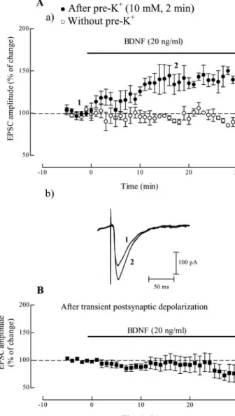
Bath application of BDNF (20 ng/ml), 46 min after the
high-K
⫹pulse enhanced the peak amplitude of EPSCs (35
⫾
7.3% increase; n
⫽ 7; p ⬍ 0.05). The maximal effect was already
attained at
⬃15 min after beginning BDNF application and it
lasted up to the end of the recording period (Fig. 2 A). In slices
that had not been exposed to the K
⫹-depolarizing pulse, BDNF
(20 ng/ml) was virtually devoid of effect on EPSC amplitude (n
⫽
3) (Fig. 2 Aa).
From the above results we could conclude that postsynaptic
depolarization was not required to trigger the BDNF action. We
4
are mean⫾ SEM; 100% (averaged fEPSP slopes at times ⫺10–0): ⫺0.64 ⫾ 0.05 mV/msec, n⫽ 7 (A), ⫺0.70 ⫾ 0.03 mV/msec, n ⫽ 2 (B), ⫺0.57 ⫾ 0.03 mV/msec, n ⫽ 9 (C), and ⫺0.50 ⫾ 0.10 mV/msec, n ⫽ 3 (D). Note that BDNF increased fEPSP slope only in pre-depolarized slices, an action prevented by the treatment with K252a.
Figure 1. Predepolarization induced by high K⫹facilitates BDNF excitatory action on
hip-pocampal synaptic transmission through Trk receptors. In A and B are shown the averaged time courses of changes in fEPSP slope induced by application of 20 ng/ml (corresponding to⬃0.8 nM) ( A), or 100 ng/ml ( B) BDNF alone. Ca illustrates the averaged time course of changes in fEPSP slope caused by BDNF (20 ng/ml), which was perfused 46 min after treatment with high-K⫹(10mM)for2min.CbshowstracesobtainedinarepresentativeexperimentinCa;each
trace is the average of eight consecutive responses obtained immediately before (1) and during (2) BDNF application, and is composed of the stimulus artifact, followed by the presynaptic volley and the fEPSP. D shows the averaged time course of the effect of BDNF (20 ng/ml) applied 46 min after treatment with high-K⫹(10 mM) for 2 min in the presence of an inhibitor of TrkB
receptors, K252a (200 nM), which was added to the slices 30 min before the pulse of high-K⫹.
Values obtained in individual experiments at time 0 and 60 min are shown as a scatter repre-sentation in A and Ca. In all panels, the arrows represent the beginning of the 2 min high-K⫹
application, and the horizontal bars represent the application of the different drugs. All values
Figure 2. The excitatory action of BDNF on synaptic transmission does not depend on postsynaptic depolarization. Aa shows the averaged time course of changes in EPSC amplitude recorded from CA1 pyramidal cells under voltage clamp conditions (Vh⫽ ⫺60 mV). BDNF (20
ng/ml) was applied alone (E, without pre-K⫹, n⫽ 3) or 46 min after a depolarizing pulse of
10 mMKCl for 2 min (F, after pre-K⫹, n⫽ 7), and remained in the bath up to the end of the
experiments, as indicated by the horizontal bar. Ab shows traces obtained in a representative experiment in Aa (F); each trace is the average of four consecutive responses obtained imme-diately before (1) and during (2) BDNF (20 ng/ml), and is composed of the stimulus artifact followed by the EPSC; resting membrane potential of this cell (measured in the current-clamp modeattheendoftheexperiment):⫺60mV;inputresistance:180M⍀.Bshowstheaveraged time course of the effect of BDNF, which was added to the slices 46 min after transient depolar-ization of postsynaptic neurons by changing, for 6 min, the holding potential in the voltage-clamp mode from⫺60to⫺40mVandthenagainto⫺60mV(n⫽4).Valuesintheordinates are mean⫾ SEM, in which 100% was taken as the averaged EPSCs amplitudes recorded for 5 min immediately before BDNF application (100% values ranged from⫺50 to ⫺370 pA in the different experiments).
next examined whether postsynaptic depolarization was
suffi-cient to unmask the effect of BDNF. When recording in the
current-clamp configuration it was observed that cells
depolar-ized, as expected from the Nernst equation, from
⫺60 to ⫺40 mV
during 6 min because of the pulse of high-K
⫹(10 m
Mfor 2 min)
(n
⫽ 2). We therefore mimicked the action of the pulse of
high-K
⫹on postsynaptic cell by changing the holding potential
in the voltage-clamp mode from
⫺60 to ⫺40 mV and then again
to
⫺60 mV, all during 6 min. BDNF (20 ng/ml) applied 46 min
after this postsynaptic depolarization did not induce an increase
in EPSC amplitude (n
⫽ 4) (Fig. 2B).
Presynaptic theta burst stimulation paired with BDNF can
elicit synaptic potentiation
The K
⫹depolarizing stimulus could still be effective if applied
after BDNF administration. Indeed, when BDNF (20 ng/ml) was
added to the slice at least 30 min before the K
⫹depolarizing pulse
(10 m
M, 2 min) and remained in the bath up to the end of the
experiment, an increase (81
⫾ 2.7%; n ⫽ 2) in fEPSP slope was
observed (Fig. 3A), which is clearly different from what occurred
when the BDNF (20 ng/ml) was applied without the K
⫹pulse, or
when the K
⫹pulse was applied without BDNF (see above).
Be-cause the K
⫹pulse causes a broad depolarization in all cells,
including glial cells and interneurons, we evaluated whether a
focal depolarization delivered through the stimulation electrode
placed at the synaptic afferents (Schaffer collaterals) was able to
elicit the BDNF effect. Weak theta burst stimulation (three bursts
of three pulses each at 100 Hz, delivered 100 msec apart) was
applied to slices that had been previously perfused with BDNF
(20 ng/ml) for at least 10 min, and fEPSPs were increased by 91
⫾
9.5% at 60 min after stimulus (n
⫽ 3, p ⬍ 0.05 as compared with
values before stimulation) (Fig. 3B); as expected (de Mendonc¸a
and Ribeiro, 2000), the same stimulation applied in the absence
of BDNF caused a smaller and non sustained increase in fEPSP
slope (20
⫾ 16.1% increase 60 min after stimulation; n ⫽ 3; p ⬎
0.05).
BDNF can facilitate LTP in particular under strong theta burst
stimulation conditions (Chen et al., 1999). We next examined
whether the facilitatory action of BDNF was also present in
con-ditions in which LTP-like phenomena could not occur, i.e., when
the postsynaptic membrane potential was clamped at
⫺60 mV
throughout the experiment, including during the theta burst
stimulation of the Schaffer collaterals. Under these conditions,
weak theta burst stimulation in the presence of BDNF (20 ng/ml)
led to a gradual increase in the amplitude of EPSCs (19
⫾ 8.8; n ⫽
2) (Fig. 3C), which is significantly different ( p
⬍ 0.05) from what
occurred in slices stimulated in the absence of BDNF, where
syn-aptic currents even decreased (20
⫾ 8.6; n ⫽ 2).
Figure 3. Presynaptic theta burst stimulation paired with BDNF can elicit synaptic potenti-ation. A shows the averaged time course of changes in fEPSP slope observed when the high-K⫹
pulse was applied to slices in the presence of BDNF (20 ng/ml). Ba illustrates the averaged time course of changes in fEPSP slope observed after a weak theta burst stimulation (3 bursts at 5 Hz, each composed of three pulses at 100 Hz) in the presence of 20 ng/ml BDNF (F), which was addedatleast10minbeforeelectricalstimulation,ascomparedwiththechangesoffEPSPslope in the same conditions of stimulation but without administration of the neurotrophin (E). Bb shows traces obtained in representative experiments in Ba; each trace is the average of eight
4
consecutive responses obtained immediately before (1) and after (2) theta burst stimulation in the absence (left) or in the presence (right) of BDNF, and is composed of the stimulus artifact, followed by the presynaptic volley and the fEPSP; recordings obtained in the same experiment aresuperimposed.CrepresentstheaveragedtimecourseofchangesinEPSCamplitudeinduced under voltage-clamp conditions (Vh⫽ ⫺60 mV) by theta burst stimulation in the presence
(F) and in absence (E) of BDNF (20 ng/ml). In all panels the arrow represents the beginning of stimulation and the horizontal bar in A indicates the presence of BDNF. All values are mean⫾ SEM; 100% (averaged fEPSP slopes at times⫺10–0):⫺0.61⫾0.07mV/msec,n⫽2(A),⫺ 0.70⫾ 0.02 mV/msec, n ⫽ 3 (Ba, F), ⫺0.66 ⫾ 0.01 mV/msec, n ⫽ 3 (Ba,E); 100% values in C (n⫽ 2 for both F and E) represent EPSCs amplitudes recorded at times ⫺5–0) and ranged from⫺110 to ⫺550 pA.
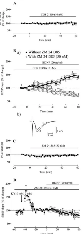
Adenosine A
2Areceptors activation facilitates BDNF
excitatory action on synaptic transmission
To evaluate if adenosine A
2Areceptors activation could influence
the action of BDNF on synaptic transmission we first tested
whether a selective agonist of the adenosine A
2Areceptor CGS
21680 (Jarvis et al., 1989) affected BDNF action. CGS 21680 (10
n
M) was added to the slices at least 30 min before BDNF
applica-tion and was virtually devoid of effect on fEPSP slope (Fig. 4 A). In
the presence of CGS 21680 (10 n
M), BDNF increased (Fig. 4 B) the
slope of fEPSPs by 43
⫾ 6.7% (n ⫽ 6; p ⬍ 0.05) in slices that had
not been predepolarized by the high-K
⫹pulse. Blockade of
aden-osine A
2Areceptors with the selective antagonist (Poucher et al.,
1995), ZM 241385 (50 n
M), which was added 30 min before CGS
21680 (10 n
M), prevented this excitatory effect of BDNF (n
⫽ 3;
p
⬍ 0.05 as compared with absence of ZM 241385) (Fig. 4B).
Indeed, in the presence of ZM 241385 (50 n
M) plus BDNF (20
ng/ml) even decreased (26
⫾ 6.5%) rather than increased, fEPSP
slope (Fig. 4 B). ZM 241385 (50 n
M) was virtually devoid of effect
on fEPSP slope (Fig. 4C).
To know whether the adenosine A
2Areceptor could play a role
in the excitatory action of BDNF observed after K
⫹depolariza-tion, we studied how adenosine A
2Areceptor blockade could
in-fluence that effect of BDNF. In the presence of the adenosine A
2Aa receptor antagonist, ZM 241385 (50 n
M), which was applied for
at least 30 min before the K
⫹pulse, the excitatory action of BDNF
(20 ng/ml) was completely prevented (n
⫽ 3; p ⬍ 0.05) (Fig. 4D).
Under these conditions BDNF (20 ng/ml) even decreased (17
⫾
8.0%) fEPSP slope as it occurred in the presence of CGS 21680
(10 n
M) plus ZM 241385 (50 n
M).
The excitatory action of BDNF is facilitated by a selective
adenosine kinase inhibitor 5-iodotubercidin
Our findings that a brief depolarization by a pulse of high-K
⫹(10
m
M) and subsequent application of BDNF resulted in a
signifi-cant increase in fEPSP slope, together with previous findings that
treatment of slices with high-K
⫹Krebs’ solution results in an
increase of adenosine release (Pazzagli et al., 1993), prompted us
to investigate if an enhancement of the extracellular adenosine
levels could mimic the action of the pulse of high-K
⫹. We used a
selective adenosine kinase inhibitor, 5-iodotubercidin (ITU),
which enhances the concentration of endogenous extracellular
adenosine at hippocampal slices (Pak et al., 1994). The
applica-tion of ITU (100 n
M) caused a decrease in synaptic transmission,
which may be attributed to activation of adenosine A
1receptors
(the predominant adenosine receptors in the hippocampus)
be-cause it was prevented by the specific adenosine A
1receptor
an-tagonist (Lohse et al., 1987), DPCPX (50 n
M) (Fig. 5A). In the
Figure 4. Adenosine A2Areceptors activation facilitates BDNF excitatory action on synaptic
transmission. A shows the averaged time course of changes in fEPSP slope induced by applica-tion of 10 nMCGS 21860 alone. Ba shows the averaged time courses of the effect of BDNF (20 ng/ml) in the presence of the A2Areceptor agonist CGS 21680 (10 nM) (F) or in the presence of
both CGS 21680 (10 nM) and the A2Areceptor antagonist ZM 241385 (50 nM) (E). CGS 21680 was applied at least 30 min before BDNF application, and ZM 241385 was applied 30 min before CGS21680application.BbshowstracesobtainedinarepresentativeexperimentinBa(F),each
4
trace is the average of eight consecutive responses obtained immediately before (1) and during (2) BDNF application, and is composed of the stimulus artifact, followed by the presynaptic volley and the fEPSP. C shows the averaged time course of changes in fEPSP slope induced by application of 50 nMZM 241385 alone. D shows the averaged time course of the effect of BDNF after a depolarizing stimulus of K⫹(10 mM) in the presence of ZM 241385; slices were
super-fused with BDNF (20 ng/ml) 46 min after treatment with high-K⫹(10 mM) for 2 min and were
inthepresenceofZM241385(50nM),whichwasaddedforatleast30minbeforethepotassium pulse. In all panels the horizontal bars represent drug application, and the arrow in D represents the beginning of the 2 min high-K⫹application. All values are mean⫾ SEM; 100% (averaged
fEPSPslopesattimes⫺10–0):⫺0.60⫾0.09mV/msec,n⫽3(A),⫺0.61⫾0.01mV/msec, n⫽6(Ba,F),⫺0.53⫾0.14mV/msec,n⫽3(Bb,E),⫺0.54⫾0.090mV/msec,n⫽3(C), ⫺0.58⫾0.06mV/msec,n⫽3(D).NotethatactivationofadenosineA2Areceptors facilitates
the excitatory action of BDNF on synaptic transmission, an action prevented by A2Areceptor
experiments where the action of BDNF in the presence of ITU
was tested, the neurotrophin was applied when the full effect of
ITU was achieved, and the slope values of the fEPSPs recorded
under these conditions were taken as 100%. As shown in Figure
5B, perfusion of BDNF (20 ng/ml) under these conditions
re-sulted in a significant increase in fEPSP slope (44
⫾ 8.7%
in-crease; n
⫽ 3; p ⬍ 0.05), that did not occur when A
2Areceptors
were blocked with ZM 241385 (50 n
M) before ITU application
(n
⫽ 2) (Fig. 5B). When ZM 241385 (50 n
M) was added only 50
min after ITU, BDNF (20 ng/ml) caused the expected
enhance-ment (51%) of synaptic transmission (one experienhance-ment). This
may suggest that A
2Areceptors are required to trigger the action
of BDNF but they do not need to remain activated during the
BDNF action.
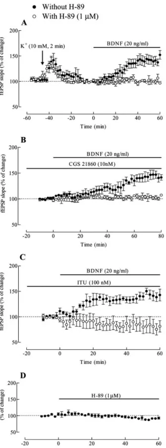
The activation of the cAMP–PKA transducing system is a
critical step in the excitatory action of BDNF
Because ITU and the pulse of high K
⫹mimicked the action of the
adenosine A
2Areceptor agonist CGS 21680 in what respects the
ability to trigger BDNF actions, and activation of A
2Areceptors
stimulates the formation of cAMP–PKA transducing system
(Fredholm et al., 2001), we evaluated if a selective PKA inhibitor
H-89 (Chijiwa et al., 1990) could modify the action of BDNF.
H-89 (1
M) was added for at least 30 min before K
⫹pulse
application (Fig. 6 A), CGS 21680 perfusion (Fig. 6 B), or ITU
administration (Fig. 6C) and remained in the bath up to the end
of the experiments. In all cases, H-89 abolished ( p
⬍ 0.05) the
excitatory action of BDNF on fEPSPs (n
⫽ 3 for each
experimen-tal condition), but by itself H-89 was virtually devoid of effect on
fEPSPs (Fig. 6 D).
To further evaluate the involvement of cAMP-dependent
PKA on the synaptic action of BDNF, we tested whether a
membrane-permeable cAMP analog, dibutyryl cAMP (dbcAMP)
(Henion et al., 1967), influences BDNF action. By itself dbcAMP
(0.5 m
M) was virtually devoid of effect on fEPSP slope (Fig. 7A),
and it was added to the slices at least 30 min before BDNF
appli-cation. In the presence of dbcAMP (0.5 m
M), BDNF caused a
significant increase (49
⫾ 13.6%; n ⫽ 4; p ⬍ 0.05) in fEPSP slope
(Fig. 7B), an effect fully blocked by H-89 (1
M; n
⫽ 2; p ⬍ 0.05)
(Fig. 7B). In slices predepolarized by a pulse of high-K
⫹(10 m
M),
dbcAMP caused only a small increase on fEPSP slope (17
⫾ 1.4%;
n
⫽ 2; p ⬍ 0.05 as compared with dbcAMP alone), suggesting
that cAMP-mediated actions are slightly influenced by
predepolarization.
To investigate whether the excitatory action of BDNF depends
on the increase in postsynaptic levels of cAMP, postsynaptic CA1
pyramidal neurons were loaded with cAMP (0.5 m
M) added to
the filling solution of the patch pipette. After membrane rupture,
EPSCs amplitudes gradually increased, probably because of
cAMP-induced alterations in the metabolic state of the cell. In
those cells that stabilized after the initial increase, bath
applica-tion of BDNF (20 ng/ml) did not cause consistent changes in
EPSC amplitudes (Fig. 7C). Thus, an increase of postsynaptic
cAMP levels seems not sufficient to trigger the excitatory action
of BDNF.
Discussion
The present results show that the excitatory action of BDNF on
hippocampal synaptic transmission is facilitated by
predepolar-ization and that this effect is dependent on adenosine A
2Arecep-tor activation. These actions were revealed by using a BDNF
con-centration that on its own did not significantly modify the slope
of fEPSPs and was mediated by a receptor of the tyrosine kinase
receptor family, because pretreatment with the Trk receptor
in-hibitor K252a (Klein et al., 1990), prevented the excitatory effect
of this neurotrophin on fEPSPs. BDNF possesses a greater affinity
for TrkB receptors than for TrkA or TrkC receptors (Lewin and
Barde, 1996) and, therefore, the observed actions of BDNF are
Figure5. TheexcitatoryactionofBDNFisfacilitatedbyaselectiveadenosinekinaseinhibitor 5-iodotubercidin. A shows averaged time courses of changes of fEPSP slope in the presence of 5-iodotubercidin (ITU; 100 nM) (E) and in the presence of both ITU (100 nM) plus DPCPX (50 nM) (F), an A1adenosine receptor antagonist. Ba shows the averaged time courses of changes in
fEPSP slope induced by BDNF (20 ng/ml) in the presence of ITU (100 nM), (F) or in the presence of both ITU (100 nM) plus the A2Aadenosine receptor antagonist, ZM 241985 (50 nM) (E). Bb shows traces obtained in a representative experiment in Ba (F). Each trace is the average of eight consecutive responses obtained immediately before (1) and during (2) BDNF application, and is composed of the stimulus artifact, followed by the presynaptic volley and the fEPSP. In all panels the horizontal bars represent drug application. All values are mean⫾ SEM; 100% (av-eraged fEPSP slopes obtained at times⫺10–0: ⫺0.65 ⫾ 0.06 mV/msec, n ⫽ 3 (A, F), ⫺0.57 ⫾ 0.03 mV/msec, n ⫽ 3 (A, E), ⫺0.52 ⫾ 0.02 mV/msec, n ⫽ 2 (Ba, E) and ⫺0.50 ⫾ 0.10 mV/msec, n ⫽ 3 (Ba, F)
probably caused by activation of TrkB receptors, which are
present in the hippocampus (Klein et al., 1990; Lewin and Barde,
1996).
The findings that after the brief K
⫹-depolarizing pulse, BDNF
could facilitate synaptic transmission to pyramidal neurons
un-der voltage-clamp conditions and that BDNF was unable to
facil-itate synaptic transmission to neurons that had been transiently
depolarized by changing the holding potential indicate that
pre-synaptic, rather than postpre-synaptic, depolarization is required to
gate the BDNF action. Also in accordance with the need of
pre-synaptic depolarization to trigger BDNF excitatory action is the
finding that theta burst stimulation of the Schaffer collateral
af-ferents paired with BDNF facilitated synaptic transmission.
In accordance with our results, are the results of Boulanger
and Poo (1999a) at developing neuromuscular synapses showing
that presynaptic depolarization is a critical factor in causing the
synaptic action of neurotrophins. This could result from the
fol-lowing mechanisms: (1) the prepulse of K
⫹triggers secretion of
endogenous neurotrophin that could act synergistically with
ap-plied BDNF to potentiate synapses; (2) potassium treatment
could induce the expression of more TrkB receptors, and (3)
treatment with high-K
⫹induces release of adenosine (Pazzagli et
al., 1993), which could gate the excitatory action of BDNF on
synaptic transmission. Our results with the adenosine A
2Aselec-tive agonist, CGS 21680 and with ITU, a selecselec-tive inhibitor of
adenosine kinase that enhances the extracellular amount of
aden-osine (Pak et al., 1994), support this last hypothesis. Thus, in the
presence of CGS 21680, BDNF increased the slope of fEPSP, even
in slices that had not been predepolarized by the high-K
⫹pulse.
Moreover the adenosine A
2Areceptor antagonist ZM 241385
pre-vented the excitatory effects of BDNF in conditions of
predepo-larization, as well as in the presence of CGS 21680. Finally, ITU
was also able to unmask the BDNF excitatory action, as did CGS
21860 and the brief K
⫹pulse, an action also prevented by the A
2Aantagonist ZM 241385.
There are two sources of extracellular adenosine: release of
adenosine by facilitated diffusion, through membrane adenosine
transporters that are equilibrative and bidirectional (Gu et al.,
1995), and extracellular conversion of released adenine
nucleo-tides into adenosine through a series of ectoenzymes, the last one
in the cascade and the rate-limiting step for adenosine formation
being ecto-5⬘-nucleotidase (Zimmermann and Braun, 1999).
ATP is co-stored with most neurotransmitters, and therefore
neuronal activity causes the release of ATP and extracellular
for-mation of adenosine in most brain areas, including the
hip-pocampus (Wieraszko et al., 1989; Cunha et al., 1996).
Depolar-ization by K
⫹predominantly induces the release of adenosine as
such (Latini and Pedata, 2001). The intracellular adenosine levels
are kept low mainly because of the activity of adenosine kinase
(Arch and Newsholme, 1978) and when this enzyme is inhibited
(e.g., by ITU) there is a marked increase in the release of
adeno-sine (Pak et al., 1994). One may question why K
⫹-induced
aden-osine release did not cause an A
1-receptor mediated decrease in
synaptic transmission, as ITU did. This most probably results
from the time course of A
1-receptor mediated actions (Sebastia˜o
4obtainedattimes⫺10–0):⫺0.57⫾0.03mV/msec,n⫽9(A,F),⫺0.48⫾0.23mV/msec, n⫽3(A,E),⫺0.61⫾0.01mV/msec,n⫽6(B,F),⫺0.47⫾0.06mV/msec,n⫽3(B,E), ⫺0.50⫾0.10mV/msec,n⫽3(C,F),⫺0.43⫾0.17mV/msec,n⫽3(C,E),⫺0.59⫾0.08 mV/msec, n⫽ 3 (D). Note that the PKA inhibition prevented the action of BDNF in all experi-mental conditions.
Figure 6. The activation of the cAMP–PKA transducing system is a critical step for the exci-tatory action of BDNF. Averaged time course of changes in fEPSP slope induced by BDNF in the absence (F) and in the presence (E) of the PKA inhibitor H-89 (1M) under the following experimental conditions: ( A) after a pre-depolarizing pulse of high-K⫹(10 mM, 2 min); ( B) in
the presence of CGS 21860 (10 nM); ( C) in the presence of 5-iodotubercidin (ITU; 100 nM). H-89 (1M) was added to the slices 30 min before the application of the K⫹pulse ( A), CGS 21680 ( B),orITU( C)andalonewasvirtuallydevoidofeffectonfEPSPslope,asshowninD.Inallpanels the horizontal bars represent drug application, and the arrow in A represents the beginning of the 2 min high-K⫹application. All values are mean⫾ SEM; 100% (averaged fEPSP slopes
et al., 1990; Lupica et al., 1992): any fast inhibitory actions,
be-cause of adenosine released during the 2 min application of K
⫹,
should be masked below the transient excitation induced by
de-polarization. In contrast, activation of A
2Areceptors for a similar
period of time might lead to long lasting enhancement in
intra-cellular cAMP caused by adenylate cyclase activation (Fredholm
et al., 2001), which could gate BDNF actions (Boulanger and Poo,
1999b). In line with this possibility are our observations that the
PKA inhibitor, H-89, prevented the excitatory action of BDNF in
the presence of K
⫹pulse, CGS 21680, or ITU. Moreover in the
presence of the cAMP analog dbcAMP, BDNF enhanced synaptic
transmission, an effect blocked by H-89. Presynaptic, rather than
postsynaptic enhancement of cAMP might be required to trigger
the action of BDNF because the neurotrophin was unable to
en-hance synaptic currents in cAMP loaded pyramidal cells.
It is known that cAMP can induce a variety of cellular
pro-cesses in different systems, including expression of mRNA for
Trk receptors and neurotrophins in primary astroglial cultures
(Condorelli et al., 1994) and recruitment of TrkB receptors to the
plasma membrane of CNS neurons (Meyer-Franke et al., 1998).
Cytosolic cAMP can be positively modulated by depolarization
and synaptic activity (Ferrendelli et al., 1980), and activation of
cAMP signaling has been shown to potentiate facilitatory actions
of BDNF at the developing neuromuscular junction (Boulanger
and Poo, 1999b). A cAMP—PKA—CREB-dependent pathway is
also shown to be involved in the ability of adenosine A
2Areceptor
agonists to potentiate nerve growth factor-induced neurite
out-growth in cultured PC12 cells (Chen et al., 2002). The results now
described show that the interactions between neurotrophins and
adenosine A
2Areceptors can occur in CNS neurons that were not
in culture, therefore not at an active developing stage. The finding
that blockade of adenosine A
2Areceptors prevents the action of
BDNF after a K
⫹predepolarizing pulse, suggests that the
prede-polarization pulse per se is not an essential requisite to unmask
BDNF excitatory effects, eventually by a K
⫹-induced increase in
cAMP levels (Ferrendelli et al., 1980). In contrast, activation of
adenosine A
2Areceptors either by released adenosine or by a
selective agonist (CGS 21680) appears to be an essential step to
trigger BDNF excitatory action on synaptic transmission.
Clear excitatory actions of BDNF on hippocampal synaptic
transmission have been observed by some authors (Kang and
Schuman, 1995, 1996), whereas others reported minimal or no
effects of this neurotrophin in the same brain area (Figurov et al.,
1996; Gottschalk et al., 1998). Whether this discrepancy results
from different levels of endogenous activation of adenosine A
2Areceptors, awaits further investigation.
Neurotrophins have been suggested to have an important role
in protecting mature neurons from neuronal atrophy in the
de-generating human brain. A decrease in BDNF levels might be
involved in neurodegenerative disorders such as Alzheimer’s
dis-ease, Parkinson’s disdis-ease, Huntington’s disease (Connor and
Dragunow, 1998), or diabetic neuropathies (Nitta et al., 2002),
making the use of the naturally occurring neurotrophic factors
very promising for treatment of these disorders. However, until
now the pharmacological administration of in vivo BDNF has not
been easy. One of the reasons is that these molecules are unable to
cross the blood– brain barrier, making invasive application
strat-egies like intracerebroventricular infusion necessary.
Intrave-nous administration of BDNF has been attempted, but it involves
a complex molecular reformulation of the neurotrophin (Wu
and Pardridge, 1999). The results now described open a new
prospective to potentiate BDNF actions on CNS neurons, i.e., by
co-activation of a specific type of adenosine receptors, the A
2Areceptors. This possibility expands the pathophysiological
impli-cations of adenosine receptor functioning in the brain (Ribeiro et
al., 2003) and points toward new strategies to interfere with
neu-Figure 7. Presynaptic cAMP mimics the effect of high-K⫹, ITU, or CGS 21680. A shows the
averagedtimecourseofchangesinfEPSPslopeinducedbyapplicationofdbcAMP(0.5mM),and BashowstheaveragedtimecourseofchangesinfEPSPslopeinducedbyBDNF(20ng/ml)inthe presence of dbcAMP (0.5 mM), either in the absence (F) and or the presence (E) of the PKA inhibitor H-89 (1M). Slices were superfused with dbcAMP 30 min before BDNF application. Bb shows traces obtained in a representative experiment in Ba (F); each trace is the average of eight consecutive responses obtained immediately before (1) and during (2) BDNF application, and is composed of the stimulus artifact, followed by the presynaptic volley and the fEPSP. C illustratestheaveragedtimecourseofchangesinducedbyBDNF(20ng/ml)ontheamplitudeof EPSCs recorded from postsynaptic pyramidal neurons loaded with cAMP, which was added (0.5 mM) to the whole-cell recording pipette. In all panels the horizontal bar represents the applica-tion of the different drugs. All values are mean⫾SEM;100%inAandB[averagedfEPSPslopes obtainedattimes⫺10–0:⫺0.70⫾0.2,n⫽2(A),⫺0.56⫾1.60mV/msec,n⫽4(Ba,E), and⫺0.64 ⫾ 0.04 mV/msec, n ⫽ 2 (Ba, F]; 100% values in C (EPSC amplitudes recorded at times⫺5–0) ranged from ⫺500 to ⫺1400 pA; n ⴝ 2. Note that BDNF enhanced synaptic transmission in the presence of the cell-permeant cAMP analog, dbcAMP, but when cAMP was present only postsynaptically ( C) it did not trigger the action of BDNF.
rotrophic factors in the therapeutics of some neurodegenerative
disease.
In conclusion, our results suggest a way to enhance the
exci-tatory action of BDNF on synaptic transmission via activation of
adenosine A
2Areceptors. Through activation of these receptors it
would be possible to overcome some difficulties with the use and
delivery of neurotrophic factors when neuroprotection by these
agents is required.
References
Anderson WW, Collingridge GL (2001) A data acquisition program for on-line analysis of long-term potentiation and other synaptic events. J Neu-rosci Methods 108:71– 83.
Arch JR, Newsholme EA (1978) The control of the metabolism and the hormonal role of adenosine. Essays Biochem 14:82–123.
Berg MM, Sternberg DW, Parada LF, Chao MV (1992) K-252a inhibits nerve growth factor-induced trk proto-oncogene tyrosine phosphoryla-tion and kinase activity. J Biol Chem 267:13–16.
Boulanger L, Poo M (1999a) Presynaptic depolarization facilitates neurotrophin-induced synaptic potentiation. Nat Neurosci 2:346 –351. Boulanger L, Poo M (1999b) Gating of BDNF-induced synaptic
potentia-tion by cAMP. Science 284:1982–1984.
Chen G, Kolbeck R, Barde Y-A, Bonhoeffer T, Kossel A (1999) Relative contribution of endogenous neurotrophins in hippocampal long-term potentiation. J Neurosci 19:7983–7990.
Chen H-C, Shih H-M, Chern Y (2002) Essential Role of cAMP-response element-binding protein activation by A2Aadenosine receptors in rescu-ing the nerve growth factor-induced neurite outgrowth impaired by blockage of the MAPK cascade. J Biol Chem 277:33930 –33942. Chijiwa T, Mishima A, Hagiwara M, Sano M, Hayashi K, Inoue T, Naito K,
Toshioka T, Hidaka H (1990) Inhibition of forskolin-induced neurite outgrowth and protein phosphorylation by a newly synthesized selective inhibitor of cyclic AMP-dependent protein kinase, N-[2-( p-bromocinnamylamino)ethyl]-5-isoquinolinesulfonamide (H-89), of PC12D pheochromocytoma cells. J Biol Chem 265:5267–5272. Condorelli DF, Dell’Albani P, Mudo` G, Timmusk T, Belluardo N (1994)
Expression of neurotrophins and their receptors in primary astroglial cultures: induction by cyclic AMP-elevating agents. J Neurochem 63:509 –516.
Connor B, Dragunow M (1998) The role of neuronal growth factors in neu-rodegenerative disorders of the human brain. Brain Res Rev 27:1–39. Cunha RA, Vizi ES, Ribeiro JA, Sebastia˜o AM (1996) Preferential release of
ATP and its extracellular catabolism as a source of adenosine upon high-but not low-frequency stimulation of rat hippocampal slices. J Neuro-chem 67:2180 –2187.
de Mendonc¸a A, Ribeiro JA (2000) Long-term potentiation observed upon blockade of adenosine A1receptors in the hippocampus is N-methyl-D-aspartate receptor dependent. Neurosci Lett 291:81– 84.
Ferrendelli JA, Blank AC, Gross RA (1980) Relationships between seizure activity ad cyclic nucleotide levels in brain. Brain Res 200:93–103. Figurov A, Pozzo-Miller LD, Olafsson P, Wang T, Lu B (1996) Regulation of
synaptic responses to high-frequency stimulation and LTP by neurotro-phins in the hippocampus. Nature (Lond) 381:706 –709.
Fredholm BB, Ijzerman AP, Jacobson KA, Klotz K-N, Linden J (2001) In-ternational union of pharmacology. XXV. Nomenclature and classifica-tion of adenosine receptors. Pharmacol Rev 53:527–552.
Gottschalk W, Pozzo-Miller LD, Figurov A, Lu B (1998) Presynaptic mod-ulation of synaptic transmission and plasticity by Brain-derived neuro-trophic factor in the developing hippocampus. J Neurosci 18:6830 – 6839. Gu JG, Foga IO, Parkinson FE, Geiger JD (1995) Involvement of bidirec-tional adenosine transporters in the release ofL-[3H]adenosine from rat brain synaptosomal preparations. J Neurochem 64:2105–2110. Henion WF, Sutherland EW, Posternak TH (1967) Effects of derivatives of
adenosine 3⬘,5⬘-phosphate on liver slices and intact animals. Biochem Biophys Acta 148:106 –113.
Jarvis MF, Schulz R, Hutchison AJ, Do UH, Sills MA, Williams M (1989) [3H]CGS 21680, a selective A
2adenosine receptor agonist, directly labels A2receptors in rat brain. J Pharmacol Exp Ther 251:888 – 893. Kang H, Schuman EM (1995) Long-lasting neurotrophin-induced
en-hancement of synaptic transmission in the adult hippocampus. Science 267:1658 –1662.
Kang H, Schuman EM (1996) A requirement for local protein synthesis in neurotrophin-induced hippocampal synaptic plasticity. Science 273: 1402–1406.
Klein R, Conway D, Parada LF, Barbacid M (1990) The TrkB tyrosine pro-tein kinase gene codes for a second neurogenic receptor that lacks the catalytic kinase domain. Cell 61:647– 656.
Kovalchuk Y, Hanse E, Kafitz KW, Konnerth A (2002) Postsynaptic induc-tion of BDNF-mediated long-term potentiainduc-tion. Science 295:1651–1653. Latini S, Pedata F (2001) Adenosine in the central nervous system: release mechanisms and extracellular concentrations. J Neurochem 79:463– 484. Lee FS, Chao MV (2001) Activation of TrK neurotrophin receptors in the
absence of neurotrophins. Proc Natl Acad Sci USA 98:3555–3560. Levine ES, Crozier RA, Black IB, Plummer MR (1998) Brain-derived
neu-rotrophic factor modulates hippocampal synaptic transmission by in-creasing N-methyl-D-aspartic acid receptor activity. Proc Natl Acad Sci USA 95:10235–10239.
Lewin GR, Barde Y-A (1996) Physiology of the neurotrophins. Annu Rev Neuroscience 19:289 –317.
Lohse MJ, Klotz KN, Lindenborn-Fotinos J, Reddington M, Schwabe U, Ol-sson RA (1987) 8-Cyclopentyl-1,3-dipropylxanthine (DPCPX)- a selec-tive high affinity antagonist radioligand for A1 adenosine receptors. Nau-nyn Schmiedebergs Arch Pharmacol 336:204 –210.
Lupica CR, Proctor WR, Dunwiddie T-U (1992) Presynaptic inhibition of excitatory synaptic transmission by adenosine in rat hippocampus: anal-ysis of unitary fEPSP variance measured by whole-cell recording. J Neu-rosci 12:3753–3764.
Messaoudi E, Ying SW, Kanhema T, Croll SD, Bramham CR (2002) Brain-derived neurotrophic factor triggers transcription-dependent, late phase long-term potentiation in vivo. J Neurosci 22:7453–7461.
Meyer-Franke A, Wilkinson GA, Kruttgen A, Hu M, Munro E, Hanson MG, Reichardt LF, Barres BA (1998) Depolarization and cAMP elevation rapidly recruit TrkB to the plasma membrane of CNS neurons. Neuron 21:681– 693.
Nitta A, Murai R, Suzuki N, Ito H, Nomoto H, Katoh G, Furukawa Y, Fu-rukawa S (2002) Diabetic neuropathies in brain are induced by defi-ciency of BDNF. Neurotoxicol Teratol 24:695–701.
Pak MA, Haas HL, Decking UKM, Schrader J (1994) Inhibition of adeno-sine kinase increases endogenous adenoadeno-sine and depresses neuronal ac-tivity in hippocampal slices. Neuropharmacology 33:1049 –1053. Pazzagli M, Pedata F, Pepeu G (1993) Effect of K⫹depolarization,
tetrodo-toxin, and NMDA receptor inhibition on extracellular adenosine levels in rat striatum. Eur J Pharmacol 234:61– 65.
Poucher SM, Keddie JR, Singh P, Stoggall SM, Caulkett PW, Jones G, Coll MG (1995) The in vitro pharmacology of ZM 241385, a potent, non-xanthine A2a selective adenosine receptor antagonist. Br J Pharmacol 115:1096 –1102.
Ribeiro JA, Sebastia˜o AM, de Mendonc¸a A (2003) Adenosine receptors in the nervous system: pathophysiological implications. Prog Neurobiol 68:377–392.
Sebastia˜o AM, Ribeiro JA (2000) Fine-tuning neuromodulation by adeno-sine. Trends Pharmacol Sci 21:341–346.
Sebastia˜o AM, Stone TW, Ribeiro JA (1990) The inhibitory adenosine re-ceptor at the neuromuscular junction and hippocampus of the rat: antag-onism by 1,3,8-substituted xanthines. Br J Pharmacol 101:453– 459. Sebastia˜o AM, de Mendonc¸a A, Moreira T, Ribeiro JA (2001) Activation of
synaptic NMDA receptors by action potential-dependent release of trans-mitter during hypoxia impairs recovery of synaptic transmission on reoxygenation. J Neurosci 21:8564 – 8571.
Tyler WJ, Pozzo-Miller LD (2001) BDNF enhances quantal neurotransmit-ter release and increases the number of docked vesicles at the active zones of hippocampal excitatory synapses. J Neurosci 21:4249 – 4258. Wieraszko A, Goldsmith G, Seyfried TN (1989) Stimulation-dependent
re-lease of adenosine triphosphate from hippocampal slices. Brain Res 485:244 –250.
Wu D, Pardridge WM (1999) Neuroprotection with non-invasive neuro-trophin delivery to the brain. Proc Natl Acad Sci USA 96:254 –259. Zimmermann H, Braun N (1999) Ecto-nucleotidases: molecular structures,
catalytic properties, and functional roles in the nervous system. Prog Brain Res 120:371–385.