Transactions
PERSPECTIVE
Cite this: DOI: 10.1039/c3dt50462j
Received 20th February 2013, Accepted 4th April 2013 DOI: 10.1039/c3dt50462j www.rsc.org/dalton
Ion pumps as biological targets for decavanadate
Manuel Aureliano,*
a,bGil Fraqueza
b,cand C. André Ohlin
dThe putative applications of poly-, oligo- and mono-oxometalates in biochemistry, biology, pharmacology and medicine are rapidly attracting interest. In particular, these compounds may act as potent ion pump inhibitors and have the potential to play a role in the treatment of e.g. ulcers, cancer and ischemic heart disease. However, the mechanism of action is not completely understood in most cases, and even remains largely unknown in other cases. In the present review we discuss the most recent insights into the inter-action between mono- and polyoxometalate ions with ion pumps, with particular focus on the interinter-action of decavanadate with Ca2+-ATPase. We also compare the proposed mode of action with those of established ion pump inhibitors which are currently in therapeutic use. Of the 18 classes of compounds which are known to act as ion pump inhibitors, the complete mechanism of inhibition is only known for a handful. It has, however, been established that most ion pump inhibitors bind mainly to the E2 ion pump conformation within the membrane domain from the extracellular side and block the cation release. Polyoxometalates such as decavanadate, in contrast, interact with Ca2+-ATPase near the nucleotide binding site domain or at a
pocket involving several cytoplasmic domains, and therefore need to cross through the membrane bilayer. In contrast to monomeric vanadate, which only binds to the E2 conformation, decavanadate binds to all protein conformations, i.e. E1, E1P, E2 and E2P. Moreover, the specific interaction of decavanadate with sarco-plasmic reticulum Ca2+-ATPase has been shown to be non-competitive with respect to ATP and induces protein cysteine oxidation with concomitant vanadium reduction which might explain the high inhibitory capacity of V10(IC50= 15μM) which is quite similar to the majority of the established therapeutic drugs.
Dr Manuel Aureliano
Dr Manuel Aureliano is an Associ-ate Professor of Biochemistry and Director of the Biochemistry degree at the University of Algarve, Faro, Portugal. Besides Biochemistry, he teaches disci-plines related to Metallomics and Muscle Contraction. Till now, he was supervisor and/or co-supervisor of several post-doc, PhD, MSc and undergraduate students (over 90), and has published about 60 peer-reviewed journal articles, reviews and book chapters. He is editor of the book “Vanadium Biochemistry”, Research SignPost, 2007, Kerala, India. His research topics include (i) the role of deca-vanadate in biology; (ii) vanadium and diabetes; (iii) antioxidants: toxic and/or beneficial effects; (iv) ion pumps as toxins/drugs targets.
Dr Gil Fraqueza
Dr Gil Fraqueza is an Assistant Professor at the Instituto Superior de Engenharia (ISE) of the University of Algarve, Faro, Portugal, where he has been on the faculty staff since 1989. He was former director of the Food
Engineering degree at the
Department of Alimentary Engin-eering, where he taught
disci-plines such as Organic
Chemistry, Enzymology, Chemi-cal Analysis, and Nutrition and Food Toxicology. His areas of research are related to investigations on the clotting of milk, bio-logical agriculture and, in the work of PhD programs, on the effects of oxometalates on ion pumps.
a
DCBB, FCT, University of Algarve, 8005-139 Faro, Portugal. E-mail: [email protected]
b
CCMar, University of Algarve, 8005-139 Faro, Portugal
c
Department of Food Engineering, IST, University of Algarve, 8005-139 Faro, Portugal
d
School of Chemistry, Monash University, Clayton, Vic 3800, Australia
Downloaded by Universidade do Algarve (UALG) on 03/05/2013 22:25:34. Published on 04 April 2013 on http://pubs.rsc.org | doi:10.1039/C3DT50462J
View Article Online
1.
Introduction
The serendipitous discovery three decades ago of vanadium as an inhibitor of ion pumps such as Na+/K+-ATPase (IC50= 40 nM)1 has encouraged the investigation of mono and polyoxo-metalates as putative drugs in the treatment of several diseases in which the molecular target has been established to be an ion pump such as Na+/K+-ATPase, H+/K+-ATPase, or Ca2+ -ATPase.2 These enzymes are all classified as P-type, or E1-E2, ATPases, in which the ion pump process is coupled to ATP hydrolysis. The mechanism is well-known and involves at least four steps and two distinct protein conformations, E1 and E2.3 These conformations are usually highly selective towards specific substrates, where the E1 state has a greater affinity for the exported ion and ATP, and a low affinity for the imported ion, while the E2 conformation has a higher affinity for the imported ion and a low affinity for the exported ion, thus releasing it into the extracellular environment. ATP provides the driving force in this process, by inducing the structural changes through the phosphorylation of an aspartate residue.
The E1–E2 cycle consists of four steps. For instance, in the process of calcium transport across the membrane, the E2 form of the calcium ATPase binds two calcium ions, thus forming the E1 conformation. This conformation is phos-phorylated by ATP forming an E1P intermediate, where the P indicates that the enzyme is phosphorylated, which leads to ion translocation and release of the calcium. This is followed by the formation of the calcium-free proton-carrying E2P con-formation which undergoes hydrolysis of the phosphate –aspar-tate bond to regenerate E2 and release the protons, and the cycle is complete.3It is in fact the phosphorylation step that gives the P-type ATPases their name, and they present a common target for inhibitors which can interfere with this particular step.
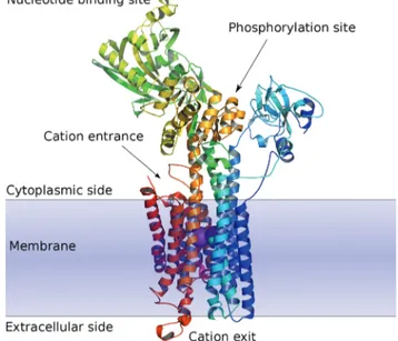
In terms of structural features these P-type ATPases each possess a nucleotide binding domain and a phosphorylation binding site on the cytoplasmic side, and a cation binding site located inside the membrane region.3,4 These three distinct domains (Fig. 1) can all serve as potential targets for different types of E1–E2 ATPase inhibitors.
A range of established drugs, such as ouabain, omeprazole and thapsigargin, are known to act as ion pump inhibitors, but the mechanism and protein binding sites, or even main target enzyme, have only been clearly established for a few of these compounds (described in Tables 1–3).2For example, the cardiotonic steroids (CTS) mainly inhibit Na+/K+-ATPase, whereas the proton pump inhibitors (PPI) and potassium blockers (also known as potassium competitive acid blockers; PCAB) are named for their inhibition of H+/K+-ATPase. Thapsi-gargin (TG) and cyclopiazonic acid (CPA) are examples of Ca2+ -ATPase inhibitors.
The principal role of the sarcoplasmic reticulum (SR) Ca2+ -ATPase is to remove cellular Ca2+ from the cytoplasm to the SR, which it does as part of the process of muscle relaxation. In addition, SR Ca2+-ATPase is an important actor in the process of calcium homeostasis.3,5This makes it a particularly attractive target as it may play important roles in the control of heart disease and cancer.
It is well-established that the process of calcium transport and ATP hydrolysis can be inhibited by monomeric vanadate (V1), VO43−, particularly under conditions where the E2 confor-mation is favoured, leading to the forconfor-mation of a conformer analogous to E2P, but involving vanadate instead of phos-phate.6,7 Therefore, the vanadate mode of action may involve formation of an E2V analogue after condensation of vanadate to the enzyme phosphorylation site, at aspartate residue 351. Analysis of the E2 conformation clearly shows structural changes relative to conformation E1 (Fig. 1). In particular, the phosphorylation site is now sterically hindered.2,4
This is not very surprising given the similarities and differ-ences between the aqueous chemistry phosphorous and vanadium, which may help explain some of the observed toxi-city of the latter, in particular compared with other group 5 and 6 metals. In particular, both vanadate and phosphate Dr C. André Ohlin
Dr C. André Ohlin is an Australian Research Council-funded senior research fellow at Monash Univer-sity in Australia. His research interests include the reaction dynamics of polyoxometalates by NMR and mass spectrometry, and the catalytic activation of small
molecules by metal oxide
complexes.
Fig. 1 Schematic view of the E1 form of Ca2+SR ATPase. Note the two calcium
ions ( purple) in the cation channel.4Figure based on RCSB deposition 1KJU.
readily form linear oligomers in the tetrahedral state, in con-trast with e.g. molybdate and tungstate. However, conden-sation occurs spontaneously for vanadates in acidic solution
and has several easily accessible oxidation states, whereas phosphate condensation is thermally driven in the solid phase and phosphorous is most likely to be found in the +V state
Table 1 IC50values and/or dissociation constants and therapeutical applications of known Na+/K+-ATPase inhibitors
Class of inhibitor Compounds IC50[μM] Kd[nM] Therapeutical applications References
Na+/K+-ATPase inhibitors
Cardiotonic steroids Ouabain >10 Treatment of congestive heart failure and cardiac arrhythmia
10 0.01–0.5 10 0.600 11 2.2 12 0.08–0.32 12 1.8 13 1–100 14 60 15 27.7 16 28 3.62 16 1 17 Dihydro-ouabain >1000 15 Digoxin 0.910 11 20 15 Digoxigenin >100 15 Strophanthidin 160 15 Ouabagenin >1000 15
Chlorpromazine Chlorpromazine (CPZ) Kd1= 32 500 Phototoxic antipsychotic drug 18
Kd2= 142 500 18
62.5 20
Chloroquine Chloroquine (CLQ) Kd1= 20 000 Anti-malarial drug 18
Kd2= 115 000 18
Procaine and derivatives Procaine 17 700 Local anesthetics 20
Chloroprocaine 13 000 20
Bupivacaine 6700 20
Mepivacaine >10 000 20
Lidocaine 30 400 20
Palytoxin 0.8 Tumor promoter 12
Palytoxin (PTX) 0.2–0.45 12
0.0035 16
0.4–3.1 20 000 21–23
Gramicidin A Gramicidin A 8.1 Antibiotic 24
Tungsten compounds (W12) 12-tungstosilicic acid
H4SiW12O40(WSiA) 3.4–4.3 Insulin-mimetic properties 25 (W12) 12-tungstophosphoric acid, H3PW12O40(WPA) 2.9–3.1 25 (W1) sodium tungstate 1300–1500 25
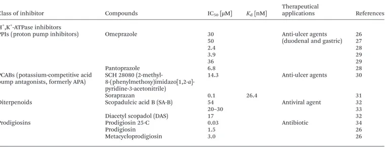
Table 2 IC50values and/or dissociation constants and therapeutical applications of known H+/K+-ATPase inhibitors
Class of inhibitor Compounds IC50[μM] Kd[nM]
Therapeutical
applications References H+,K+-ATPase inhibitors
PPIs (proton pump inhibitors) Omeprazole 30 Anti-ulcer agents (duodenal and gastric)
26 50 27 2.4 28 3.9 29 36 29 Pantoprazole 6.8 28
PCABs (potassium-competitive acid pump antagonists, formerly APA)
SCH 28080
(2-methyl- 8-(phenylmethoxy)imidazo[1,2-a]-pyridine-3-acetonitrile)
14.3 Anti-ulcer agents 30
Soraprazan 0.1 26.4 31
Diterpenoids Scopadulcic acid B (SA-B) 54 Antiviral agent 32
20–30 33
Diacetyl scopadol (DAS) 17 32
Prodigiosins Prodigiosin 25-C 0.03 Antibiotic 34
Prodigiosin 1.5 26
Metacycloprodigiosin 3.0 26
under physiological conditions. There is also no shortage of polyoxovanadates consisting of octahedral building blocks while phosphates typically are found as tetrahedral units. In addition, vanadium is considerably more electrophilic than phosphorous and V–O bonds are less covalent than P–O bonds.
The situation is, however, rendered more complex by the availability of three distinct binding domains in the P-ATPases, only one of which is involved in the phosphorylation step. For example, decavanadate (V10), [V10O28]6−, is a more potent SR Ca2+-ATPase inhibitor than monomeric vanadate6,8 and inter-acts with other conformations besides E2. In this case the binding site is not the same as the ATP binding site.8 More-over, recently it was shown that during the mechanism of deca-vanadate-mediated SR Ca2+-ATPase inhibition, a protein cysteine unit is oxidized through reduction of the vanadate, although the full implications of this process are not yet clear.9Therefore, a better understanding of the mechanism of decavanadate calcium pump inhibition will help in evaluating the potential of other members of the rich polyoxometalate family as therapeutic agents for a range of diseases.
In this Perspective we aim to analyse and compare several ion pump inhibitors, their putative mode of action and protein binding sites, with particular focus on the role of vanadium and other transition metal oxide ions. The majority of these ion pump inhibitors are in current use as thera-peutic drugs and we compare their inhibitory capacity with mono- and polyoxometalates, which have activities of similar magnitude.
Decavanadate has already been established as a potent ion pump inhibitor and has several attractive properties. It is a non-competitive inhibitor with respect to ATP, it has a high inhibitory capacity (IC50 = 15 μM), and it is not selective towards a specific protein interaction and can interact with the calcium-induced E1 conformation rather than just the E2 con-formation. V10 also binds to a cytoplasmic pocket involving several domains and it induces cysteine oxidation and vanadium reduction to yield V(IV) species. In contrast, the
majority of the established drugs bind to the protein only from the extracellular side within the membrane domain, and block the cation release, without interfering with the nucleotide binding site.
2.
Na
+/K
+-ATPase inhibitors
Na+/K+-ATPase is a target in the treatment of a wide range of conditions, and inhibitors of this target include compounds used to treat diseases such as heart failure, psychosis and malaria. In addition, this pump is also a target for anae-sthetics, antibiotics and insulin mimetic agents (Table 1).
Cardiotonic steroids such as ouabain are strong inhibitors of Na+/K+-ATPase, with IC50 values below 1 μM (Table 1). Ouabain inhibits the pump through binding to the extracellu-lar domain of the pump near the cation entrance site when the pump is in the E2P conformation, thereby inducing an increase of intracellular Na+ concentration that leads to an
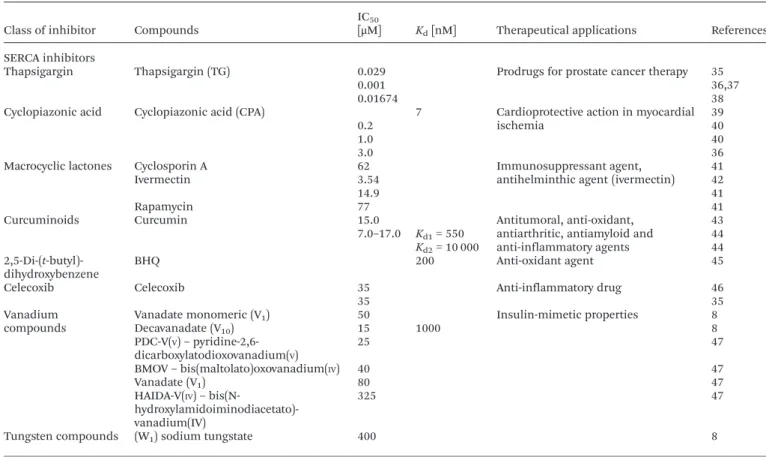
Table 3 IC50values and/or dissociation constants and therapeutical applications of known Ca2+-ATPase (SERCA) inhibitors
Class of inhibitor Compounds
IC50
[μM] Kd[nM] Therapeutical applications References
SERCA inhibitors
Thapsigargin Thapsigargin (TG) 0.029 Prodrugs for prostate cancer therapy 35
0.001 36,37
0.01674 38
Cyclopiazonic acid Cyclopiazonic acid (CPA) 7 Cardioprotective action in myocardial ischemia
39
0.2 40
1.0 40
3.0 36
Macrocyclic lactones Cyclosporin A 62 Immunosuppressant agent, antihelminthic agent (ivermectin)
41
Ivermectin 3.54 42
14.9 41
Rapamycin 77 41
Curcuminoids Curcumin 15.0 Antitumoral, anti-oxidant, antiarthritic, antiamyloid and anti-inflammatory agents 43 7.0–17.0 Kd1= 550 44 Kd2= 10 000 44 2,5-Di-(t-butyl)-dihydroxybenzene BHQ 200 Anti-oxidant agent 45
Celecoxib Celecoxib 35 Anti-inflammatory drug 46
35 35
Vanadium compounds
Vanadate monomeric (V1) 50 Insulin-mimetic properties 8
Decavanadate (V10) 15 1000 8 PDC-V(V)– pyridine-2,6-dicarboxylatodioxovanadium(V) 25 47 BMOV– bis(maltolato)oxovanadium(IV) 40 47 Vanadate (V1) 80 47 HAIDA-V(IV)– bis(N- hydroxylamidoiminodiacetato)-vanadium(IV) 325 47
Tungsten compounds (W1) sodium tungstate 400 8
increase of intracellular calcium concentration through the Na+/Ca2+ exchanger. This in turn leads to an increase in muscle contraction which is the origin of the classification of ouabain as a cardiotonic drug. That it is also being used in the treatment of breast cancer emphasises the importance of the ion pumps in the regulation of calcium homeostasis and cell survival.10–17It is not clear if different-sized CTS also bind to the same site or as strongly, and whether they block the trans-location of intracellular sodium with the same potency.2
Other Na+/K+-ATPase inhibitors include the anti-psychotic drug chlorpromazine, the anti-malarial agent chloroquine and the antibiotic gramicidine A (IC50 = 8 μM) (Table 1).10–25 These drugs also affect other proteins, however, and the effects on the Na+/K+-ATPase are only observed for higher concentrations.18,19
Monomeric vanadate is a well-known Na+/K+-ATPase inhibi-tor which is selective towards the E2P conformation. Besides vanadate, tungstate also is known to inhibit the Na+/K+ -ATPase, with dodecatungstate (W12) exhibiting a higher capacity of inhibition (IC50ca. 3μM) than the tungstate ion, WO42−(IC50= 1.5 mM).25However, monomeric tungstate has been demonstrated to be a more potent inhibitor than dodeca-tungstate towards SR Ca2+-ATPase (IC50= 400μM).8Moreover, it has been suggested that for Na+/K+-ATPase tungstate pre-sents a competitive type of inhibition towards ATP, and blocks the enzyme at the nucleotide domain.8
3.
H
+/K
+-ATPase inhibitors
H+/K+-ATPase inhibitors include anti-ulceral, anti-viral and antibiotic agents (Table 2). H+/K+-inhibition is normally a strat-egy in preventing acidic segregation since gastric diseases are very painful and can lead to hepatic ulceration. Proton pump inhibitors (PPI), such as the drug omeprazole (IC50 ca. 3–4 μM), present lower inhibitory capacity than the potassium competitive acid blockers (PCAB; IC50= 0.1μM) or prodigiosin (IC50= 0.03μM).26–34
Omeprazole irreversibly inhibits H+/K+-ATPase through the formation of a metabolite which reacts with the cysteine resi-dues of the protein and causes pump inhibition. It was shown that cysteine residue 813 is oxidized upon PPI interaction, but other cysteine residues may also be involved such as cysteine residues 321, 822 or 892, which are part of several trans-membranal helices.
Vanadate is shown to be a potent H+/K+-ATPase inhibitor as it is for the Na+/K+-ATPase.62It reacts with the E2 form of the enzyme and when the E1 form is favoured the inhibition was not observed.63
4.
Ca
2+-ATPase inhibitors
The Ca2+-ATPase inhibitors include compounds used for cancer therapy, cardioprotection, immunosuppression and as antitumoral agents (Table 3). Thapsigargin (TG) is currently
the most potent calcium pump inhibitor known, and presents an inhibitory value in the nM range (IC50 = 1 nM)8,35,45–47 (Table 3). The inhibition leads to increasing cytoplasmic calcium levels, which leads to the activation of apoptotic factors which trigger cell death, making TG an active anti-tumoral agent.
Both TG and cyclopiazonic acid (CPA; IC50ca. 1μM) bind to the E2 conformation of the enzyme, but at different binding sites.2 As described for other P-type inhibitors, these com-pounds bind to the protein in the membrane domain, at the entrance of the calcium release channel close to the mem-brane surface. However, TG and CPA do not bind to the same exact site, and therefore the protein can bind simultaneously to both inhibitors.2Benzohydroquinone (BHQ) inhibitors bind to the same binding site as CPA.
Whereas TG blocks the transmembranal helices TM3, TM5 and TM7, CPA moves TM1, TM2 towards to TM4. In both types of interaction the drugs prevent the release of calcium. Although the majority of the Ca2+-ATPase inhibitors in Table 3 seem to block the E2 conformation of the enzyme, a few, such as curcumine, also interact with the E1P conformation.2
Vanadate species, in particular decavanadate (V10), are potent inhibitors (IC50= 15μM) of the hydrolytic activity of the sarcoplasmic reticulum Ca2+-ATPase, with activities similar to that observed for decaniobate, [Nb10O28]6−, and higher than for WO42− and MO42−.8 SR Ca2+-ATPase activity is also inhi-bited by vanadium complexes such as vanadium-citrate and bis(maltolato)oxovanadium(IV) (BMOV) (Table 1).47,48 These
and other vanadium compounds have been described in the treatment of diabetes mellitus.49Furthermore, decavanadate,50
and compounds containing decavanadate51 have been
reported to lower glycemic levels,50,51and to normalize plasma lipid profiles.51
Due to the ability of V10to interfere with several biological processes, such as calcium homeostasis, muscle contraction and oxidative stress,8,54,55not only in vitro but also in vivo,52,53 decavanadate has recently been associated with the treatment of Leishmaniosis.56In this and probably in many other cases the mechanism of action of decavanadate progresses via the inhibition of the E1–E2 Ca2+-ATPases. However, mitochondria may be an additional target as several mitochondrial processes have been shown to be affected by decavanadate at nanomolar concentrations.57,58
Decavanadate can also induce protein cysteine oxidation with concomitant V(V) reduction to V(IV). Thus we cannot
exclude the potential activity of V(IV) species in inducing
changes in the Ca2+-ATPase structure and function.8,9 Both decaniobate and decavanadate have recently been shown to interact non-competitively with SR Ca2+-ATPases with respect to ATP. The V10binding site on the E2 conformer is located at the intersection of the three cytoplasmic protein domains that includes the nucleotide, the phosphorylation and theβ-strand domains.4 Twenty years ago decavanadate was demonstrated to be specific in inducing crystallization of SR Ca2+-ATPase.59 These and other recent studies emphasize that decavanadate interactions with the calcium pump are clearly different from
those of monomeric vanadate by inducing different changes in protein structure and function, and indicate different biologi-cal roles for the two oxovanadate species. These differences may explain the different biological effects described in several studies. For instance, at near-physiological conditions with calcium and without a calcium ionophore only decavanadate inhibits calcium translocation whereas vanadate does not.6 Still, although there has been some progress in improving the understanding of the interaction and the effect of V10on the structure and function of the Ca2+-ATPases, the mechanism of action on the Ca2+-ATPase is still not fully understood, parti-cularly under physiological conditions.
Given that the decavanadate-specific Ca2+-ATPase binding site is located on the cytoplasmic side, one would expect that V10 needs to cross the cell membrane in order to be active. Decavanadate can, however, also affect the cell membrane itself,60 for example by decreasing lipid packing which may induce changes in the composition of proteins in membrane microdomains.61This offers an indirect mode of action which does not require transmembranal transport. By changing membrane structure and dynamics, alterations on the struc-ture and function of the E1–E2 ATPases can also occur. Finally, we cannot exclude that V10might interact with the ion pumps from the extracellular space, as is the case with many drugs (Fig. 1).
Polyoxotungstates have also been shown to inhibit not only ion pumps such as Na+/K+-ATPase, H+/K+-ATPase, and Ca2+ -ATPase but also ecto--ATPases.25In fact, polyoxotungstates are 10–100 times more potent as inhibitors25than monovanadium coordination complexes,47,48 while decavanadate IC50 values are one to two orders of magnitude lower than those of the polyoxotungstates.8,9
The mechanism of inhibition and interaction of decavana-date on the sarcoplasmic reticulum calcium pump ATPase is clearly different from that of other polyoxometalates and oxo-metalates. It involves protein cysteine oxidation and vanadate reduction processes, which lead to non-competitive ATPase inhibition. This inhibition is not prevented by antioxidant agents that can reverse the cysteine oxidation.9Moreover, deca-vanadate interacts with ATPase in the same fashion for all the conformations that occurs during the ATP-hydrolysis coupled calcium translocation. These recent findings emphasise the crucial need to properly understand and compare the ion pump inhibition processes of several types of inhibitors, since they may well turn out to be significantly different.
Given the specific activity of these structures towards the ion pumps, polyoxometalates are promising as drugs for anti-cancer activity, protection against viral, bacterial and protozoa infections, and antidiabetic activity.49–51,56
5.
Conclusions
Although some 18 classes of ion pump inhibitors are known, the biochemical mechanisms are only known for a handful of compounds. The majority of these compounds – such as
ouabain, omeprazole, thapsigargin, CPA and BHQ – seem to interact selectively with the E2 or E2P conformations of the target enzyme, which can be understood in light of the binding site being located in the transmembranal region, at or close to the calcium binding site. The binding is normally reversible, but, in the case of omeprazole, nearby cysteine units are also directly affected, which leads to the irreversible inhibition of the pump.
Thapsigargin is one of the most potent inhibitors of the Ca2+-ATPases, and of all the E1–E2 ion pump inhibitors dis-cussed in the present Perspective, with an IC50 in the nM range of concentration. While vanadium and tungsten mono-and polyoxometalates have IC50values which are three orders of magnitude (IC50= 2–15 μM) higher than TG, they still have activities comparable with other relevant ion pump inhibitors.
The mode of action of decavanadate and the other oxometa-lates implies that the main focus of the interaction is on the cytoplasmic domain of the protein, more specifically at the nucleotide or phosphorylation domains or at a pocket invol-ving several cytoplasmic domains. Although decavanadate interacts most strongly with the E2 conformation of the Ca2+ -ATPase, as do some of the other drugs described above, deca-vanadate also interacts with other conformations.6,8Therefore, decavanadate exhibits a particular feature that may be useful for the understanding of the mode of action of ion pump inhibitors in general. Besides, the specific behaviour of decava-nadate towards ion pumps may guide the development of new drugs that would have these pumps as therapeutic targets. Finally, it is believed that the present observations will contri-bute to the understanding of oxometalate interactions with the
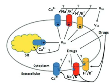
Fig. 2 E1–E2 ATPases, a family of cation pump enzymes, are known targets for specific drugs including decavanadate (V10). The majority of the drugs bind
within the bilayer near the entrance of the cation exit channel whereas decava-nadate binds to a pocket formed by protein domains at the cytoplasmic side. Decavanadate and other polyoxometalates need to cross the bilayer before tar-geting the ion pumps, something which probably occurs through transport membrane systems such as channels, or specific transporters and/or pumps. In contrast, established organic drugs do not apparently need to cross the mem-brane, and probably for V10this mechanism of action cannot be completely
excluded. Na+/K+, H+/K+, and Ca2+stand for Na+, K+-ATPase, H+, K+-ATPase, and
Ca2+-ATPase, respectively. SR denotes sarcoplasmic reticulum.
calcium pump and the role of decavanadate in biology, particularly the contribution of decavanadate to calcium homeostasis.
In summary, E1–E2 ATPases are targets for several classes of compounds, including decavanadate, and present several potential binding sites. The majority of the established drugs bind near the entrance of the cation binding sites within the bilayer, whereas decavanadate binds to a pocket formed by protein domains at the cytoplasmic side. Decavanadate and other polyoxometalates need to cross the bilayer before target-ing the ion pumps, somethtarget-ing which probably occurs through transport membrane systems such as channels, or specific transporters and/or pumps. In contrast, a majority of estab-lished organic drugs do not appear to need to cross the mem-brane. However, the former mechanism of action cannot be completely excluded for decavanadate (Fig. 2).
Abbreviations
BHQ Benzohydroquinone
CPA Cyclopiazonic acid
CTS Cardiotonic steroids
E1 Protein conformation E1
E1P Protein conformation E1 phosphorylated
E2 Protein conformation E2
E2P Protein conformation E2 phosphorylated PCAB Potassium competitive acid blockers PPIs Proton pump inhibitors
SR Sarcoplasmic reticulum
SERCA Sarco(endo) reticulum calcium ATPase
TG Thapsigargin
TM Transmembranal helices
V10 Decavanadate, a vanadate oligomer containing 10 vanadate units
W12 Polyoxotungstate containing 12 tungstate units
Acknowledgements
We thank the Portuguese Research Council (FCT) for funding via project PTDC/QUI-BIQ/112943/2009, and FCT grant (SFRH/ PROTEC/67781/2010) to GF which also supported the studies in ref. 8 and 9. MA and GF also acknowledge financial support from CCMAR at the University of Algarve. CAO thanks the Australian Research Council for grants DP110105530 and DP130100483.
References
1 L. C. Cantley Jr., L. Josephson, R. Warner, M. Yanagisawa, C. Lechene and G. Guidotti, J. Biol. Chem., 1977, 252, 7421–7423.
2 L. Yatime, M. J. Buch-Pedersen, M. Musgaard, J. P. Morth, L. Anne-Marie, B. Winther, P. Pedersen, C. Olesen, J. P. Andersen, B. Vilsen, B. Schiøtt, M. G. Palmgre,
J. V. Møller, P. Nissen and N. Fedosova, Biochim. Biophys. Acta, Bioenerg., 2009, 1787, 207–220.
3 L. de Meis and A. L. Vianna, Annu. Rev. Biochem., 1979, 48, 275–292.
4 C. Toyoshima, M. Nakasako, H. Nomura and H. Ogawa, Nature, 2004, 405, 647–655.
5 M. Aureliano, World J. Biol. Chem., 2011, 2, 215–238. 6 M. Aureliano and V. M. C. Madeira, Biochim. Biophys. Acta,
Mol. Cell Res., 1994, 1221, 259–271.
7 P. L. Pedersen and E. Carafoli, Trends Biochem. Sci., 1987, 12, 146–150.
8 G. Fraqueza, C. A. Ohlin, W. H. Casey and M. Aureliano, J. Inorg. Biochem., 2012, 107, 82–89.
9 G. Fraqueza, L. A. E. Batista de Carvalho, M. Paula, M. Marques, L. Maia, C. André Ohlin, W. H. Casey and M. Aureliano, Dalton Trans., 2012, 41, 12749–12758. 10 M. Juhaszova and M. P. Blaustein, Proc. Natl. Acad.
Sci. U. S. A., 1997, 94, 1800–1805.
11 K. Ihenetu, H. M. Qazzaz, F. Crespo, R. Fernandez-Botran and R. Valdes Jr., Clin. Chem., 2007, 53, 1315–1322. 12 H. Bottinger and E. Habermann, Naunyn-Schmiedebergs
Arch. Pharmacol., 1984, 325, 85–87.
13 D. N. Almotrefi, Gen. Pharmacol., 1991, 22, 403–406. 14 A. N. Wansapura, V. Lasko, Z. Xie, O. V. Fedorova,
A. Y. Bagrov, J. B. Lingrel and J. N. Lorenz, Am. J. Physiol.: Heart Circ. Physiol., 2009, 296, 1833–1839.
15 J. Joep, H. H. M. De Pont, H. G. P. Swarts, A. Karawajczyk, G. Schaftenaar, P. H. G. M. Willems and J. B. Koenderink, Pfluegers Arch., 2009, 457, 623–634.
16 C. Scheiner-Bobis and H. Schneider, Eur. J. Biochem., 1997, 248, 717–723.
17 Z. Zhang, Z. Li, J. Tian, W. Jiang, Y. Wang, X. Zhang, Z. Li, Q. You, J. I. Shapiro, S. Si and Z. Xie, Mol. Pharmacol., 2010, 77, 961–967.
18 D. Bhattacharyya and P. C. Sen, Mol. Cell. Biochem., 1999, 198, 179–185.
19 P. R. S. Kodavanti, W. R. Mundy, H. A. Tilson and G. Jean Harry, Fundam. Appl. Toxicol., 1993, 21, 308–316.
20 H. Kutchai and L. M. Geddis, Pharmacol. Res., 2001, 43, 399–403.
21 R. Coca, F. Soler and F. Fernández-Belda, Arch. Biochem. Biophys., 2008, 478, 36–42.
22 Y. Ishida, K. Takagi, M. Takahashi, N. Satake and S. Shibata, J. Biol. Chem., 1983, 258, 7900–7902.
23 H. Chau Wu, Toxicon, 2009, 54, 1183–1189.
24 Y. Takada, K. Matsuo and T. Kataoka, Mol. Cell. Biochem., 2008, 319, 99–103.
25 M. B. Colović, D. V. Bajuk-Bogdanovic, N. S. Avramovic, I. D. Holclajtner-Antunovic, N. S. Bošnjaković-Pavlovic, V. M. Vasić and D. Z. Krstić, Bioorg. Med. Chem., 2011, 19, 7063–7069.
26 H. Matsuya, M. Okamoto, T. Ochi, A. Nishikawa,
S. Shimizu, T. Kataoka, K. Nagai, H. H. Wasserman and S. Ohkuma, Biochem. Pharmacol., 2000, 60, 1855–1863. 27 D. Seto-Young, B. Monk, A. B. Mason and D. S. Perlin,
Biochim. Biophys. Acta, Biomembr., 1997, 1326, 249–256.
28 W. Beil, U. Staar and K. F. Sewing, Eur. J. Pharmacol., 1992, 218, 265–271.
29 D. J. Keeling, C. Fallowfield, K. J. Milliner, S. K. Tingley, R. J. Ife and A. H. Underwood, Biochem. Pharmacol., 1985, 34, 2967–2973.
30 R. A. Farley, S. Schreiber, S.-G. Wang and G. Scheiner-Bobis, J. Biol. Chem., 2001, 276, 2608–2615.
31 W. A. Simon, M. Herrmann, T. Klein, J. M. Shin, R. Huber, J. Senn-Bilfinger and S. Postius, J. Pharmacol. Exp. Ther., 2007, 321, 866–874.
32 S. Asano, M. Mizutani, T. Hayashi, N. Morita and N. Takeguchi, J. Biol. Chem., 1990, 265, 22167–22173. 33 T. Hayashi, K. Okamura, M. Kakemi, S. Asano, M. Mizutani,
N. Takeguchi, M. Kawasaki, Y. Tezuka, T. Kikuchi and N. Morita, Chem. Pharm. Bull., 1990, 38, 2740–2745. 34 T. Kataoka, M. Muroi, S. Ohkuma, T. Waritani, J. Magae,
A. Takatsuki, S. Kondo, M. Yamasaki and K. Nagai, FEBS Lett., 1995, 359, 53–59.
35 A. J. Johnson, A.-L. Hsu, H.-P. Lin, X. Song and C.-S. Chen, Biochem. J., 2002, 366, 831–837.
36 Y. Geng and M. Lotz, J. Cell Biol., 1995, 129, 1651–1657. 37 Y. Sagara and G. Inesi, J. Biol. Chem., 1991, 266,
13503–13506.
38 I. Moreno, L. Norambuena, D. Maturana, M. Toro, C. Vergara, A. Orellana, A. Zurita-Silva and V. R. Ordenes, J. Biol. Chem., 2008, 283, 9633–9641.
39 S. F. Plenge-Tellechea, I. Fortea and F. Fernandez-Belda, Biochemistry, 1998, 37, 4266–4274.
40 N. J. Yard, M. Chiesi and H. A. Ball, Br. J. Pharmacol., 1994, 113, 1001–1007.
41 J. G. Bilmen, L. L. Wootton and F. Michelangeli, Biochem. J., 2002, 366, 255–263.
42 G. P. Ahern, P. R. Junankar, S. M. Pace, S. Curtis, J. A. Mould and A. F. Dulhunty, J. Physiol., 1999, 514, 313–326.
43 M. J. Logan-Smith, P. J. Lockyer, J. M. East and A. G. Lee, J. Biol. Chem., 2001, 276, 46905–46911.
44 J. G. Bilmen, S. Z. Khan, M. H. Javed and F. Michelangeli, Eur. J. Biochem., 2001, 268, 6318–6327.
45 M. J. Logan-Smith, P. J. Lockyer, J. M. East and A. G. Lee, J. Biol. Chem., 2001, 276, 46905–46911.
46 S. Grösch, T. J. Maier, S. Schiffmann and G. Geisslinger, J. Natl. Cancer Inst., 2006, 98, 736–747.
47 M. Aureliano, F. Henao, T. Tiago, R. O. Duarte,
J. J. G. Moura, B. Baruah and D. C. Crans, Inorg. Chem., 2008, 47, 5677–5684.
48 M. Aureliano, T. Tiago, R. M. C. Gândara, A. Sousa, A. Moderno, M. Kaliva, A. Salifoglou, R. O. Duarte and J. J. G. Moura, J. Inorg. Biochem., 2005, 99, 2355–2361. 49 K. H. Thompson and C. Orvig, Met. Ions Biol. Syst., 2004,
41, 221–252.
50 M. J. Pereira, E. Carvalho, J. W. Eriksson, D. C. Crans and M. Aureliano, J. Inorg. Biochem., 2009, 103, 1687–1692. 51 F. Yraola, S. Garcia-Vicente, L. Marti, F. Albericio,
A. Zorzano and M. Royo, Chem. Biol. Drug Des., 2007, 69, 423–428.
52 M. Aureliano and R. M. C. Gândara, J. Inorg. Biochem., 2005, 99, 979–985.
53 M. Aureliano and D. C. Crans, J. Inorg. Biochem., 2009, 103, 536–546.
54 M. Aureliano, Dalton Trans., 2009, 9093.
55 R. M. C. Gândara, S. S. Soares, H. Martins, C. Gutiérrez-Merino and M. Aureliano, J. Inorg. Biochem., 2005, 99, 1238–1244.
56 T. L. Turner, V. H. Nguyen, C. C. McLauchlan, Z. Dymon, B. M. Dorsey, J. D. Hooker and M. A. Jones, J. Inorg. Biochem., 2012, 108, 96–104.
57 S. S. Soares, C. Gutiérrez-Merino and M. Aureliano, J. Inorg. Biochem., 2007, 101, 789–796.
58 S. S. Soares, F. Henao, M. Aureliano and C. Gutierrez-Merino, Chem. Res. Toxicol., 2008, 21, 607–618.
59 A. Maurer and S. Fleischer, J. Bioenerg. Biomembr., 1984, 16, 491–505.
60 B. Baruah, L. A. Swafford, D. C. Crans and N. E. Levinger, J. Phys. Chem. B, 2008, 112, 10158–10164.
61 P. W. Winter, A. Al-Quati, A. L. Wolf-Ringwall, S. Schoeberl, P. B. Chatterjee, B. G. Barisas, D. A. Roess and D. C. Crans, Dalton Trans., 2012, 41, 6419–6430.
62 L. D. Faller, E. Rabon and G. Sachs, Biochemistry, 1983, 22, 4676–4685.
63 K. L. Dürr, N. N. Tavraz and T. Friedrich, PLoS One, 2012, 7, 33645.