INTRODUCTION
The principle of osseoint egrat ion is based on int imat e bone- implant cont act and t he bone volume and qualit y are f undament al f act ors t o achieve osseoint egrat ion during t he healing and maint ained along t he years under load condit ions (1).
Syst emic diseases such as diabet es mellit us (2,3) and ost eoporosis (4, 5, 6), radiot herapy (6), smoking habit s (7) and some drugs t herapy (8, 9) can induce alt erat ions on bone met abolism leading t o a poor bone qualit y and impairing t he healing of bone t issue. Cyclosporine A (CsA) is an immunosuppressive agent commonly used t o prevent organ t ransplant at ion reject ion and t reat ot her immunologic diseases (10). CsA act s on immune syst em inducing T- helper lymphocyt es suppression. This mechanism may af f ect bone t issue, since immune syst em, part icularly T-lymphocyt es, play a crit ical role on bone remodeling (11- 14). Some animal st udies have demonst rat ed t hat t his drug leads t o a high bone t urnover, result ing in unbalance of resorpt ion and f ormat ion, leading t o ost eopenia (11- 13). St udies in t ransplant ed pat ient s receiving CsA t herapy showed high incidence of ost eoporosis conf irming t his delet erious ef f ect on bone met abolism in human (15- 17).
The inf luence of CsA in osseoint egrat ion has been st udied by Duart e et al. (2001) (8) and Sakakura et al. (2003) (9) and it has been shown t hat CsA may negat ively af f ect osseoint egrat ion, reducing t he bone- t o- implant cont act and bone f ormat ion around dent al implant s. On t he ot her hand, t here are no st udies report ing t he inf luence of CsA on t he preexist ing bone densit y, which may be considered an import ant indicat or of t he qualit y of bone t issue f ormed around dent al implant s. Theref ore, t he aim of
CELSO EDUARDO SAKAKURA, DDS, MSC, PHD
2, BEATRIZ MARIA VALÉRIO LOPES, DDS, MSC, PHD
1,
ROGÉRIO MARGONAR DDS, MSC, PHD
3, THALLITA PEREIRA QUEIROZ, DDS, MSC, PHD
4,
FRANCISCO HUMBERTO NOCITI JÚNIOR, DDS, MSC, PHD
5, ELCIO MARCANTONIO JÚNIOR, DDS, MS, PHD
1>
1
Department of Periodontology, Araraquara Dental School, São Paulo State University (UNESP) Araraquara, São Paulo, Brazil
2
Department of Periodontology, Barretos Dental School, Education Foundation of Barretos, FEB, Barretos, SP, Brazil
3
Department of Health Sciences, Disciplines of Periodontology, Integrated Clinic, and Implantology Post Graduation Course, Dental School,
University Center of Araraquara, UNIARA, Araraquara, São Paulo, Brazil
4
Department of Health Sciences, Disciplines of Oral and Maxillofacial Surgery, Integrated Clinic and Implantology Post Graduation Course,
Dental School, University Center of Araraquara, UNIARA, Araraquara, São Paulo, Brazil
5
Department of Prosthodontics and Periodontics, Division of Periodontics, Piracicaba Dental School, UNICAMP, São Paulo, Brazil
Cyclosporine-a and bone density around titanium
implants: a histometric study in rabbits
KEY WORDS
Bone Density; Cyclosporine; Dental Implants.
ABSTRACT
Aim
Cyclosporine A (CsA) is an immunosuppressive agent
common-ly used to prevent organ transplantation rejection. It has been
demonstrated that CsA may negatively affect osseointegration
around dental implants. Therefore, the aim of this study was to
eva-luate the effect of CsA administration on bone density around
tita-nium dental implants.
Materials and Methods
Fourteen New Zealand rabbits were
ran-domly divided into 2 groups with seven animals each. The test group
(CsA) received daily subcutaneous injection of CsA (10mg/kg body
weight) and the control group (CTL) received saline solution by the
same route of administration. Three days after the beginning of
immunosuppressive therapy, one machined dental implant (7.00 mm
in lenght and 3.75 mm in diameter) was inserted bilaterally at the
region of the tibial methaphysis. After 4 and 8 weeks the animals were
sacrificed and the histometrical procedures were performed to
analy-se the bone density around the first four threads of the coronal part of
the implant.
Results
A significant increase in the bone density was observed from
the 4- to the 8 week-period in the control group (37.41% + 14.85
versus 58.23% + 16.38 – p < 0.01). In contrast, bone density
con-sistently decreased in the test group overtime (46.31% + 17.38
ver-sus 16.28 + 5.08 – p <0.05). In the 8-week period, there was a
significant difference in bone density between the control and the
test groups (58.23 + 16.38 eand16.28 + 5.08 – p= 0.001).
t his st udy was t o evaluat e t he inf luence of CsA administ rat ion on bone densit y around t it anium dent al implant s.
M ATERIALS AND M ETHODS
Animals
Fourt een New Zealand whit e rabbit s, 9 t o 12 mont hs old (3500- 4500 g), were used in t he st udy. The animals were housed in individual cages, f ed by a st andard laborat ory diet and given t ap wat er ad libit um. The experiment w as approved by t he Inst it ut ional Experiment at ion Commit t ee of t he Araraquara Dent al School, São Paulo, Brazil.
Experiment al prot ocol
Af t er a 2- week acclimat izat ion period, t he animals were randomly divided int o t wo groups, t est (CsA) and cont rol (CTL) groups, wit h seven animals each. CsA was daily administ ered by a subcut aneous (10 mg/kg bodyweight ), whereas t he same rout e was used t o administ er saline solut ion (NaCl 0.9%) t o t he CTL group. The administ rat ion of t he drugs began t hree days bef ore t he implant s placement and last ed f or 4 and 8 weeks post operat ively.
Implant surgery
The animals were anest het ized by int ramuscular inject ions of a combinat ion of ket amine (Francot ar®; Virbac do Brasil Lt da, Brazil) (0.35 mg/kg bodyweight ) and xylazine (Rumpum® Bayer S.A. São Paulo, Brazil) (0.5mg/kg bodyweight ). The region of t he t ibial met aphysis was cleansed wit h iodine surgical soap. Incisions of approximat ely 3 cm in lengt h were perf ormed bilat erally at t he int ernal side of t he hind-leg, just below t he knee. Af t er gent le dissect ion, t he bone surf ace of t he t ibial met aphysis was exposed. Unicort ical implant beds were prepared by using a progressive sequence of spiral drills under generous saline cooling.
One machine surf ace t it anium implant s, (7 mm lengt h and 3.75 mm in diamet er), was placed in each leg (Fig. 1). The sof t t issues were sut ured in separat e layers and t he animals received a single int ramuscular inject ion of ant ibiot ic (Pent abiót ico®, Wyet h- Whit ehall Lt da, São Paulo, Brazil) (0.1 ml/kg bodyweight of an associat ion of Penicillin wit h St rept omycin) post operat ively.
Hist omet ric procedure
Af t er 4 and 8 weeks, t he animals were sacrif iced and t he hist omet rical procedures were perf ormed t o obt ain t he bone densit y around t he f irst f our t hreads apical t o t he implant plat f orm, according t o met hods published by Feit osa et al. 2008 (18) and Correa et al. 2010 (19).
The convent ional implant s wit h surrounding t issue in each t ibia were removed and f ixed in 4% neut ral f ormalin f or 48 hours. Non decalcif ied sect ions were prepared by a t echnique previously described by Donat h and Breuner (1992) (20) Subsequent ly, t he sect ions were st ained as f ollows:
>
t he slide- cont aining specimen was placed in avessel cont aining St evenel’s blue preheat ed t o, and maint ained at , 60 °C f or 15 minut es;
>
t he specimen was rinsed in dist illed wat er at 60 °Cand air dried;
>
a small amount of alizarin red was placed ont o t hespecimen surf ace at room t emperat ure f or 5 minut es.
Then, it was washed t horoughly in running dist illed wat er t o remove excess st ain and air dried.
The bone densit y (i.e, proport ion of mineralized bone in a 500 µm- wide zone lat eral t o t he implant and not inside t he t hreads) was measured at bot h sides of t he implant , at t he f irst f our t hreads (Fig. 2). The analysis was perf ormed by a single examiner, t rained and calibrat ed. Theref ore, t his area lat eral t o t he implant w as enclosed by a rect angle w hose t ot al area corresponded t o 100%.
Then, t he area of bone f ill wit hin t he rect angle was calculat ed and t he percent age value was obt ained by simple rule of t hree. The mean of dat a obt ained f or bot h sides of t he implant w as considered f or st at ist ical analysis.
Fig. 1
Machined surface titanium implant placed in each tibial
metaphysis. A. Receptor site; B. Preparation of receptor site; C. Implant
placement.
Fig. 2
St at ist ical analysis
Since dat a were normally dist ribut ed, as demonst rat ed by Kolmogorov and Smirnov t est , unpaired t t est was used t o access dif f erence in bone densit y bet ween t he groups in each experiment al period (4- week and 8-week period). Paired t t est was used t o compare dif f erences in bone densit y bet ween t he 4- week and t he 8- week period in each experiment al group separat ely. P value was set at 0.05.
RESULTS
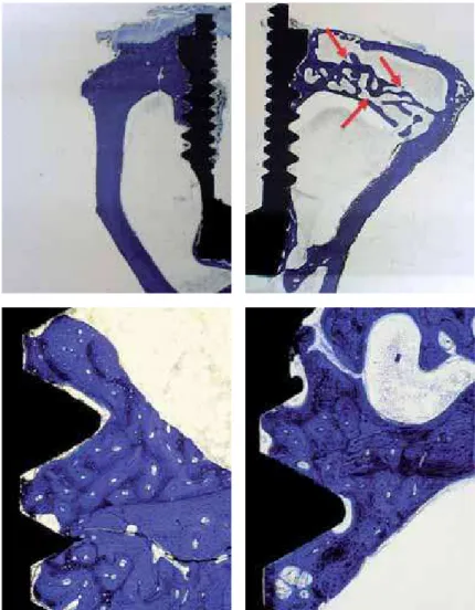
Table 1 shows t he mean and st andard deviat ion of each group at dif f erent periods of analysis. A st at ist ically signif icant increase in bone densit y bet ween t he 4 and 8- week period was observed f or t he cont rol group (37.41% + 14.85 versus 58.23% + 16.38 – p < 0.01). In cont rast , bone densit y consist ent ly decreased in t he t est group overt ime (46.31% + 17.38 versus 16.28 + 5.08 – p <0.05) (Fig. 3). In t he 8- week period, t here was a st at ist ically signif icant dif f erence in bone densit y bet ween t he cont rol and t he t est group (58.23 + 16.38 versus
16.28 + 5.08 – p= 0.001) (Fig. 4 A, B, C and D). No st at ist ically signif icant dif f erence could be f ound bet ween t est and cont rol groups in t he 4- week period (46.31% + 17.38 versus 37.41% + 14.85).
PERIODS CONTROL TEST
4 weeks 33,11 73,06
50,94 60,74
33,77 28,96
57,88 37,25
32,46 32,05
16,30 45,81
37,41 46,31
Mean Standard 37,41 46,31
Deviation 13,56 15,86
8 weeks 60,60 24,16
78,01 19,83
52,55 15,57
76,14 10,22
40,16 12,16
41,95 15,76
58,24 16,28
Mean Standard 58,23 16,28
Deviation 14,95 4,64
Table 1
Bone density values from each
animal, and mean and standard deviation
from both groups and periods.
Fig. 3
Mean (%) and standard deviation for bone density around
implants for control and test groups at 4 and 8 weeks post-surgery.
areas where t rabecular bone predominat es, such as in t he maxilla, bone densit y around dent al implant s may be severely damaged. Fu et al. (1999) (28) f ound an increased alveolar bone resorpt ion in rat s receiving CsA, wit h great er bone loss in sit es af f ect ed by periodont it is.
Alt hough it could be demonst rat ed a negat ive side ef f ect of CsA on cort ical bone healing around dent al implant s, f urt her st udies should be developed in order t o invest igat e t he ef f ect s of t his drug on t rabecular bone and around osseoint egrat ed f unct ionally loaded dent al implant s. Wit hin t he limit s of t his st udy, long- t erm CsA immunesuppression may reduce bone densit y around t it anium dent al implant s during t he osseoint egrat ion process.
REFERENCES
1. Albrekt sson T. Bone t issue response. In: Brånemark P- I, Zarb G, Albrekt sson T, eds. Tissue int egrat ed prost hesis osseoint egrat ion in clinical dent ist ry. Chicago: Quint essence, 1985: 129- 144.
2. M argonar R, Sakakura CE, Holzhausen M , Pepat o M T, Alba Jr RC, M arcant onio Jr E. The Inf luence of Diabet es M ellit us and Insulin Therapy on Biom echanical Ret ent ionAround Dent al Implant s: A St udy in Rabbit s. Implant Dent (in press)
3. Takeshit a F, M urai K, Iyama S, et al. Uncont rolled diabet es hinders bone f ormat ionaround t it anium implant s in rat t ibiae. A light and f luorescence microscopy,and image processing st udy. J Periodont ol.1998;69:314–320. 4. Dao TT, Anderson JD, Zarb GA. Is ost eoporosis a risk f act or
f or osseoint egrat ion of dent al implant s? Int J Oral M axillof ac Implant s. 1993: 8; 137- 144.
5. Duart e PM , Cesar- Net o JB, Sallum AW, Sallum EA, Nocit i FH Jr. Ef f ect of est rogen and calcit onin t herapies on bone densit y in a lat eral area adjacent t o implant s placed in t he t ibiae of ovariect om ized rat s. J Periodont ol. 2003;74(11):1618- 24.
6. Granst röm G. Radiot herapy, osseoint egrat ion and hyperbaric oxygen t herapy. Periodont ol 2000 2003: 33; 145- 162.
7. Cesar- Net o JB, Duart e PM , Sallum EA, Barbieri D, M oreno H Jr, Nocit i FH Jr. A comparat ive st udy on t he ef f ect of nicot ine administ rat ion and cigaret t e smoke inhalat ion on bone healing around t it anium implant s. J Periodont ol. 2003 Oct ;74(10):1454- 9.
8. Duart e PM , Nogueira Filho GR, Sallum EA, Sallum AW, Nocit i Junior FH. The ef f ect of an immunosuppressive t herapy and it s w it hdraw al on bone healing around t it anium implant s. A hist omet ric st udy in rabbit s. J Periodont ol 2001; 72:1391- 7.
9. Sakakura CE, M argonar R, Holzhausen M , Nocit i Jr FH, Alba Jr RC, M arcant onio Jr E. inf luence of cyclosporin a t herapy on bone healing around t it anium implant s. A hist om et ric and biom echanic st udy in rabbit s. J Periodont ol. 2003; 74: 974- 979.
10. Ruegger A, Kuhn M , Licht i H, et al. Cyclosporin A, a Pept ide M et abolit e f rom Trichoderma polysporum (Link ex
DISCUSSION AND CONCLUSION
The increasing of bone densit y around dent al implant s along t he t ime may occur due t o t he bone remodeling processes (Davies et al., 2003) (21). In our st udy, it was also f ound a st at ist ically signif icant increase in bone densit y f rom t he 4- week t o t he 8-week period in t he cont rol group (37.41% + 14.85 versus 58.23% + 16.38 – p < 0.01).
However, in t he CsA group, a st at ist ically signif icant decrease in bone densit y was observed around t he implant s f rom t he 4- week t o t he 8- week period (46.31% + 17.38 and 16.28 + 5.08 – p <0.05). This suggest s t hat CsA administ rat ion during 8- week period may have negat ively af f ect ed t he bone mineralizat ion around dent al implant . The possible reasons f or explain t hese result s are relat ed wit h t he mechanism of imunossupression caused by CsA. It is known t hat t he immune syst em act ively part icipat es in bone mineral met abolism (13, 14, 17) and t hat t he T lymphocyt es play a crit ical role in t he development of CsA- induced ost eopenia (22). This is not surprising as t he T cell is t he t radit ional t arget of CsA, and nat urally occurring T lymphocyt e pert urbat ions are implicat ed in t he development of primary ost eoporosis in humans (8). Besides, an in vit ro st udy (23) corroborat es wit h t his result , by describing t he necessit y of t hymus- derived lymphocyt es presence f or t he product ion of t he ost eoclast - act ivat ing f act or.
The lymphocyt es suppression result s in a high bone met abolism st at e, where t he bone f ormat ion is supplant ed by t he bone resorpt ion, leading t o a decrease in t he t rabecular bone volume (12, 13, 15, 16). Under t hese condit ions, a decrease in bone densit y around dent al implant was observed despit e of t he ost eoinduct ion propriet ies of t it anium oxide on t he implant surf ace. The precise mechanism of act ion of CsA on bone t issue is st ill not well underst ood. It is known t hat t hese bone alt erat ions correlat e wit h immunosuppressive mechanisms and are mediat ed by cyt okines (14, 22). M oreover, possible CsA ef f ect s on ost eoblast s and ost eoclast s should not be disregarded, which may result in a secondary phenomenon, leading t o a high bone remodeling st at e wit h exceeding bone resorpt ion (14). The side ef f ect s of CsA on bone t issue seem t o be t ime and dose- dependent . Higher doses and long t ime administ rat ion lead t o severe alt erat ions in bone met abolism (13).
Pers.) Rif ai, w it h a rem arkable im m unosuppressive act ivit y Helv Chim Act a. 1976; 59:1075- 92.
11. M ccauley LK, Rosol TJ, Capen CC. Ef f ect s of cyclosporin A on rat ost eoblast s (ROS 17/2.8 Cells) in vit ro. Calcif Tissue Int 1992; 51: 291- 297.
12. Movsowit z C, Epst ein S, Ismail F, Fallon M, Thomas S. Cyclosporin A in t he oophorect omized rat : unexpect ed severe bone resorpt ion. J Bone Miner Res 1989; 4: 393- 398. 13. M ovsow it z C, Epst ein S, Fallon M , Ismail F, Thomas S. Cyclosporin- A in vivo procedures severe ost eopenia in t he rat : ef f ect of dose and durat ion of administ rat ion. Endocrinol 1988; 123: 2571- 2577.
14. Rucinski B, Liu CC, Epst ein S. Ut ilizat ion of cyclosporine H t o elucidat e t he possible mechanisms of cyclosporine A–induced ost eopenia in t he rat . M et abolism 1994; 43:1114–18.
15. Cayco A, Wysolmerski J, Simpson C, et al. Post t ransplant bone disease: evidence f or a high bone resorpt ion st at e. Transplant at ion 2000; 70: 1722- 1728.
16. Julian BA, Laskow DA, Dubovsky J, Dubovsky EV, Curt is JJ, Quarles LD. Rapid loss of vert ebral mineral densit y af t er renal t ransplant at ion. N Engl J M ed 1991; 325: 544- 50. 17. Vedi S, Greer S, Skingle SJ, et al. M echanism of bone loss
af t er liver t ransplant at ion: a hist omorphomet ric analysis. J Bone M iner Res 1999; 14: 281- 287.
18. Feit osa D da S, Bezerra B de B, Ambrosano GM , Nocit i FH, Casat i M Z, Sallum EA, de Toledo S. Thyroid hormones may inf luence cort ical bone healing around t it anium implant s: a hist omet ric st udy in rat s. J Periodont ol 2008; 79:881- 7.
19. Correa M G, Gomes Campos M L, César- Net o JB, Casat i M Z, Nocit i FH, Sallum EA. Hist omet ric evaluat ion of bone around t it anium im plant s w it h dif f erent surf asse t reat ment s in rat s exposed t o cigaret t e smoke inhalat ion.
Clin Oral Implant s Res 2009; 20: 588- 93.
20. Donat h K, Breuner G. A met hod f or st udy of undecalcif ied bones and t eet h w it h at t ached sof t t issue. The sage- Sclif f (saw ing and grinding) t echnique. J Oral Pat hol 1982; 11: 318- 326.
21. Davies, JE. Underst anding peri- im plant endosseous healing. J Dent Edu. 2003: 67 (8): 932- 949
22. Buchinsky FJ, M a Y, M ann GN, et al. T lymphocyt es play a crit ical role in t he development of cyclosporin A–induced ost eopenia. Endocrinol 1996;137:2278- 2285.
23. Horow it z M , Vignery A, Gershon RK, Baron R. Thymus-derived lym phocyt es and t heir int eract ion w it h macrophages are required f or product ion of ost eoclast -act ing f -act or in mouse. Proc Nat l Acad Sci USA 1984; 81: 2181- 2185.
24. Grizon F, Aguado E, Huré G, Baslé M F, Chappard D. Enhanced bone int egrat ion of implant s w it h increased surf ace roughness: a long t erm st udy in t he sheep. J Dent 2002; 30: 195- 203.
25. Albrekt sson T, Wennerberg A. Oral implant surf aces: Part 1 – review f ocusing on t opographic and chem ical propert ies of dif f erent surf aces and in vivo responses t o t hem. Int J Prost hodont 2004; 17: 536- 43.
26. Schneider GB, Zaharias R, Seabold D, Keller J, St anf ord C. Dif f erent iat ion of preost eoblast s in af f ect ed by implant surf ace microt opographies. J Biomed M at er Res A 2004; 69: 462- 8.
27. Shane E, Rodino M A, M cM ahon DJ. et al. Prevent ion of bone loss af t er heart t ransplant at ion w it h ant iresorpt ive t herapy: a pilot st udy. J Heart Lung Transplant 1998; 17: 1089- 1096.