I
UNIVERSIDADE DA BEIRA INTERIOR
Ciências da Saúde
Efeito do Consumo do Chá Branco no Coração de
Ratos Diabéticos Tipo 2
Nelson Augusto Ferreira Teixeira
Dissertação para a obtenção de grau de mestre em
Ciências Biomédicas
(2º ciclo de estudos)
Orientadora: Prof.ª Doutora Branca M. Silva (CICS-UBI)
Co-Orientador: Prof. Doutor Marco G. Alves (CICS-UBI)
II
UNIVERSIDADE DA BEIRA INTERIOR
Ciências da Saúde
Effect of White Tea Consumption on the Heart of
Type 2 Diabetic Rats
Nelson Augusto Ferreira Teixeira
Master degree Thesis in Biomedical Science
Ciências Biomédicas
(2
ndcycle of studies)
Supervisor: Prof.ª Doutora Branca M. Silva (CICS-UBI)
Co-Supervisor: Prof. Doutor Marco G. Alves (CICS-UBI)
III
IV
O conteúdo do presente trabalho é da exclusiva responsabilidade do
autor:
______________________________________________________________
___
V
Acknowledgments
I would like to express my deepest and sincere gratitude to my supervisor Branca
M. Silva and co-supervisor Marco G. Alves for their patience, immense knowledge,
critical review, opinions, suggestions and guidance during the whole completion of
my work. Without them this dissertation would not have been possible
I also would like to express my sincere gratitude to my girlfriend, Filipa Pires for
her willingness to help, immense support, comprehension and when everything seems
to fade she pull me back up.
I also like would like to thanks professor Pedro F. Oliveira for the information,
opinions, and suggestions provided the whole years
A special thanks to Luis Rato, Sara, Cátia Vaz and Ana Martins for their laboratory
help and for their experience transmission.
I acknowledge my friends Tito, Gonçalo, Mário, Raquel, Inês, Ricardo, Tiago Roxo
for making the lab a place easy to work and fun.
Also I would like to thanks my parent, Maria Coromoto and Nelson Ferreira for
their support and believing in me.
Last but not least, thank to Tiago Salgueiro, Maike Gomes because they show me
there are worse thing that i could be doing.
VI
Resumo
De acordo com a Organização Mundial de Saúde (WHO), a diabetes mellitus (DM) é a nona causa de morte a nível mundial e cerca de 80% destas mortes são devido a doenças cardiovasculares. O coração funciona como uma bomba e tem uma necessidade contínua de energia, sendo muito susceptível a alterações metabólicas e/ou oxidativas. É bem conhecido que a DM é responsável por alterações metabólicas importantes que resultam num aumento de stress oxidativo (OS). Muitas plantas medicinais são usadas por todo o mundo para contrariar os efeitos deletérios da DM. De facto, alguns efeitos prejudiciais da DM são prevenidos pelo uso dessas plantas, mas os mecanismos pelos quais essa prevenção ocorre, permanecem desconhecidos. Nos últimos anos tem-se assistido a um aumento significativo de estudos científicos baseados no consumo de chá, uma das bebidas mais consumidas no mundo. Existem vários estudos que demonstram que o consumo de chá pode melhorar a função cardíaca de indivíduos diabéticos. No entanto, a maioria destes estudos está focada no chá verde (GT) e os mecanismos de acção do chá permanecem desconhecidos. Neste trabalho colocámos a hipótese de que o consumo de chá branco (WTEA), que é o chá menos estudado, poderia melhorar o funcionamento do coração de ratos diabéticos. Para testar a nossa hipótese usámos um modelo de rato para a diabetes tipo 2 (T2D). Os animais foram divididos em 3 grupos: grupo controlo, grupo T2D induzido por estreptozotocina (STZ) e grupo T2D induzido por STZ ao qual foi administrado chá branco (STZ+WTEA). Antes do sacrifício, os animais foram sujeitos aos testes de tolerância à glucose e resistência à insulina. Após recolha do tecido cardíaco, os níveis de peroxidação lipídica e de oxidação proteica foram determinados. O conteúdo de glicose, lactato, alanina e acetato nos corações foi quantificado. Os níveis de mRNA do transportador de glicose 1 (GLUT1), lactato desidrogenase (LDH) e transportador de monocarboxilatos 4 (MCT4) foram igualmente determinados, assim como a actividade da LDH.
Os resultados obtidos demonstram que o consumo regular de WTEA permitiu a recuperação da sensitividade à insulina e da tolerância à glicose nos ratos T2D. Além disso, preveniu a peroxidação lipídica e diminuiu a oxidação proteica nos corações de ratos T2D. Esses ratos apresentaram distúrbios severos no processo glicolítico que foram normalizados pelo consumo de WTEA. Para isso contribuiu um efeito importante na regulação da expressão do GLUT1 e na actividade da LDH.
Mais estudos são necessários para confirmar os efeitos benéficos do consumo regular de WTEA mas os nossos resultados demonstram que a ingestão de chá branco pode ser uma boa estratégia para diminuir os efeitos nefastos da T2D na função cardíaca.
VII
Palavras-Chave:
Diabetes Mellitus, Chá Branco, Camellia sinensis, Doenças cardiovasculares, Stress
VIII
Resumo Alargado
De acordo com a Organização Mundial de Saúde (WHO), a diabetes mellitus (DM) é a nona causa de morte no mundo e cerca de 80% destas mortes são devidas a doenças cardiovasculares. Actualmente, o número de indivíduos com diabetes tipo 2 (T2D) tem vindo a aumentar para números pandémicos, especialmente devido aos hábitos alimentares e ao estilo de vida. Vários investigadores têm apontado como principal causa de morte nas sociedades modernas e em desenvolvimento, as doenças metabólicas relacionadas com distúrbios na manutenção dos níveis plasmáticos de glicose. O coração funciona como uma bomba e tem uma necessidade energética contínua, necessitando de uma quantidade basal de energia elevada para manter a actividade contráctil. Por esse motivo, é um órgão altamente susceptível a alterações metabólicas e/ou oxidativas. É bem conhecido que a DM é responsável por alterações metabólicas importantes, nomeadamente pelo aumento dos níveis de glicose na corrente sanguínea que, tal como acontece em outros órgãos, chega ao coração através de difusão resultando num aumento da actividade metabólica e, consequentemente, num aumento do stress oxidativo (OS). Em todo o mundo, muitas são as plantas medicinais usadas para contrariar os efeitos deletérios da DM. De facto, algumas características prejudiciais da DM são prevenidas pelo uso dessas plantas, embora os mecanismos pelos quais são conseguidos esses melhoramentos permaneçam desconhecidos. Nos últimos anos tem-se assistido a um aumento significativo de estudos científicos baseados no consumo de chá. O chá é usado como um produto medicinal há centenas de anos e é, ainda nos dias de hoje, uma das bebidas mais populares e consumidas no mundo. Existem vários estudos que demonstram que o consumo de chá, devido à sua actividade antioxidante, pode melhorar a tolerância à glucose e a sensibilidade à insulina em indivíduos diabéticos. Tem ainda sido sugerido que o consumo de chá pode melhorar a função cardíaca desses indivíduos. No entanto, muitos destes estudos são focados no chá verde (GT) e os mecanismos envolvidos na prevenção dos efeitos nefastos da DM permanecem desconhecidos. Neste trabalho, começámos por determinar o poder antioxidante de extractos de GT e chá branco (WTEA), através do teste do potencial antioxidante férrico-redutor (FRAP), e concluímos que o WTEA possuía um potencial antioxidante muito superior. Assim, colocámos a hipótese de que o consumo de chá branco, sendo o chá menos processado e com maior potencial antioxidante, poderia melhorar alguns parâmetros metabólicos corporais e do tecido cardíaco em ratos diabéticos. Para testar a nossa hipótese usámos um modelo de rato para a diabetes tipo 2 (T2D). Os animais foram divididos em 3 grupos: grupo controlo, grupo T2D induzido por estreptozotocina (STZ) e grupo T2D induzido por STZ ao qual foi administrado WTEA (STZ+WTEA). Antes do sacrifício, os animais foram sujeitos aos testes de tolerância à glucose e resistência à insulina. O tecido cardíaco foi então sujeito ao teste FRAP para determinar o potencial antioxidante nos corações dos animais dos vários grupos. A peroxidação lipídica e a oxidação de proteínas foram determinadas quantificando as espécies reactivas ao ácido tiobarbitúrico (TBARS) e os
IX
níveis de grupos carbonilo, respectivamente. O conteúdo de glicose, lactato, alanina e acetato nos corações foi determinado por ressonância magnética nuclear (NMR). Os níveis de mRNA do transportador de glicose 1 (GLUT1), lactato desidrogenase (LDH) e transportador de monocarboxilatos 4 (MCT4) no tecido cardíaco foram determinados por RT-PCR. A actividade da LDH foi medida espectrofotometricamente.
Os nossos resultados demonstram que o consumo regular de WTEA permitiu a recuperação da sensitividade à insulina e da tolerância à glucose nos ratos com T2D. Os corações dos ratos com T2D apresentavam menor poder antioxidante que os do grupo controlo e o consumo de WTEA permitiu recuperar o poder antioxidante. Além disso, o consumo de WTEA preveniu a peroxidação lipídica e diminuiu a oxidação proteica nos corações de ratos com T2D. Esses ratos apresentaram ainda distúrbios severos no processo glicolítico que foram prevenidos pelo consumo de WTEA, nomeadamente no que diz respeito ao conteúdo de lactato e de acetato, que foram restaurados para valores mais próximos dos normais. Para isso contribuiu um efeito importante na expressão do GLUT1 e na actividade da LDH, ambas normalizadas pelo consumo de WTEA, no tecido cardíaco de ratos com T2D.
Mais estudos serão então necessários para confirmar os efeitos benéficos do consumo regular de WTEA, mas os nossos resultados demonstram que beber WTEA pode ser uma estratégia eficaz, segura e económica para diminuir os efeitos nefastos da T2D na função cardíaca.
X
Abstract
According to the World Health Organization (WHO), diabetes mellitus (DM) is the ninth leading cause of death worldwide, and 80% of those deaths are due to cardiovascular disease. The heart functions as a pump with a continuous need for energy and therefore is very susceptible to metabolic and/or oxidative alterations. It is well known that DM induces important metabolic alterations that result in increased oxidative stress (OS). Many medicinal plants are used worldwide to counteract the deleterious effects of DM. Although it is a fact that some harmful characteristics of DM are indeed ameliorated, the mechanisms by which medicinal plants improve the body response to this disease remain unknown. In the last few years, scientific studies concerning the effect of tea consumption, one of the most consumed beverages in the world, significantly increased. There are several works reporting that tea consumption may improve heart function in diabetic individuals. However, much of these studies are focused in green tea (GT) and most of the mechanisms of tea action remain unknown. Herein, we hypothesized that the consumption of the less studied tea, the white tea (WTEA), improves the heart functioning of diabetic individuals. To test our hypothesis, we used a STZ-induced Type 2 diabetes (T2D) rat model. The animals were divided in 3 groups: control, STZ-induced T2D (STZ) and WTEA drinking STZ-induced T2D rats (STZ+WTEA). Before sacrifice, the animals were subjected to a glucose tolerance and insulin sensitivity tests. Heart lipid peroxidation and protein oxidation were determined. Glucose, lactate, alanine and acetate contents in the hearts were quantified. mRNA expression levels of glucose transporter-1 (GLUT1), lactate dehydrogenase (LDH) and monocarboxylate transporter 4 (MCT4) were also determined, as well as LDH activity, in the heart.
Our results show that WTEA consumption restored insulin sensitivity and glucose tolerance in STZ-induced T2D rats. Besides, WTEA consumption restored lipid peroxidation to control values and decreased protein oxidation in hearts from induced T2D rats. Finally, STZ-induced T2D rats presented an impaired glycolysis that appears to be ameliorated by WTEA consumption. In these processes, regulation of GLUT1 and LDH activity proved to be essential in the protective effect shown by WTEA consumption.
More studies are needed to confirm the beneficial effects of WTEA consumption but our results provide clear evidence that WTEA ingestion can be a good, safe and inexpensive strategy to decrease the deleterious effects of T2D to the heart.
XI
Keywords:
Diabetes Mellitus, White tea, Camellia sinensis, Cardiovascular diseases, Oxidative
stress.
XII
Table of Contents
Acknowledgments
V
Resumo
VI
Palavras-Chave:
VII
Resumo Alargado
VIII
Abstract
X
Table of Contents
XII
List of Figures
XV
List of Tables
XVII
List of Abbreviations
XVIII
I . Introduction
1
1.
Diabetes Mellitus
2
1.1
Management of diabetes
3
1.2
Diabetes and oxidative stress
4
1.3
Diabetes and myocardial substrate metabolism
5
2
Tea
9
2.1
Types of Tea
9
2.1.1
White tea
10
2.2
Chemical composition
10
2.3
Metabolism, bioavailability and elimination of Catechins
11
2.4
Antioxidant potential and health benefits of white tea
12
2.5
Influence of tea on diseases
13
2.6
Antidiabetic potential
13
2.7
Anti-CVD
14
II . Aim of the present study
18
XIII
3.1. Chemicals
21
3.2. White tea infusion
21
3.3. Animal model and experiment design
21
3.4. Insulin and glucose tolerance tests
22
3.5. Ferric reducing antioxidant power assay
22
3.6. Thiobarbituric acid reactive species assay
22
3.7. Analysis of carbonyl groups
23
3.8. Metabolites extraction for NMR analysis
23
3.9. NMR spectroscopy
24
3.10.
RT-PCR
24
3.11.
LDH enzymatic activity
25
3.12.
Statistical analyses
25
IV. Results
26
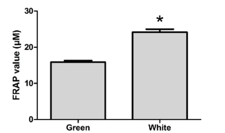
4.1. White tea possesses a higher antioxidant power than green tea
27
4.2. General characteristics of the STZ-induced diabetes model and effect of
White tea consumption
27
4.3. Heart tissue antioxidant potential of STZ-induced diabetic rats is increased
by white tea ingestion
30
4.4. White tea consumption decreases lipid peroxidation in hearts of STZ-induced
diabetic rats
31
4.5. White tea consumption slightly decreases carbonyl group formation in hearts
from STZ-induced diabetic rats.
32
4.6. White tea consumption did not altered the glucose content in hearts of
STZ-induced diabetic rats but increased lactate content
33
4.7. White tea consumption did not alter the alanine content in hearts of
STZ-induced diabetic rats but decreased the lactate/alanine ratio
33
4.8. White consumption increases the transcriptional levels of GLUT1, LDH and
MCT4 in the STZ-induced diabetic rats
36
4.9. White tea consumption restores the LDH activity in the STZ-induced diabetic
rats 39
XIV
V. Discussion
40
VI. Conclusions
46
XV
List of Figures
Figure 1.3.1. The myocardial substrate metabolism, pathways and regulatory
points.………6
Figure 1.3.2. The regulation of glycolysis.……….7
Figure 2.2.1. Main chemical structure of tea catechins..………11
Figure 4.1.1. Antioxidant power of green tea and white tea measured by
FRAP assay………27
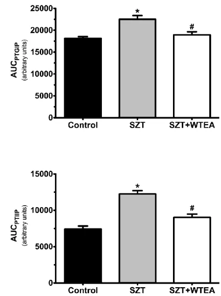
Figure 4.2.1. Effect of white consumption by STZ-induced T2DM rats in the
glucose tolerance and insulin resistance test. Graph A: shows the AUC of
Control, STZ-induced diabetic rats and STZ-induced diabetic rats consuming
WTEA, during the i.p. glucose tolerance test. Graph B: shows the AUC blood
glucose levels in control, STZ-induced diabetic rats and STZ-induced diabetic
rats consuming WTEA, measured during the i.p. insulin resistance test……….29
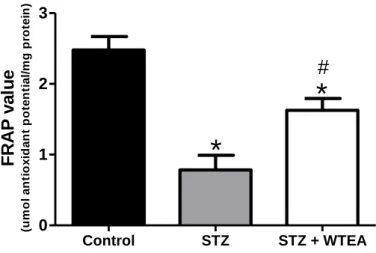
Figure 4.3.1. Effect of White tea consumption by STZ-induced diabetes in the
antioxidant power of heart tissue………30
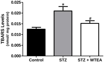
Figure 4.4.1. Effect of white tea consumption on lipid peroxidation of the
heart tissue in the STZ-induced diabetic rats……….………31
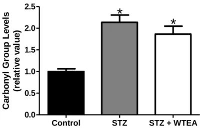
Figure 4.5.1. Effect of white tea consumption on protein oxidation of the
heart tissue in the STZ-induced diabetic rats……….………32
Figure 4.6.1. Panel A: Heart glucose content expressed in nmol/mg tissue in
the control, STZ-induced diabetic (STZ) and STZ-induced diabetic rats that
consumed white tea (STZ+WTEA). Panel B: Heart Lactate content expressed
in nmol/mg tissue on the control, STZ-induced diabetic (STZ) and STZ-induced
diabetic rats that consumed white tea (STZ+WTEA)……….34
Figure 4.7.1. Panel A: Heart alanine content expressed in nmol/mg tissue in
the control, STZ-induced diabetic rats (STZ) and STZ-induced diabetic rats
that consumed white tea (STZ+WTEA). Panel B: ratio between the lactate and
the alanine content of the heart tissue of rats from control, STZ-induced
diabetic (STZ) and STZ-induced diabetic that consumed white tea
(STZ+WTEA)………35
XVI
Figure 4.7.2. Acetate content in heart tissue (nmol/mg tissue) of control,
STZ-induced diabetic rats (STZ) and STZ-induced diabetic rats that consumed
White tea (STZ-WTEA)……….…….36
Figure 4.8.1. mRNA expression of GLUT1 in the heart tissue of Control,
STZ-induced diabetic rats (STZ) and STZ-STZ-induced diabetic rats consuming WTEA
(STZ+WTEA)……….………….36
Figure 4.8.2. mRNA expression of LDHa in the heart tissue of Control,
STZ-induced diabetic rats (STZ) and STZ-STZ-induced diabetic rats consuming WTEA
(STZ+WTEA)……….……….37
Figure 4.8.3. mRNA expression of MCT4 in the heart tissue of Control,
STZ-induced diabetes rats (STZ) and STZ-STZ-induced diabetic rats consuming WTEA
(STZ+WTEA)………38
Figure 4.9.1.Effect of white tea on the heart LDH activity of the STZ-induced
XVII
List of Tables
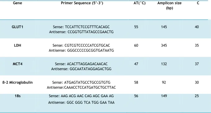
Table 1. Primer sequences, optimal annealing temperature, number of cycles
required for exponential amplification phase of fragments and fragment sizes.
……….25
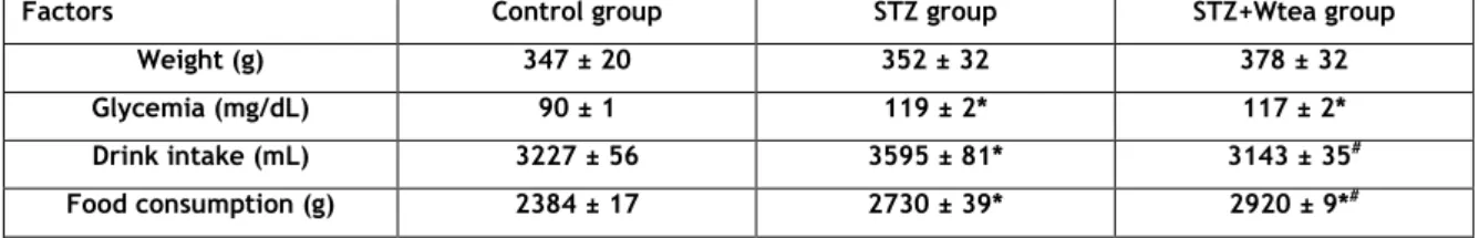
Table 2. Mean values of weight, blood glycaemia, water intake and food
XVIII
List of Abbreviations
AGE - Advanced glycation end-products
ADP - Adenosine diphosphate
ATP - Adenosine triphosphate
AUC - Area under the curve
BMI - Body mass index
Cs - Catechins
CAC - Citric acid cycle
CO
2- Carbon dioxide
COMT - Catechol-O-methyltransferase
CVD - Cardiovascular diseases
DM - Mellitus diabetes
DPPH - Diphenylpicrylhydrazyl
DNPH - 2,4-Dinitrophenylhydrazine
DNA - Deoxyribonucleic acid
EC - Epicatechin
ECG - Epicatechin gallate
EGC - Epigallocatechin
EGCG - Epigallocatechin 3-gallate
FADH
2- Flavin adenine dinucleotide
FRAP - Ferric reducing antioxidant power
GLUT1 - Glucose transporter type 1
GT - Green tea
HDL – high density lipoprotein
HDL
c– cholesterol High density lipoprotein
IDF - International Diabetes Federation
INT – tretrazolium salt
LDL – Low density lipoprotein
LDL
c- Cholesterol low density lipoprotein
LDH - Lactate dehydrogenase
LDHA - Lactate dehydrogenase A
MCT4 - monocarboxilase transporter 4
XIX
MDA - Malondialdehyde
NAD
+- Nicotinamide adenine dinucleotide
NADPH - Nicotinamide adenine dinucleotide phosphate
NMR – nuclear magnetic resonance
NO - Nitric oxide
OADs - Oral Anti-diabetic drugs
OS - Oxidative stress
PBS - Phosphate buffered saline
RT-PCR – Real time polymerase chain reaction
PEP - Phosphoenolpyruvate
PFK - Phosphofructokinase
PK – Piruvate kinase
PPO - Polyphenol oxidase
PST - Phenolsulfotransferase
RNA - Ribonucleic acid
ROS - Reactive oxygen species
RONS – Reactive oxygen and nitrogen species
SDS - Sodium dodecyl sulfate
STZ - Streptozotocin
T1D - Type 1 diabetes mellitus
T2D - Type 2 diabetes mellitus
TBA - Thiobarbituric acid
TBARS - Thiobarbituric acid reactive substances
TPTZ - 2,4,6-tripyridyl-s-Triazine
UGT - UDP-glucuronosyltransferase
WHO - World Health Organization
WHR - Waist – to hip-ratio
1
2
1.
Diabetes Mellitus
Diabetes mellitus (DM) is a life lasting, devastating, expensive, manageable but incurable disease that is very common nowadays. There was an estimated 285 million adults with DM in 2010 and by 2030 this number will raise to 439 million, due to aging and growth of the population, increase in obesity and sedentary lifestyle (Shaw, Sicree, & Zimmet, 2010). However, the diagnostic of obesity and DM is raising in young individuals (Blanck et al., 2006; Wild, Roglic, Green, Sicree, & King, 2004), thus these numbers may be underestimated. According to (Zhang et al., 2010), the global expenditure of DM in 2010 was of USD 376 billion, imposing a large economic burden on the national health care systems.
This disease consists in a chronic metabolic disorder, characterized by a state of insulin deficiency that leads to hyperglycemia (Gupta et al., 2005) inducing changes in the metabolism of carbohydrates, lipids and proteins (Da Poian & de Carvalho-Alves, 2005; Negri, 2005), defects in the reactive oxygen species (ROS) scavenging enzymes (Kesavulu, Giri, Rao, & Apparao, 2000), and increases the oxidative stress (OS) impairing pancreatic beta cells (Baliga & Sapsford, 2009; Hamden, Jaouadi, Carreau, Aouidet, & Elfeki, 2011). Increased OS has been related with the pathogenesis of DM (Kaushik, Satya, Khandelwal, & Naik, 2010). Moreover, hyperglycemia-induced protein glycation generates high amounts of superoxide free radicals (Atalay & Laaksonen, 2002; Cunningham, Leffell, Mearkle, & Harmatz, 1995; Lipinski, 2001; Memısoğullari, Taysı, Bakan, & Capoglu, 2003; Raskin et al., 2000). The generation of active ROS may lead to lipid peroxidation and the creation of reactive products, which induces damages in cell molecules and structures (Kaushik et al., 2010). These processes are responsible for increasing chances of developing cardiovascular diseases (CVD). There are mainly two types of DM: type 1 (T1D) and type 2 (T2D). T1D results from the autoimmune destruction of the insulin-producing β pancreatic cells, and therefore there is a complete lack of insulin that leads to the increase of glucose levels in blood and urine. Thus, T1D patients need exogenous insulin administration and are well known for being insulin dependent. T2D is characterized by a progressive impairment of insulin secretion by the β-cells from the pancreas and by a relative decline sensitivity of target tissues to the action of the hormone (Remy Burcelin et al., 1999; Cleeman, Grundy, Becker, & Clark, 2001). These patients are not insulin dependent and face pharmacological treatment. They represent almost 90% of DM cases worldwide. DM is characterized by high blood glucose, polyuria (frequent urination), polydipsia (increased thirst), polyphagia (increased hunger), weight loss, blurred vision, nausea and vomiting, weakness and tiredness, irritability, mood changes (Modak, Dixit, Londhe, Ghaskadbi, & Devasagayam, 2007), reduced plasma antioxidant levels (Facchini, Hua, Reaven, & Stoohs, 2000), among other characteristics.
One of the major causes of mortality in diabetic patients are the CVD. Several reports suggest that the majority of people with T2D may die from CVD (Boccara & Cohen, 2004; Morrish, Wang, Stevens, Fuller, & Keen, 2001). Hypertension is almost twice as frequent in diabetic
3
individuals comparatively to non-diabetic (Sowers, Epstein, & Frohlich, 2001). Other important risk factor for CVD of these individual, beside insulin resistance, are: obesity, atherosclerosis, dyslipidemia, low plasma levels of HDL, microalbuminuria, endothelial dysfuntion, platelet hyperaggregability and coagulation abnormalities (Sowers et al., 2001; Wolfram, 2007). These parameters often appear clustered in diabetic patients so that several international organizations such as the World Health Organization (WHO) and the, International Diabetes Federation (IDF) defined this cluster of risk factors, as the “metabolic syndrome” (Alberti & Zimmet, 1998; Cleeman et al., 2001; Detection, 2001; Zimmet, Alberti, & Shaw, 2005). The WHO briefly defined, “metabolic syndrome” as: insulin resistance and/or impaired fasting glucose (110-125 mmol/L) plus at least 2 of the following factors: obesity (body mass index (BMI)> 30 kg/m2 and/or waist-to-hip ratio (WHR)>0.9 for men and >0.85 for women), dyslipidemia (HDLc< 0.35 for men and <0.40 for women; and/or triglycerides >150
mg/dL), hypertension (>140/90 mmHg and/or antihypertensive medication) or microalbuminuria (albumin/creatine ratio 25-250 mg/g). According to IDF, people suffer of “metabolic syndrome” if: central obesity with waist circumference >94 cm for men and >80 cm for women, plus at least 2 of the following factors: hypertriglycerimia (>150 mg/dL or specific treatment), low HDL cholesterol (<40 mg/dL for men and <50 mg/dL for women or specific treatment), hypertension (>130/85 mmHg or treatment of previously diagnosed hypertension), impaired fasting glycemia (>100 mg/dL or previously diagnosed T2D).
1.1 Management of diabetes
There are no therapies that can cure DM (Maiti, Jana, Das, & Ghosh, 2004) but there are many strategies available for the treatment such as: stimulation of endogenous insulin secretion, enhancement of insulin action at the target tissues, inhibition of dietary starch lipid degradation and pharmacological treatments with oral antidiabetic drugs (OADs) like biguanides and sulfonylureas (García-Pérez, Álvarez, Dilla, Gil-Guillén, & Orozco-Beltrán, 2013). However, these OADs can cause side effects such as major and minor hypoglycemia, gastrointestinal problems, peripheral edema, body weight gain, liver diseases and, over time, they lose their efficacy (García-Pérez et al., 2013).
The overall management of DM not only assures on achieving a normoglycemic state (HbA1c≤
6.5 mg/100 ml, fasting blood glucose ≤ 110 mg/100 ml) but also on reducing the risk for other metabolic diseases. To prevent metabolic diseases, visceral serum cholesterol should be ≤ 150 mg/100 ml, serum triglyceride ≤ 140 mg/ 100 ml, cholesterol low density lipoprotein (LDLc) ≤
70mg/100 ml and high density lipoprotein (HDL) ≥ 60 mg/100 ml (Kaushik et al., 2010). The limitation of these therapies has boosted the search of more efficient and cost-effective alternatives, recurring to dietary and lifestyle changes. In recent years, there is an increased interest in functional and nutraceutical food for pharmacological purposes, in order to complement or replace current therapies. In the case of DM, it has been reported that numerous extracts obtained from plants can effectively reduce glycaemia (Gupta et al., 2005;
4
Lee & Sohn, 2009; Pushparaj, Tan, & Tan, 2000; Sohn et al., 2010). However little is known on the molecular mechanisms of protection activated when using such extracts.
In fact, the best way to “cure” T2D is “not getting” T2D, and this may be achieved by living an active life, practicing physical activity, reducing caloric intake and eating healthy food. Physical activity is negatively associated with risk factors for CVD, such as waist circumference, BMI, WHR and triglycerides and is positively associated with HDL (Aadahl, Kjaer, & Jorgensen, 2007). Nutritional guidelines include reduction of saturated fat and trans fatty acids, cholesterol <200 mg and the intake of n-3 polyunsaturated fatty acids from fish or seeds oils (Expert Panel on detection, 2002). Carbohydrate intake should be limited (60 % of total calories or 50 % in the presence of high triglycerides or low HDLc) and the consume of carbohydrate sources with high-fiber content is preferential rather than refined carbohydrates sources (Wolfram, 2007). Limiting the sodium intake to < 1.5 g/d (sodium chloride < 3.8 g/d) and increasing potassium intake (approximately to 4.7 g/d) (Appel et al., 2006) significantly reduces blood pressure and thus reduces cardiovascular risk (Appel et al., 2006; Sacks et al., 2001). All these parameters should be taken into account to avoid T2D. Diabetes can lead to long-term complications such as atherosclerosis, hypertension, hypertriglyceridemia, hypercholesterolemia, myocardial infarction, ischemic attacks, impotence (Stadler, Jenei, von Bölcsházy, Somogyi, & Jakus, 2003), retinopathy (blindness), nephropathy (kidney damage), neuropathy (nerve damage) and diabetic foot (Islam, 2011). Diabetic patients possess a higher risk of death, together with lower survival rates and lower life expectancy than non-diabetic adults (Gu, Cowie, & Harris, 1998). Amongst all the comorbidities associated with T2D, the importance of CVD in diabetic individuals has been highlighted in recent years since CVD accounts for up to 80% of the death in T2D patients (Haffner, Lehto, Rönnemaa, Pyörälä, & Laakso, 1998). A population-based study showed that CVD mortality was 7.5-fold higher among T2DM individuals without a previous myocardial infarction than those without DM. The mortality was 3 times higher among diabetic patients who had suffer previous myocardial infarction than among nondiabetic individuals (Haffner et al., 1998). Interestingly, the risk of onset CVD due to diabetes was found to be greater in women than in men (Sowers, 1998).
The development of CVD in diabetic individuals can be related to several factors such as: enhanced platelet aggregation, relatively greater coagulation and decreased fibrinolytic activity, lipoprotein abnormalities, endothelial dysfunction, enhanced OS, vascular protein glycation and enhanced growth factor stimulation (Sowers, 1998).
1.2 Diabetes and oxidative stress
OS plays an important role in the development of vascular complications in diabetic individuals (Lipinski, 2001).
5
In diabetic individuals, free radicals are formed through several ways such as glucose oxidation, nonenzymatic glycation of proteins and the oxidative degradation of glycated proteins (Maritim, Sanders, & Watkins, 2003). The nonradical oxidants such as hydrogen peroxide, hypochlorous acid, singlet oxygen and radical oxygen species like superoxide anion and hydroxyl radicals, can attack the double bounds of unsaturated fatty acids promoting the formation of lipid peroxides (Lipinski, 2001).
This abnormal enhancement of free radicals and decline of antioxidant defense mechanisms lead to the damage of cellular organelles and enzymes, the increase in the lipids peroxidation and the increase of insulin resistance (Maritim et al., 2003). Thus, it is important to measure lipids peroxidation. The 2-ThioBarbituric acid reactive substances (TBARS) assay is a well-recognized method for quantifying the lipid peroxides (Devasagayam, Boloor, & Ramasarma, 2003). This method is based on the ability of malondialdehyde (MDA), which is one of the secondary products of lipid peroxidation, to react with thiobarbituric acid in acidic conditions an at high temperatures to form a pink MDA-(TBA) complex (Sochor et al., 2012).
Protein oxidation originates carbonyl groups and their level in tissues and plasma is a stable marker of oxidative stress (Odetti et al., 1999). When protein side chains (especially arginine, lysine, proline and threonine) are oxidized, carbonyl groups are produced. Usually the content of these carbonyl groups is used as marker of protein oxidation and carbonyl group accumulation has been observed in several diseases such as diabetes, arthritis and Alzheimer´s (Dalle-Donne, Rossi, Giustarini, Milzani, & Colombo, 2003).
The study of OS biomarkers like superoxide dismutase, catalase, glutathione reductase, glutathione peroxidase, glutathione levels, lipid peroxidation, nonenzymatic glycosylated proteins and vitamins are essential in way to explore the mechanisms by which the increase of free radicals accelerates the development of diabetic complications (Maritim et al., 2003).
1.3 Diabetes and myocardial substrate metabolism
The heart is known to be a pump that converts the chemical energy into mechanical work. To support its contractile activity, the heart requires a continuous supply of energy. To satisfy its energy requirements, the heart is capable of consuming a variety of exogenous substrates such as lactate, ketone bodies, glucose and fatty acids (Figure 1.3.1.). Mitochondrial oxidative phosphorylation is fueled with energy from electrons that are transferred from carbon fuels by dehydrogenation reactions that generate NADH and FADH2 produced primarily
in the fatty acid β-oxidation pathway, the citric acid cycle (CAC), from the pyruvate dehydrogenase reaction and glycolysis (William C Stanley et al., 2005). Nevertheless, glucose is a well-known reliable substrate, especially when the heart is subjected to unfavorable conditions (Marco G Alves, Oliveira, & Carvalho, 2011).
6
Intense exercise promotes a large increase in cardiac power but, in healthy heart, ATP content remains constant because the rate of oxidative phosphorylation is linked to the rate of ATP hydrolysis (Balaban, 1990). Indeed, the oxidative phosphorylation provides the greatest percentage (~95%) of ATP formation in the heart in normal healthy conditions. The remaining ATP comes from glycolysis and guanosine triphosphate formation in the CAC. The NADH and FADH2 formed in glycolysis, fatty acid oxidation, and the CAC are energy-rich
molecules because each contains a pair of electrons having a high transfer potential. When these electrons are used to reduce molecular oxygen to water, a large amount of free energy is liberated, which can be used to generate ATP. In oxidative phosphorylation, ATP is formed as a result of the transfer of electrons from NADH or FADH2 to O2 by a series of electron
carriers (Figure 1.3.1).
The heart metabolic machinery responsible for generating large amounts of ATP can be turned on or off by allosteric modification of regulatory enzymes, translocations of proteins to their site of function or changing the concentration of stimulatory or inhibitory metabolites. These mechanisms are responsible for the rapid adaptation to stress situations such as exercise, ischemia and metabolic diseases. As discussed earlier, in these conditions, glucose becomes the most reliable substrate for the heart maintenance of the contractile activity. The overall rate of glucose uptake is limited since the rate of glucose transport is slower than its phosphorylation (Depre et al., 1998). Thus, inside of cardiomyocytes the concentration of free glucose is lower than in blood, so that a gradient that favor the glucose entry is maintaining (Fischer, Becker, & Löken, 1999).
In glycolysis, the six-carbon sugar glucose is oxidized and split to create two molecules of pyruvate and two ATP are generated by each molecule of glucose. Products resulting of glycolysis, like pyruvate and NADH, are transported to mitochondria to generate NAD+ and CO
2
Figure 1.3.1. The myocardial substrate metabolism, pathways and regulatory points. The heart
contractile force is regulated by the Ca2+ions that enter the myocardial cells via Ca2+ channels
triggering Ca2+ release from the sarcoplasmic reticulum, which allows contraction. Energy for
contraction comes from the hydrolysis of ATP. The NADH and FADH2 generated by either the
dehydrogenases of glycolysis, the oxidation of lactate and pyruvate and fatty acid β-oxidation, or the citric acid cycle deliver of electrons to the electron transport chain, resulting in ATP formation by oxidative phosphorylation. Lactate transport across the cardiac sarcolemma is facilitated by the monocarboxylic acid transporters (William C Stanley, Recchia, & Lopaschuk, 2005).
7
or go to cytosol where they are converted to lactate and NAD+. The plasma membrane of cells
possesses a set of transporters that allow the movement of glucose either into or out of cells (Gould & Holman, 1993). The rate of entry of glucose into a cell is limited by the number of glucose transporters on the cell surface and the affinity of the transporters for glucose. Due to insulin stimulation, occurs the translocation of glucose transporters from intracellular vesicles to sarcolemma membrane which increases the rate of glucose uptake and the ability of membrane to transport glucose (William C Stanley et al., 2005). When glucose transport is stimulated, hexokinase phosphorylate glucose originating glucose-6-phosphate (Fig.1.3.2). In mammalian cells the rate of formation of glucose-6-phosphate is reduced by the regulation of breakdown of glycogen. In addition to originate pyruvate, glucose-6-phosphate can be used to synthetize ribose for DNA and RNA nucleotides.
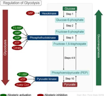
The reaction catalyzed by phosphofructokinase (PFK) is usually described as the most important regulatory step in glycolysis. PFK is regulated by the cell fraction of the adenosine nucleotides that contains high-energy bonds (energy charge of cell) thus, AMP and fructose-2,6-biphosphate are activators of PFK. PFK activity is essentialsince it is responsible for the conversion of fructose 6-phosphate to fructose 1,6-bisphosphate, the first irreversible step of glycolysis, and a limiting step of glycolytic flux (Underwood & Newsholme, 1965)
.
The myocardium becomes a lactate producer only when glycolysis is accelerated in face of impaired oxidation of pyruvate, such as in ischemia or poorly controlled diabetes (William C Stanley et al., 2005). When the heart is perfused, then pyruvate becomes the main substrate (Marco G Alves et al., 2011).
Figure 1.3.2. Regulation of glycolysis. The glycolytic pathway is shown on the left. The enzymes hexokinase,
phosphofructokinase and pyruvate kinase catalyze three important exergonic steps. For each of these pathways, the allosteric activators (labeled in green) and allosteric inhibitors (labeled in red) are indicated. Gluconeogenesis (shown on the right) is the reverse of glycolysis, with the exception of steps 1, 3, and 10 and the enzymes that catalyze these steps. (https://wikispaces.psu.edu/download/attachments/46924785/image-1.jpg)
8
The three main fates to pyruvate formed from glycolysis are: 1) conversion to lactate, 2) carboxylation to oxaloacetate/malate or 3) decarboxylation to acetyl-CoA. In this system, the pyruvate kinase (PK) and the lactate dehydrogenase (LDH) play key roles. The PK catalyzes the transfer of a phosphate group from phosphoenolpyruvate (PEP) to ADP, yielding one molecule of pyruvate and one molecule of ATP. It is inhibited by acetyl-CoA, ATP and allosterically by fatty acids serving as an indicator that alternative energy sources are available for the cell. The activity of PK is increased when glycolysis is activated. In addition to carbon entry into the CAC via acetyl-CoA, carbon entry can also occur via so-called anapleurotic pathways (Lloyd, Brocks, & Chatham, 2003). Anapleurotic reaction is a metabolic reaction that replenishes intermediates of pathways. Due to carboxylation to malate or oxaloacetate (an “anaplerotic” reaction), pyruvate enters in CAC (Lloyd et al., 2003). The transamination of glutamate to α-ketoglutarate and the formation of succinyl-CoA from propionyl-CoA are examples of others anaplerotic reactions (Lloyd et al., 2003).
The formation of CAC intermediate as α-ketoglutarate and alanine by transamination of glutamate is also an important contribution of pyruvate to anaplerosis (William C Stanley et al., 2005). In cases where glycolysis is accelerated like hyperglycemia, the rate of alanine output is usually unaffected (William C Stanley et al., 2005).
In addition to being oxidized, pyruvate can also be converted to alanine via alanine aminotransferase and to lactate via LDH (Lloyd et al., 2003).
9
2 Tea
Tea is, next to water, one of the most consumed beverages in the world (Cheng, 2006). This beverage popularity is mainly associated with its taste and aroma, relative low retail prices, stimulating effects and known and possible health benefits (Baptista, da P Tavares, & Carvalho, 1999; Baptista, Tavares, & Carvalho, 1998).
Tea has a long history as a folk remedy, but his beneficial properties have mainly been elucidated in the past two decades (Anderson & Polansky, 2002). Tea is one of the oldest known medicine and has been used in China for the last 5000 years for its detoxifying properties as the elimination of alcohol and toxins, to improve blood circulation and urine flow, to relieve joint pains and to improve disease resistance (Balentine, Wiseman, & Bouwens, 1997).
Tea is an infusion prepared with the leaves of the plant Camellia sinensis (L.) from the Theaceae family (López & Calvo, 2011). There are two main varieties of tea plants: C.
sinensis var. sinensis and C. sinensis var. assamica. The C. sinensis var. sinensis is a small
leaved, bushlike plant indigenous from China, which is widespread through all the Southeast Asia more adapt to a cold climate, and C. sinensis var. assamica, that is a large-leaved tree found in the Assam region, India, which is more adapt to semitropical climates (de Mejia, Ramirez-Mares, & Puangpraphant, 2009).
Tea was introduce to Europe from China by the Dutch and the Portuguese merchants (Hollman, Hertog, & Katan, 1996). The origins of modern tea industry happened between 1818–1834 in India through the importation of the var. sinensis and the discovery of native var. assamica (Harbowy, Balentine, Davies, & Cai, 1997). With the advances in technology and in the “know how”, new plantations were made across tropical areas like Africa, South America, Russia (Georgia) (Eden, 1976) and also in the island of S. Miguel, Azores Archipelago (Portugal) (Baptista et al., 1999).
2.1 Types of Tea
Tea diversity is due to the botanical variety, geographical origin (de Mejia et al., 2009) starting material, harvesting and processing method. The basic differentiation of tea, is related with the degree of “fermentation”: as white tea (WTEA) and green tea (GT) (not fermented), oolong (semi-fermented), and black tea (completely fermented) (M. P. Almajano, Carbo, Jiménez, & Gordon, 2008). The level of fermentation influences the composition (phenolic profile) and organoleptic properties (appearances and taste) of the tea (Moderno, Carvalho, & Silva, 2009). However this process is wrongfully defined as “fermentation”, the more correct term is oxidation (Bartlett, 2004), a reaction which is catalyzed by polyphenol oxidase (PPO).
10
There are two seasons for tea harvest: the “First Flush” in early spring and the “second flush” in summer. Once the tea leaves (leaf buds or tips, for WTEA) are picked, if they are not dried afterward, they turn progressively darker because chlorophylls break down and tannins are release. The oxidation process can, and is, interrupted by tea industry at a predetermined stage by heating, inhibiting PPO. This enzyme converts catechins (Cs) to theaflavins and their polymers, thearubigins, mainly present in fermented teas as oolong and black tea (Subramanian, Venkatesh, Ganguli, & Sinkar, 1999).
2.1.1 White tea
WTEA is prepared from very young tea leaves or leaf buds covered with tiny, silvery hairs, which are picked up before fully opened only once a year in the early spring (Espinosa et al., 2012). WTEA is steamed and dried quickly after harvest, preventing whitering and inactivating the PPO thus becoming the least processed of all teas (Mao et al., 2010).
2.2 Chemical composition
The manufacturing method influences the overall chemical composition of the tea, that is very complex and includes proteins, chlorophyll, organic and amino acids, polysaccharides, vitamins, carbohydrates, lignins, methylxanthines (caffeine, theophylline and theobromine), minerals and trace elements, volatile oils and flavonoids (Moderno et al., 2009; Seeram et al., 2006). Flavonoids are phenolic compounds that have unique biological properties that may be responsible for many health benefits attributed to tea (Rietveld & Wiseman, 2003). There are over 4000 different flavonoids described in the literature, and they are categorized in flavonols, flavones, catechins, flavanones, anthocyanidins and isoflavonoids (Firenzuoli, Gori, Crupi, & Neri, 2004). The major class of phenolic compounds present in the tea leaves are catechins (Cs) (also known as flavan-3-ols), they represent 30-40 % of their dry weight (Karakaya & Kavas, 1999; Nihal, Ahmad, Mukhtar, & Wood, 2005; Wheeler & Wheeler, 2004). The main Cs of WTEA are: (-)-epicatechin (EC), (-)-epigallocatechin (EGC), defined as flavanol monomers, (-)-epicatechin 3-gallate (ECG), (-)-epigallocatechin 3-gallate (EGCG), which are known as flavanol gallates (Figure. 2.2.1) (Hara, Luo, Wickremasinghe, & Yamanishi, 1995a). The most abundant catechin in tea leaves is EGCG, 50-80% of total catechins, and is thought to exert the beneficial health effect ascribed to tea (Khan & Mukhtar, 2007; Ortsater, Grankvist, Wolfram, Kuehn, & Sjoholm, 2012)
Quercetin, kaempferol, myricetin, rutin and their glycosides are the main flavonols of tea (C. Dufresne & Farnworth, 2000; C. J. Dufresne & Farnworth, 2001). Also methylxantines are present in tea, 2-4% as caffeine and small amounts of theophylline and theobromine (Hara et al., 1995a). From the amino acids present in tea, theanine is specific of the tea plant,
11
accounting for 50 % of the total amino acid content (C. Dufresne & Farnworth, 2000). Tea also contains carbohydrates, vitamins such as E, K, A, B and C (C. Dufresne & Farnworth, 2000). Large amounts of potassium, manganese and fluoride ions are provided by tea (Hara, Luo, Wickremasinghe, & Yamanishi, 1995b).
2.3 Metabolism, bioavailability and elimination of Catechins
Studies on bioavailability of phenolic compounds and their body distributions have been carried using sensitive and accurate methods to analyze metabolites in biological fluids and tissues (Hirayama, Takagi, Hukumoto, & Katoh, 1997; Hollman et al., 1996). The potential health effects of Cs depend on the amount ingested and on their bioavailability. (Okushio, Matsumoto, Korhi, & Susuki, 1996) identified the four major Cs (EC, ECG, EGC and EGCG) in the portal vein, following the oral administration, indicating that tea Cs are absorbed intestinally.In a human study, (C. S. Yang, Lee, & Chen, 1999) reported that saliva possess a catechin esterase activity, suggesting that EGCG may be degalloylated in the mouth and esophagus. Furthermore Cs have been identified to suffer glucoronidation, sulfation, and O-methylation reaction by the enzimes UDP-glucuronosyltransferase (UGT), phenolsulfotransferase (PST) and catechol-O-methyltransferase (COMT) respectively, and also ring fission metabolism. The metabolic fate of tea constituents depends on their structure. Studies with radioactively
12
labeled Cs in humans shown that they are efficiently metabolized (Hollman et al., 1996). A study on the activity of conjugative enzymes in rat tissue found the highest UGT activity in the small´s and large´s intestine mucosa, the PST was in the liver and COMT was in the liver but also in the kidney (Piskula & Terao, 1998).
Polyphenols have shown a strong affinity for proteins, particularly when proteins have a high proline content such as casein, gelatin and salivary proteins (C. Dufresne & Farnworth, 2000). This protein-binding capacity may reduce the digestibility of alimentary proteins and rise feacal nitrogen excretion in humans, according to results observed in herbivorous animals (Hollman et al., 1996). Tea polyphenols had also demonstrated a strong interaction with transition metals and formed insoluble complexes with iron which reduce the bioavailability of non-heme iron. This bind inhibits iron gastrointestinal adsorption (Bravo, 1998; Hollman et al., 1996; Hurrell, Reddy, & Cook, 1999). Besides, L-ascorbic acid inhibits the formation of these complexes. A poor iron intake can result in increased chances of anemia. So, vegetarians may be advised of consuming tea brew during meals, because iron from plant is not available and the tea binding action may further reduce iron bioavailability. The absorption inhibition of zinc has been observed in rats, but copper results are not clear (C. J. Dufresne & Farnworth, 2001). Polyphenols can also affect the bioavailability of sodium and aluminum but not of calcium, magnesium or manganese (Bravo, 1998).
2.4 Antioxidant potential and health benefits of white tea
In the last few years, antioxidant components have aroused great interest such as phenolic compounds from plant because of their ability to reduce the harmful effects of reactive oxygen and nitrogen species (RONS) on several biologic and pathologic processes (Alarcón, Campos, Edwards, Lissi, & López-Alarcón, 2008). The majority of the living organisms have an efficient protective mechanism against the excess production of RONS through enzymatic or non-enzymatic via. Yet there are several external factors (diet, alcohol, smoke, drugs, among others) and internal factors (such as aging) that decreases the efficiency of these endogenous antioxidant defenses, creating an impairment in the redox equilibrium of a healthy condition (Rietveld & Wiseman, 2003; Willett, 1994). Long exposure to RONS can damage DNA, lipidic membranes, lipoproteins, and structural and functional proteins (Halliwell, 1997; Sohal & Weindruch, 1996). RONS have been related with the development of several chronic diseases such as: CVD, DM, chronic inflammation, neurodegenerative disorders, and some types of cancer (Valko et al., 2007; Valko, Rhodes, Moncol, Izakovic, & Mazur, 2006)
Several studies have reported that tea (poly)phenolic substances exert beneficial effects not only by scavenging RONS but also as metal chelators, preventing LDL oxidation, DNA strand scission and enhancing immune function (J. Cao, Xu, Chen, & Klaunig, 1996; Costa et al., 2009; Fattouch et al., 2007; Fiorentino et al., 2008; Giada & Filho, 2006; Magalhães et al., 2009; Oliveira et al., 2007; Silva, Valentão, Seabra, & Andrade, 2008). A number of structures
13
are reported to be important for this antioxidant activity of tea polyphenols (Figure. 2.2.1). One is the ortho-3’,4’-dihydroxyl (catechol) group in the B-ring, that promotes the formation of a stable phenoxyl radical due to effective electron delocalization (Wiseman, Balentine, & Frei, 1997); another is the 3’,4’,5’-trihydroxyl (gallate) group in the B-ring, a gallate group esterified at the 3 position of the C-ring, and hydroxyl groups at the 5 and 7 positions of the A-ring (Rice-Evans, Miller, & Paganga, 1996).
Studies in humans apparently demonstrate that the moderate consumption of GT and/or black tea (1-6 cups/day) significantly augmented the plasma antioxidant capacity after 1 hour (Rietveld & Wiseman, 2003).
The antioxidant power of Cs determined by the diphenylpicrylhydrazyl (DPPH) method was found to be: EGCG > ECG > EGC > EC > C; (Katalinic, Milos, Kulisic, & Jukic, 2006; Łuczaj & Skrzydlewska, 2005).
2.5 Influence of tea on diseases
Several scientific reports have shown that tea ingestion brings benefit to health and may play a role in the prevention of chronic diseases, such as CVD (Y. Abe et al., 1995; Cheng, 2000; Curin & Andriantsitohaina, 2005; Hodgson, Burke, & Puddey, 2005; Kono, Shinchi, Ikeda, Yanai, & Imanishi, 1992; Tokunaga et al., 2002), anti-inflammatory (Alschuler, 1998), immune response (Bhattacharyya, Mandal, Lahiry, Sa, & Das, 2004; Hu, Toda, Okubo, Hara, & Shimamura, 1992), carcinogenic effect (Mukhtar, Katiyar, & Agarwal, 1994), mutagenic (Santana-Rios et al., 2001; C. S. Yang, Chung, Yang, Chhabra, & Lee, 2000), anti-diabetic effect (Anderson & Polansky, 2002), longevity (Sadakata, Fukao, & Hisamichi, 1992), anti-neurodegenerative disorders (M. Almajano, Vila, & Ginés, 2011; Barranco Quintana, Allam, Del Castillo, & Navajas, 2009; Checkoway et al., 2002; Commenges et al., 2000; Tan et al., 2008) and antimicrobial (M. P. Almajano et al., 2008; Nakayama et al., 1993; Taguri, Tanaka, & Kouno, 2004; Weber, Ruzindana-Umunyana, Imbeault, & Sircar, 2003). EGCG, the most abundant phenolic compound in non-fermented teas, such as WTEA and GT, has been reported to possess vasculoprotective effect (Potenza et al., 2007; Wolfram, 2007).
2.6 Antidiabetic potential
There are several reports showing that plant extracts are efficient in reducing glycemia, lowering the side effects and costs presented by traditional OADs (Gupta et al., 2005; Lee & Sohn, 2009; Pushparaj et al., 2000; Sohn et al., 2010). There are some evidence that tea is an hypoglycemic agent (Todd MacKenzie, Lisa Leary, & W Blair Brooks, 2007). However, the exact mechanism by which tea ameliorates DM has not been elucidated yet. All the studies
14
suggest that tea polyphenols do not increase insulin secretion, but rather decrease insulin resistance and ameliorates insulin sensitivity (Islam, 2011).
It is well known that hyperglycemia increases the formation of RONS and decreases antioxidant endogenous mechanisms (Rahimi, Nikfar, Larijani, & Abdollahi, 2005). OS triggered by hyperglycemia seem to be one the major causes for post DM complications (Valko et al., 2007). In both T1D and T2D there is an increase of OS (Naziroğlu & Butterworth, 2005). It was shown that cytokine-induced β-cell damage was reduced by EGCG in vitro preventing the reduction of islet mass induced by treatment with streptozotocin (STZ) (Song, Hur, & Han, 2003). Yet in another study where STZ was co-injected with EGCG, it was unclear if the protective effect observed was due to direct inhibition of STZ by EGCG (I. Abe, Kashiwagi, & Noguchi, 2000). An study by (Roghani & Baluchnejadmojarad, 2010) reported that EGCG supplementation reduced serum glucose, total cholesterol and triglycerides and LDLc in
STZ-induced diabetic rats. The effect of a single catechin from tea, EGCG, can be different from a whole crude tea extract.
2.7 Anti-CVD
In a cohort study involving 40530 Japanese adults, the consumption of 5 or more cups of GT per day was found to significantly reduce mortality due to all causes (-16 %) and CVD (-26 %) compared to subjects consuming less than 1 cup per day (Kuriyama et al., 2006). Interestingly, the beneficial effect of GT on CVD mortality increased according to the overall consumption evidencing that GT may indeed have a cardioprotective effect (Wolfram, 2007). Besides, it is well known that hypertension is the major risk factor for stroke and CVD. In a cross-sectional study by (Y.-C. Yang, Lu, Wu, Wu, & Chang, 2004), the role of habitual consumption of GT was observed in 1507 Chinese subjects. Individuals ingesting 120-599 ml of GT per day for at least one year, presented reduced risk of developing hypertension in 46 % when comparing with those consuming less than 120 mL per day. Noteworthy, those who consumed more than 600 mL per day reduce their risk by 65 %.
In another study by (Sasazuki et al., 2000), it was reported an inverse association between GT consumption and coronary atherosclerosis. A subgroup of T2D patient without dietary or drug treatment presented a reduced risk of stenosis in 50% for subjects consuming 2-3 cup a day and 60% for those consuming 4 or more cups, in comparison to those consuming 1 cup or less per day.
In a retrospective cohort study among 17413 Japanese adults, the risk of developing DM drop 33% in subjects consuming 6 or more cups of GT per day, in comparison to those consuming less than 1 cup per week (Iso, Date, Wakai, Fukui, & Tamakoshi, 2006). Interestingly, in women, there is a strong dose-response relationship and it was observe for subjects consuming 1-6 cups per week, 1-2, 3-5, and 6 or more cups per day a reduction of 21%, 34%, 39% and 51% respectively (Wolfram, 2007). These studies provide clear evidence that the
15
habitual consumption of tea is inversely associated with CVD mortality, the risk of developing hypertension and DM.
In an open, uncontrolled study by (W. Kim et al., 2006) on 20 chronic smokers, the effect of GT consumption on flow-mediated dilation was investigated. During 2 week, every day, they consumed 8 g of powdered GT. Tea consumption significantly enhanced the flow-mediated dilation and the number of circulating endothelial progenitor cells. The authors suggested that GT consumption may prevent future cardiovascular events in smokers. Similar results were obtained in a cross-over study by (Nagaya et al., 2004) were the forearm blood flow was enhanced and the urinary concentration of an oxidative stress marker, 8-iso-prostaglandin-F2α, was reduced by the consumption of 400 mL GT (~247 mg EGCG in ~692 mg total catechins per day). (Widlansky et al., 2007) performed a randomized, double-blind, placebo-controlled, cross-over study in 42 subjects with endothelial dysfunction and investigated the effects of supplementation with 300 mg EGCG daily for 2 weeks on brachial artery mediated dilation. The results showed that EGCG acutely improved brachial artery flow-mediated dilation and the changes in vascular function are paralleled to the changes in EGCG plasma concentration. Later, an intervention study by (Widlansky et al., 2007) was the first to report a cardiovascular benefit from a single tea catechin.
In a randomized, controlled study in a parallel, open-label design, (Hakim et al., 2003) it was studied the antioxidant effect of decaffeinated GT or black tea consumption (4 cup per day) during 4 months in 133 smokers. When compared to water and black tea, GT consumption (~144 mg EGCG in ~294 mg total Cs per day) decreased urinary 8-hydroxydeoxyguanosin, a marker of DNA oxidative damage. Analogous results were obtained in a randomized, double-blinded, placebo-controlled trial by (Luo et al., 2006) with subjects with a high risk for hepatocellular carcinoma. The consumption of 500 or 1000 mg of GT Cs per day, for the period of 3 months, resulted in a significant decrease of the levels of 8-hydroxydeoxyguanosin compared to placebo. Besides, GT consumption increased the antioxidant capacity and decreased plasma peroxides in plasma and also reduced oxidative damage and glutathione peroxidase activity in lymphocytes (Erba et al., 2005).
The acute effects of GT on glucose tolerance were investigated in young healthy Japanese volunteers (Tsuneki et al., 2004). It was observed that the consumption of 1.5 g of GT extract, 20 min before an oral glucose load significantly reduced plasma glucose levels during the glucose tolerance test.
Hase et al. (2001) have reported that the consumption of GT Cs (300 mg EGCG in 480 mg total Cs) for 3 months, reduced glucose and insulin levels in healthy, slightly overweight, Japanese subjects. These results suggested that chronic consumption of GT Cs improves insulin sensitivity, but it also caused weight loss, which as a consequence may have decreased the levels of glucose and insulin.
Ryu et al. (2006) have studied the effect of the daily consumption of 900 mL water containing 9 g of GT, over a month period, in 55 Korean subjects with T2D. A trend towards reduce fasting glucose was observed but no other parameter was changed.
16
A study by H. L. Li et al. (2006) investigated the effects of EGG on cardiac hypertrophy in
vitro and in vivo. In rats, the cardiac hypertrophy was induced by pressure overload due to
constriction of the abdominal aorta; afterward they received oral EGCG (50 mg/kg) for 3 weeks. The EGCG treatment prevented the overload-induced cardiac hypertrophy, the increase of systolic blood pressure and the decrease in fractional shortening. EGCG reduced the generation of ROS and the expression of NADPH oxidase due to angiotensin II and the pressure overload. EGCG inhibiting angiotensin II, induced the activation of nuclear factor-κB and activator protein-1. Hotta et al. (2006) observed another cardiovascular benefit of EGCG in guinea pig hearts. The left ventricular pressure was increased as well nitric oxide (NO) and the calcium content of the heart, when EGCG was added to the perfusion medium (10 and 100 μM). Therefore, EGCG had a positive inotrophic effect without accompanying positive chronotrophic effects in an NO-dependent manner.
The effect of GT in rats with STZ-induced DM has already been documented (P. V. A. Babu, K. E. Sabitha, & C. S. Shyamaladevi, 2006c). These authors observed that oral intake of GT extract (300 mg/kg for 4 weeks) reduced lipid peroxides and the activity of antioxidant enzymes, while increased the content of glutathione in the heart and aorta of diabetic rats. In other study, the same group found reduced blood glucose, lipid peroxides, triglycerides and protein glycation in the heart of diabetic rats (P. V. A. Babu, K. E. Sabitha, & C. S. Shyamaladevi, 2006a). Furthermore, the rise in cardiac calcium and sodium concentrations were blunted by GT extract due to increased calcium-ATPase and sodium/potassium-ATPase activities in diabetic rats. In another study by P. Babu, K. Sabitha, and C. Shyamaladevi (2006), GT extract caused recovery of body weight and heart weight/body weight ratio, reduced blood glucose, serum cholesterol, triglycerides, free fatty acid, and LDLc and
increased HDLc. In myocardium, GT extracts decreased cholesterol, triglycerides and free
fatty acid and also the activity of lipoprotein lipase. Also, GT extract reduced systolic blood pressure, blood glucose, serum lipid peroxides and elevated serum glutathione and vitamin C in another study from P. V. A. Babu, K. E. Sabitha, and C. S. Shyamaladevi (2006b), with the same experimental design. The accumulation of aortic collagen, extent of glycation, advanced glycation end-products (AGE) formation and cross-linking of collagen were reduced by the GT extract. Similar results were reported, in other study, where GT extract reduced the rise in blood glucose, glycated hemoglobin and systolic blood pressure in diabetic rats (Babu, Sabitha, Srinivasan, & Shyamaladevi, 2007). It also prevented diabetes-induced increase in lactate dehydrogenase, aspartate transaminase and creatine kinase activities in serum and Maillard-type fluorescence and collagen cross-linking in myocardium.
The effect of EGCG (25, 50, 100 mg/kg for 50 days) was investigated in rats with STZ-induced diabetes and subtotal nephrectomy (Yamabe, Yokozawa, Oya, & Kim, 2006). EGCG reduced lipid peroxidation, proteinuria, hyperglycemia and the renal AGE accumulation in the kidney cortex.
Other study reported that GT Cs inhibited the production of superoxides, lipid peroxides, and oxidized protein in the STZ-induced diabetic rats kidney (Choi et al., 2004). Furthermore, it
17
was suggested that GT Cs decreased inflammatory response due to reduced leukotriene B4 production in the leukocytes. Mustata et al. (2005) studied the effects of GT consumption during 12 months on intra- and extracellular OS and glucotoxicity in STZ-induced diabetic rats, and found that it reduces the OS induced by DM. This effect was evaluated by the increase of erythrocyte glutathione content, as well as by the decrease of retinal superoxide formation, plasma peroxides and renal mitochondrial respiratory chain defects. However, tendon collagen glycoxidation and cross-linking were worsened, suggesting that GT may exert effects in the intra- and extracellular compartment. GT did not alter blood glucose and glycated hemoglobin.
Wu, Juan, Ho, Hsu, and Hwang (2004) investigated the effects of GT consumption on insulin sensitivity in rats. GT extract was administrated via drinking water (~370 mg/kg for 4 months) with a regular diet, and rats displayed significantly decreased fasting plasma levels of glucose, insulin, free fatty acids and triglycerides. Plasma insulin level was lower in the GT consuming rats during an oral glucose tolerance test. However, the adipocytes isolated from these rats showed an enhanced capacity for glucose uptake and increased specific insulin binding than the adipocytes from the control group.
Li, Douglas, Maiyoh, Adeli, and Theriault (2006) investigated in fructose-induced insulin-resistant hamster model, the effects of a GT extract (300 mg/kg for 1 month) on the glucose and lipid metabolism, and found improvements in the oral glucose tolerance and reduced serum insulin levels. Furthermore, the protein expression of PPARα and PPARγ was increased, suggesting that GT extract modified glucose and lipid metabolism partly via PPARs.