Outubro de 2011
Lídia Sofia Sequeira
Study of new methods for the
characterisation and the preservation
of diatom cultures
UMinho|20 11 Lídia Sof ia Seq ueir a Study of ne w me thods for t he characterisation and t he preserDissertação de Mestrado
Mestrado Integrado em Engenharia Biomédica
Ramo de Engenharia Clínica
Trabalho realizado sob a orientação da
Doutora Ana Nicolau
e do
Doutor Cledir Santos
Outubro de 2011
Lídia Sofia Sequeira
Study of new methods for the
characterisation and the preservation
of diatom cultures
ii
Declaração
Nome: Lídia Sofia Sequeira
Nº Cartão Cidadão: 13359955
Tel./Telem.: 00351 915123187
Correio electrónico: [email protected]
Curso: Mestrado Integrado em Engenharia Biomédica
Ano de conclusão da dissertação: 2011
Área de Especialização: Engenharia Clínica
Escola de Engenharia Departamento de Engenharia Biológica
Título da dissertação:
Título em PT: Estudo de novos métodos de caracterização e preservação de culturas de
diatomáceas
Título em EN: Study of new methods for the characterisation and the preservation of
diatom cultures
Orientadores:
Doutora Ana Nicolau
Doutor Cledir Santos
É AUTORIZADA A REPRODUÇÃO PARCIAL DESTA TESE/TRABALHO PARA EFEITOS DE
INVESTIGAÇÃO, MEDIANTE DECLARAÇÃO ESCRITA DO INTERESSADO, QUE A TAL SE
COMPROMETE.
Braga, ____/____/________
iii
Acknowledgements
I cannot finish my thesis without expressing my sincere gratitude to all who supported, encouraged and accompanied me in this project. I am, naturally, indebted to many people who directly or indirectly contributed to this thesis. Most important are Professor Manuel Mota, Doctor Ana Nicolau and Doctor Cledir Santos for the opportunity to work on this ambitious and intriguing topic. I would have not succeeded without their support, encouragement and understanding. I am particularity grateful for them sharing their scientific knowledge, giving comments and advise.
Secondly, I want to gratefully thank Professor José Maria Oliveira, Eng. Madalena Vieira, Technic Adelaide Francisco, Marta Simões, Cristiane Ottoni, and Leonel Pereira for welcoming me in their laboratory. I also want to thank to all who worked with me and supported and encouraged me in my project. I am particularly indebted with Ana Gouveia and José Serra, Ana Oliveira, Luís Melo, Graça Pinto, Ana Rodrigues and Maria Silva and I want to thank them for the support and expert help.
Thirdly I want to thank my friends for their friendship, affection and encouragement in good and bad times.
Finally I want to thank to my mammy and brother for everything.
iv
Abstract
Diatoms are microscopic, unicellular photosynthetic microrganisms which possess rigid cell walls (frustules) composed by amorphous silica. As photoautotrophic, their cultures are affected by light. Other limiting factors are nutrients content (phosphate, nitrogen and silicon), pH and temperature. Diatoms can be explored in Biotechnology (pharmaceutical industry, cosmetics and biofuel) and the frustule itself is of vast interest to Nanotechnology. However, the success of industrial applications using diatoms depends on the choice of the species with the most relevant properties for the required application. As a result, the methods of taxonomic characterisation must be improved.
The work presented in this thesis aims at investigating new methods for the characterisation and preservation of diatoms (Seminavis robusta, Cosciscodiscus, Thalassiosira, Cyclotella
meneghiniana) and, more specifically, the possibility of using the MALDI-TOF ICMS technique for
their characterisation. The immobilisation of the marine centric diatom Coscinodiscus, in gelatine and pectin beads as a method of long-term preservation was studied as a way of preserving the genomic, morphological and physiological characteristics. Finally, surfactants were used in an attempt to detach the pennate diatom Seminavis robusta through chemical action.
From these studies, it was concluded that: (1) diatoms generate MALDI-TOF ICMS fingerprints that can be used in their identification and characterisation; (2) diatoms immobilisation in pectin and gelatine beads did not have any advantages over the existing methods; (3) the chemical action proved to be an alternative method to detach diatoms. However, more studies about the characterisation and detachment are required.
v
Resumo
As diatomáceas são seres microscópicos, unicelulares, eucariontes e fotossintéticos que possuem paredes celulares rígidas (frústulas) compostas por sílica amorfa. Sendo fotoautotróficas, as diatomáceas são especialmente sensíveis ao efeito da luz. Outros factores limitantes são os nutrientes (fosfato, nitrogénio, silício), o pH e a temperatura. As diatomáceas podem ser exploradas na Biotecnologia (indústria farmacêutica, cosmética e de biocombustíveis) e a frústula tem despertado um grande interesse para aplicações nanotecnológicas. No entanto, o sucesso das aplicações industriais usando diatomáceas depende da escolha da espécie com as propriedades mais adequadas para a aplicação desejada. Deste modo, os métodos de caracterização taxonómica são fundamentais e devem ser optimizados.
O trabalho apresentado nesta tese teve como objectivo investigar novos métodos de preservação e caracterização de diatomáceas (Seminavis robusta, Cosciscodiscus sp., Thalassiosira sp., Cyclotella meneghiniana) e, mais especificamente, a possibilidade de utilizar a técnica de MALDI-TOF ICMS para a caracterização destes microrganismos. A imobilização das diatomáceas cêntricas e marinhas, no caso Coscinodiscus, em esferas de gelatina e pectina como um método de preservação a longo prazo, foi estudada como forma de preservar as características genómicas, morfológicas e fisiológicas. Finalmente, os surfactantes foram usados numa tentativa de destacar as diatomáceas
pennate da espécie Seminavis robusta por acção química.
Após o presente estudo, pôde concluiu-se que: (1) as diatomáceas são capazes de gerar
fingerprints obtidos em MALDITOF ICMS que poderão ser usados na sua identificação e
caracterização; (2) a imobilização de diatomáceas em esferas de pectina e gelatina não apresenta vantagens em relação aos métodos já existentes; (3) a acção química provou ser um método alternativo para destacar diatomáceas. No entanto, mais estudos sobre a caracterização e destacamento são necessários para se cumprirem totalmente os objectivos iniciais.
vi
Index
1 General introduction and thesis outline ... 1
2 Diatoms Classic Characterisation ... 3
2.1 Introduction ... 4
2.1.1 General Overview on Diatoms ... 4
2.1.2 Basis taxonomy of diatoms ... 6
2.1.3 Industrial applications ... 7
2.2 Materials and methods ... 7
2.2.1 Cell cultures ... 7
2.2.2 Culture conditions ... 8
2.2.3 Culture growth ... 10
2.2.4 Assessment of the viability ... 11
2.2.5 Growth curve ... 11
2.3 Results and discussion ... 12
2.3.1 Characterisation ... 12
2.3.2 Growth curve ... 17
2.4 Conclusions ... 22
3 Diatom characterisation by MALDI-TOF ICMS... 23
3.1 Introduction ... 24
3.2 Materials and methods ... 26
3.2.1 Growth conditions ... 26
3.2.2 MALDI-TOF Analysis ... 26
3.3 Results and discussion ... 27
3.4 Conclusions ... 31
4 Immobilisation of diatoms for long-term preservation ... 33
4.1 Introduction ... 34
4.2 Materials and methods ... 35
4.2.1 Cell cultures and culture conditions ... 35
4.2.2 Immobilisation ... 35
4.2.3 Viability ... 36
4.3 Results and discussion ... 36
vii 4.3.2 Crosslinking of Gelatine ... 39 4.3.3 Pectin ... 40 4.4 Conclusions ... 41 5 Diatom detachment ... 43 5.1 Introduction ... 44
5.2 Materials and methods ... 45
5.2.1 Cell cultures and culture conditions ... 45
5.2.2 Detergents ... 45
5.2.3 Viability ... 46
5.3 Results and discussion ... 46
5.4 Conclusions ... 48
viii
List of figures
Figure 2.1. Diversity of diatoms (Gordon et al. 2008). ... 4 Figure 2.2. Schematic diagram of the diatom cell wall structure a) in centric diatoms; b) in pennate diatoms (Al-Kandari et al. 2009). ... 5 Figure 2.3. “Schematic representation of diatom cell division: a) vegetative mother cell (S phase), b) after cytokinesis, showing two daughter cell protoplasts within the mother cell thecae, c) valve synthesis occurring within the daughter cell, d) daughter cell valves after exocytosis, and e) daughter cell separation” from Hildbrand (2008). ... 6 Figure 2.4. Seminavis robusta. ... 13 Figure 2.5. Living cells of Seminavis robusta. a, b, c) strain 84 A; d, e, f) strain 85 A; g, h, i) strain 85 As; j, k, l) strain 85 Bs. ... 14 Figure 2.6. Living cells of centric diatoms. a, b, c) strain Coscinodiscus sp.; d, e, f) strain Thalassiosira sp.; g, h, i) strain Cyclotella meneghiniana. a, b, c, d, e and f) using the objective of 10x magnification; g, h and i) using the objective of 60 x magnification. ... 16 Figure 2.7. All Seminavis robusta growth curve using Coulter. ... 17 Figure 2.8. All Seminavis robusta growth curve using inverted microscope. ... 18 Figure 2.9. Seminavis robusta 84A growth curve using three different methods (Coulter, inverted microscopy and fluorimetry). ... 19 Figure 2.10. Coscinodiscus sp. growth curve using two different methods (inverted microscopy and fluorimetry). ... 20 Figure 2.11. Thallassiosira growth curve using inverted microscopy. ... 21 Figure 2.12. Cyclotella growth curve using two different methods (inverted microscopy and fluorimetry). ... 21 Figure 3.1. Schematic representation of MALDI-TOF MS operation: After molecule ionisation the separation of ions occurs into the flight tube based on their molecular masses (Santos et al. 2010). 25 Figure 3.2. Duplicates MALDI-TOF ICMS mass spectra for Seminavis robusta strain 85A at 5 days old. ... 28 Figure 3.3. Reproducible MALDI-TOF ICMS mass spectra for (A1-2) Cyclotella meneghiniana and (B1-2) Seminavis robusta strains 84A at 9 days old. ... 29 Figure 3.4. Reproducible MALDI-TOF ICMS mass spectra for (A1-2) Thalassiosia sp. and (B1-2)
Coscinodiscus sp. at 13 days old. ... 29
Figure 3.5. MALDI-TOF ICMS mass spectra for Coscinodiscus sp. at 13, 18 and 30 days old from top to bottom. ... 30 Figure 3.6. MALDI-TOF ICMS spectra-based dendrogram of the diatoms isolates studied in this work. ... 31 Figure 4.1. a) Gelatine beads in a concentration of 20 % (w/v); b) Gelatine beads in a concentration of 10 % (w/v)... 37 Figure 4.2. a) Gelatine beads at time zero; b) Gelatine beads in 15 days after its production; c) Gelatine beads 31 days after its production. ... 38 Figure4.3 A – Gelatine beads in a concentration of 10 % (w/v) in f/2 medium; B - Gelatine beads in a concentration of 10 % (w/v) in .of f/2 medium + of mPBS; C – Gelatine beads in a concentration of 10 % (w/v) in mPBS. ... 38
ix
Figure 4.4. Coscinodiscus immobilisation in gelatine beads in mPBs – a) in light microscopy and b) in epifluorescence microscopy; c, d) Coscinodiscus immobilisation in gelatine beads in mPBs more f/2
medium, in light and in epifluorescence microscopy, respectively. ... 39
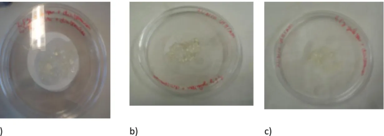
Figure 4.5. a) Beads at time zero; b) Beads in the second day. ... 40
Figure 4.6. a) Gelatine beads at time zero; b) Gelatine beads after 30 days. ... 40
Figure 4.7. a) Pectin beads at time zero; b) Pectin beads in 30 days after its production. ... 41
Figure 5.1. SDS structure. ... 44
Figure 5.2. Tris base structure. ... 45
Figure 5.3. Triton x-100 structure. ... 45
x
List of tables
Table 2.1. Marine enrichment f/2 medium (Guillard, 1975) ... 9 Table 2.2. Freshwater enrichment WC medium (Guillard and Lorenzen, 1972) ... 10 Table 5.1. Viability of diatom cultures after surfactant use ... 48
xi
List of abbreviations
AS Anionic surfactant
CaCl2 Calcium chloride
CaCl2.2H2O Calcium chloride dihydrate
CaCl2.6H2O Calcium chloride hexahydrate
CuSO4.5H2O Copper sulfate pentahydrate
CMC Critical micelle concentration
DHB 2,5 % dihydroxybenzoic acid
dH2O Distilled water
DNA Deoxyribonucleic acid
DMSO Dimethyl sulfoxide
EPS Extracellular polymeric substance
FDA Fluorescein diacetate
Fe Iron
FeCl2.6H2O Iron chloride hexahydrate
G1/2 phase Gap 1/2 phase of cell cycle
GTA Glutaraldehyde
HCl Hydrochloric acid
H3BO3 Boric acid
K2HPO4 Dipotassium phosphate
LAS Linear alkylbenzene sulfonates
LB Luria-Bertani
M phase Mitosis phase of cell cycle
MALDI-TOF Matrix Assisted Laser Desorption Ionisation – Time Of Flight
xii
Spectrometry
MALDI-TOF MS Matrix Assisted Laser Desorption Ionisation – Time Of Flight Mass Spectrometry
MgSO4.2H2O Magnesium sulfate dihydrate
MnCl2.4H2O Manganese chloride tetrahydrate
mPBS Marine phosphate buffered saline
MUM Micoteca da Universidade do Minho
m/z Mass to Charge Ratio
N2 Nitrogen
NaCl Sodium chloride
NaHCO3 Sodium bicarbonate
Na2HPO4 Sodium phosphate dibasic
NaH2PO4.2H2O Sodium dihydrogen phosphate dyhydrate
Na2EDTA Ethylenediaminetetraacetic acid disodium salt
Na2MoO4.2H2O Sodium molybdate dihydrate
NaNO3 Sodium nitrate
NaOH Sodium hydroxide
NaSiO2.9H20 Sodium metasilicate nanohydrate
Nd:YAG neodymium-doped yttrium aluminium garnet PAE Protistology and Aquatic Ecology
PUFAs Polyunsaturared fatty acids
S phase Synthesis (DNA replication) phase of cell cycle
SARAMIS™ Spectral Archiving and Microbial Identification System
SEM Scanning Electron Microscopy
SDS Sodium dodecyl sulphate
SDV Silica deposition vesicle
TFA Trifluoroacetic acid
xiii
WC Wright´s Chu
xv
“Most diatoms are beneficial to the oceans’ ecosystems and ultimately to human health”
1 General introduction
and thesis outline
2
Diatoms (class Bacillariophyceae) are unicellular photosynthetic eukaryotes (Huysman et al. 2010), which have a particular cell wall, composed of silica. These microorganisms are divided in two major architectural types: “centric” diatoms, a paraphyletic group with radially patterned valves; and “pennate” diatoms, a monophyletic group characterised by a feather-like valve structure (Scala and Bowler, 2001; Gillard et al. 2008). Diatoms are one of the predominant contributors to global carbon fixation (Parkinson et al. 1999; Scala and Bowler, 2001; Sarthou et al. 2005).
Recently, the interest in diatoms has increased since they have many possible applications (e.g. aquaculture, nanotechnology, among others). Consequently, proper diatom identification processes are necessary in order to select the best species for each given purpose. Diatom taxonomic classifications were originally based on optical microscopic analysis; this identification is currently made by Scanning Electron Microscopy (SEM) because the frustule structures are the basis for species identification. However, this technique is subject to misidentification (Scala and Bowler, 2001).
The aim of this work was the development of new methods for the characterisation and cultivation of diatoms. One of the studies performed in this work was based on the use of immobilisation in gelatine beads, for long-term preservation of diatoms as an alternative to the existing maintenance strategies, that is, obscurity in cold room or cryopreservation. Additionally, the possibility of detaching diatoms through chemical action was studied. And finally, the possibility of using the MALDI-TOF ICMS technique as a fast, low cost and reliable method for characterisation of diatom strains was evaluated.
This thesis consists of 6 chapters. In the first Chapter, the subject and the theoretical framework of the work are presented. In Chapter 2, the biology and morphology of diatoms, as well as their specified characterisation, is presented. In Chapter 3, the possibility of using the MALDI-TOF ICMS technique to characterise the diatom strains is discussed. In Chapter 4, the long-term preservation of the centric diatom Coscinodiscus sp. in gelatine and pectin beads is studied. In Chapter 5, a method to detach benthic diatoms, Seminavis robusta 84A in this case, through the action of surfactants is presented. Finally, in Chapter 6, general conclusions of the thesis are summarized and perspectives for future research are provided.
2 Diatoms Classic Characterisation
ABSTRACT. Before working with any living being it is necessary to understand its way of living. In this chapter, the unique characteristics of diatoms and their growth are described.
4
2.1 Introduction
2.1.1 General Overview on Diatoms
Diatoms (division Heterokotophyta, class Bacillariophyceae) are microscopic organisms ranging in general from 20 to 200 m (Figure 2.1) (Scala and Bowler. 2001; Gordon et al. 2008) and live in aquatic ecosystems, freshwater or marine (Trainer et al. 2008; Groger et al. 2008). These microorganisms are divided in two types: benthic attached to submerged surfaces and planktonic -free swimming in open water. Benthic diatoms can grow in sediments or on the surface of plants (epiphytic), animals (epizootic), or rocks (epilithic) (Winter and Duthie, 2000; Trainer et al. 2008). Both benthic and planktonic diatoms can be found as single cells, while others form long chains of adjacent cells, either by end to end junction or by joining their protruding spines or setae (Trainer et al. 2008).
5
Diatoms are the most diverse group within algae, reaching about 200 000 species (Holtermann et al. 2010) and, like plants, they contain chlorophyll and other pigments that capture the energy of sunlight and convert carbon dioxide and water molecules into carbohydrates via photosynthesis (Trainer et al. 2008). Thus, they are a major contributor to global carbon dioxide fixation. They are also responsible for 20 % to 25 % of the world net primary production, and roughly for 40% of annual marine biomass production supporting most of the world fisheries (Gordon et al. 2008; Trainer et al. 2008) .
The name diatom comes from the Greek: dia “through” and temein “to cut”, meaning “cut in half” referring to their highly ornamented siliceous cell wall – or frustule – divided in two twin parts (Trainer et al. 2008). The frustule is composed of two overlapping halves (or valves) that fit together like a petri dish in which the upper half – the epitheca - covers the lower half – the hypotheca (Figure 2.2). Generally, the diatoms are categorized into two major groups based on how the silica (SiO2 =
glass) ribs on the valve radiate: centrics - a paraphyletic group with discoid or cylindrical cells and radially symmetrical valves - and pennates - a monophyletic group having ‘feathery’ patterned and more or less bilaterally symmetrical valves (Round et al., 1990; Graham and Wilcox, 2000; Scala and Bowler, 2001; Trainer et al. 2008; Gillard et al. 2008; Hildebrand, 2008). These two groups consist of about 285 genera (Round et al. 1990) and 200 000 morphologically different species. It is worth noting that, on the basis of molecular genetic analyses, there are probably over 100000 (pseudo)cryptic species. Motility is different in these two groups: centric diatoms (Figure 2.2.a) have flagellated gametes but are otherwise swept passively by currents; pennate diatoms (Figure 2.2.b), on the other hand, are capable of limited motility (Trainer et al. 2008).
Figure 2.2.Schematic diagram of the diatom cell wall structure a) in centric diatoms; b) in pennate diatoms (Al-Kandari et
al. 2009).
a)
6
The frustule is composed of solid silica, being the hypotheca slightly smaller than the epitheca (Figure 2.2). This causes a cell size reduction during the vegetative cell division carried out by mitotic division. From time to time, the diatoms restore the original cell size via sexual reproduction. These two stages have different time laps: a prolonged vegetative stage (mitotic division of cells) lasting from months to years, a comparatively short stage of sexual reproduction (gametogenesis and fertilization) lasting several hours and then a complex developmental process (leading to the formation of the new vegetative cells) taking from hours to one week or more. The vegetative cell cycle (Figure 2.3) in diatoms consists of interphase (G1, S and G2 phases) and mitosis (M phase), the two daughter cell protoplasts still contained within the mother cell and not separated (Hildebrand, 2008). Cells in G2 and M cannot be distinguished on the basis of DNA content by flow cytometry; therefore, the term “G2+M” is used to describe this situation. During the G2+M step, the valves in of daughter cells are synthesized in the silica deposition vesicle (SDV) and once completed, are exocytosed, becoming extracellular. Cell separation occurs after synthesis of new valves in each of the daughter cells. As said above, an additional special feature of the diatom life cycle is the size-dependent control of sexuality in which only cells of a particular size range (viz. comparatively small cells) are able to become sexualized (Trainer et al. 2008; Hildebrand, 2008).
Figure 2.3. “Schematic representation of diatom cell division: a) vegetative mother cell (S phase), b) after cytokinesis, showing two daughter cell protoplasts within the mother cell thecae, c) valve synthesis occurring within the daughter cell, d) daughter cell valves after exocytosis, and e) daughter cell separation” from Hildbrand (2008).
2.1.2 Basis taxonomy of diatoms
Diatom taxonomy was primarily based on light microscopy observation of the morphologies of their frustule. The use of SEM resulted is a significant advance for taxonomy, since it revealed additional characteristics not observable by light microscopy. Based on their morphology, Round et al. (1990) proposed the classification of diatoms in three classes including the centric diatoms (Coscinodiscophyceae), the pennate diatoms without a raphe (Fragilariophyceae) and the pennate diatoms with a raphe (Bacillariophyceae) (Al-Kandari et al. 2009).
7
2.1.3 Industrial applications
Due to their high productivity, intra-and extracellular composition and single cell wall composition and morphology, diatoms have sparked a growing interest for several industrial applications (Lopez et al. 2005).
One example comes from aquaculture since algae are used in biomass production for food and feed. Also, diatoms are a natural source of relevant biotechnology products, such as amino-acids and polyunsaturated fatty acids (PUFAs) for, respectively, cosmetic and pharmaceutical applications (Lebeau and Robert 2003; Lopez et al. 2005). Due to the algal biomass main components, it is possible to use diatoms in the production of biofuel: carbohydrates for ethanol production via fermentation, proteins for methane production via anaerobic gasification and natural oils for biodiesel production (Bozarth et al. 2009). Diatoms have the ability to accumulate up to 60 % of their weight in cytoplasmic oil drops (Lebeau and Robert 2003). The resulted products of their metabolism are often released into the medium, such as the blue-green pigment called "marennine". Also, diatom metabolites (e.g. domoic acid) can be used in pharmaceutical applications. Finally, diatoms can also be applied in the nanotechnology industry, as a result of their ability of surpassing some of the modern engineering competences (Bozarth et al. 2009) as their formation does not require high temperatures, high pressure or the use of caustic chemicals, being accomplished under mild physiological conditions (Lopez et al. 2005). In this way, the frustules can be explored for various applications, such as abrasive products, filter agents for water purification, gel filtration for protein purification, biosensors, immunoisolation, photonics, drug delivery (Parkinson et al. 1999).
2.2 Materials and methods
2.2.1 Cell cultures
Stock cultures of the diatoms species and strains were obtained from the Culture Collection of the Laboratory of Protistology and Aquatic Ecology (PAE) in Ghent University (http://www.pae.ugent.be/collection.html).
8
2.2.2 Culture conditions
2.2.2.1 Culture media
For the marine species Seminavis robusta, Coscinodiscus sp. and Thalassiosira sp., the f/2 medium was used and for t,he freshwater species Cyclotella meneghiniana, the WC medium was used. The f/2 medium (Guillard, 1975) is a common and widely used general enriched seawater medium designed for growing coastal marine algae, especially diatoms. The concentration of the original formulation of the "f Medium" (Guillard and Ryther 1962) was reduced by half. The WC medium is derived from the Chu #10 Medium (Chu 1942). The medium was originally formulated to aid Richard Wright in his efforts to cultivate cryptophytes (WC = Wright's Chu #10), and first published by Guillard and Lorenzen (1972). Subsequently, Guillard has modified the medium being referred to, in the present work, as WC medium. Stock of all ingredients and culture media were stored in Schott® glass bottles (Duran) and kept in the refrigerator at 5°C.
2.2.2.1.1 f/2 medium (Guillard, 1975) with natural seawater
Seawater, collected from the northwestern Portuguese coast (Vila de Conde) was filtered using a filtration apparatus, consisting of an Erlenmeyer flask, a funnel, a 55 mm Ø filter paper circle (VWR® European Cat. No. 516-0870) and of a rubber tube that connected the flask to the air pump. A volume of 1 L seawater was filtered over each filter paper. The filtered seawater was transferred to Schott® (Duran) glass bottles of 1 to 2 L volume.
The f/2 medium was prepared by adding to 950 mL of filtered seawater, 1mL of major nutrients stock solutions and also 1 mL of trace metals solution (table 2.1). The f/2 medium was sterilized using an autoclave (uniclave 88) at 121 ˚C for 15 minutes. Before autoclaving, the pH was adjusted to 8 with 1 N HCL or 1 N NaOH. Autoclaved bottles were cooled and the vitamins stock solution was added (table 2.2)
9
Table 2.1. Marine enrichment f/2 medium (Guillard, 1975)
Component Stock Solution Major nutrients (mg/mL in distilled water)
NaNO3 75
NaH2PO4.H2O 5
Na2SiO3.9H2O 30
Trace metals (mg/mL in distilled water) Na2EDTA 4.36 FeCl3.6H2O 3.15 CuSO4.5H2O 0.01 ZnSO4.7H2O 0.022 CoCl2.H2O 0.01 MnCl2.H2O 0.18 Na2MoO4.2H2O 0.006
Vitamins (µg/mL in distilled water) Thiamine.HCl 100
Biotin 0.5 Cyanocobalamin 0.5
2.2.2.1.1.1 WC medium (Guillard and Lorenzen, 1972)
Stock solutions of the components for freshwater WC medium are shown in table 2.2. Similar components to the f/2 medium were prepared only once. Other major nutrients were individually prepared. Components were added to distilled water in Schott bottles (Duran) using 1 mL stock solution/L (except for the vitamins stock solution). The mixed solutions were autoclaved at 121°C for 15 minutes.
10
Table 2.2. Freshwater enrichment WC medium (Guillard and Lorenzen, 1972)
Component Stock Solution Major nutrients (mg/mL in distilled water)
NaNO3 85.01 NaHCO3 12.60 Na2SiO3.9H2O 28.42 CaCl2.2H2O 36.76 MgSO4.7H2O 36.97 K2HPO4 8.71
Trace metals (mg/mL in distilled water) Na2EDTA 4.36 FeCl3.6H2O 3.15 CuSO4.5H2O 0.01 ZnSO4.7H2O 0.022 CoCl2.H2O 0.01 MnCl2.H2O 0.18 Na2MoO4.2H2O 0.006 H3BO3 1
Vitamins (µg/mL in distilled water) Thiamine.HCl 100
Biotin 0.5 Cyanocobalamin 0.5
2.2.3 Culture growth
The Seminavis robusta cultures were re-inoculated by adding 2 mL of the old culture (gently scraping with a cell scraper) in f/2 medium, in a total volume of 50 mL. Cells were cultivated in 75 cm2 tissue culture flasks with filter cap (Orange Scientific®). Incubation was at room temperature by a 12:12 h light:dark cycle (85 µmol photons/m2/s from cool-white fluorescent lamps). Cultures were observed weekly under the inverted microscope to assess growth and possible contaminations. On the other hand, planktonic species (Thalassiosira sp, Coscinodiscus sp, Cyclotella meneghiniana) were harvested and re-inoculated by adding 12 mL to the f/2 medium, to complete a total volume of 250
11
mL. Harvesting and inoculation of cells were done in the close vicinity of a flame. The cultures used were uni-algal but not axenic, although bacterial numbers were minimal at all stages.
2.2.4 Assessment of the viability
2.2.4.1 Observation under the inverted microscope
A sample of 1 mL of recovered cells suspension was transferred to 3 wells of a 6 well-plate. Settled cells were observed under the inverted microscope (Nikon).
2.2.4.2 Observation under the light microscopy
A sample of 30 µL of recovered cells suspension was transferred to a glass slide and covered with a coverslide. Cells were observed under the light microscopy (LEITZ).
2.2.4.3 Epifluorescence microscope (diatom viability using FDA)
The viability of the diatoms was assessed using the fluorescein diacetate (FDA) staining method. One milliliter of the benthic diatoms culture and the planktonic species were removed, the first gently scraped with a cell scraper. These samples were incubated with 10 µL FDA stock solutions (Michels et a. 2010) for 20 min in the dark at room temperature. The samples were observed under an epifluorescence microscope (OLYMPUS BX51) using a fluorescence emission of fluorescein detected at 525 nm (Holm et al. 2008, Michels et a. 2010).
2.2.5 Growth curve
2.2.5.1 Inverted microscope
All Seminavis robusta cultures were directly observed every eight hours under the microscope. The planktonic species, including Thalassiosira sp, Coscinodiscus sp, Cyclotella
meneghiniana, were assessed in a different way: 1 mL of culture was removed every eight hours and
12
2.2.5.2 Use of Coulter Counter
Seminavis robusta cultures were gently scraped with a cell scraper and centrifuged (10 min at
3000xg, in centrifuge 2 HERAEUS MEGAFUGE 1.0R). The pellet was collected and resuspended in 20 mL of diluent. After this, cells were counted in a Bechman Coulter (Z2 Couter® Particle count and size
analyser) every eight hours.
2.2.5.3 Fluorimetry
The benthic Seminavis robusta was gently scraped with a cell scraper and centrifuged (10 min at 3000 g), being the pellet collected and resuspended in 2 mL of mPBS (Cerda-Cuellar et al. 1997). The centric genera were removed by a volume of 2 mL of culture. The cell suspensions were analysed by flourimetry (Fluorescence Spectrometer Jasco FP6200). For that, 2 ml of cell suspension were diluted in mPBS (to fit the calibration curve) and placed in a fluorimeter cell (cuvette with at least three polished windows and no background fluorescence). The emission at 685 nm was read after excitation at 440 nm (Ikeda et al. 2008). All readings were done in triplicate every eight hours.
2.3 Results and discussion
2.3.1 Characterisation
In order to fully characterise the diatoms, these were observed under three types of microscopes (light microscopy, inverted microscope and epifluorescence microscope).
2.3.1.1 Pennate diatoms
Seminavis robusta (Figure 2.4) is a small pennate of the marine epipelon and epiphyton
(Garcia, 2007). The most important characteristics for their identification are: the outline of the ventral margins, the shape of the apices, the length of the striae on the dorsal valve face, the presence or the absence of striation interruption on the ventral valve face and the shape and the position of the raphe in relation to the ventral margin (Garcia, 2007).
13
Figure 2.4. Seminavis robusta.
All the Seminavis strains used in this work (Figure 2.5) have the same shape but different sizes and were of different mating types. Semivavis robusta 84A (Figure 2.5 a) and 85A (figure 2.5 d) have a size of about 45 µm and Semivavis robusta 85As (Figure 2.5 g) and 85Bs (Figure 2.5 j) have a size of about 25 µm.
Under the light microscopy, it was possible to verify the elongated frustule and two chloroplasts. As described in the literature, the frustule is generally elongated with a longitudinal axis of symmetry (rib) in each valve (bilateral symmetry) (Kröger and Poulsen, 2008).
With the inverted microscope, the spatial organisation in culture was studied. As in light microscopy, it was possible to check again the elongated frustule, but not the presence of the chloroplasts.
Under the epifluorescence microscope, the FDA was used to determine if the cells were intact. With FDA, it was possible to see the chloroplasts in red and the membrane in green.
14
Light microscopy Inverted microscope Epifluorescence microscope (FDA) a) b) c) d) e) f) g) h) i) j) k) l)
Figure 2.5. Living cells of Seminavis robusta. a, b, c) strain 84 A; d, e, f) strain 85 A; g, h, i) strain 85 As; j, k, l) strain 85 Bs. a, b, c, d, e, f, g, h, I, j and l) using the objective of 40x magnification; k) using the objective of 60x magnification.
15
2.3.1.2 Centric diatoms
Three centric diatoms (Coscinodiscus sp., Thalassiosira sp. and Cyclotella meneghiniana) were studied.
Coscinodiscusis a big centric diatom with a diameter of 30 to 500 µm. These marine diatoms
have numerous chloroplasts. The cell is described as being disc-shaped, cylindrical or wedge-shaped, and solitary.
Thalassiosirais an important centric diatom, due to its presence in marine environments with
more than 100 already identified species. The first genome sequenced among diatoms was the
Thalassiosira pseudonana, which was important to enable the unravelling of the genetic basis of the
unique properties underlying the ecological and evolutionary success of diatoms. The morphological identification of the genus Thalassiosira, with diameters ranging from 3 to 186 µm, is based on ultra-structure details as the number and the location of the rimoportulae and fultoportulae processes on the valve (Garcia and Odebrecht, 2009).
The last centric diatom used in this work is the Cyclotella meneghiniana, being one of the most extensively studied freshwater diatoms species (Beszteri et al. 2005, Beszteri et al. 2007). Their cells are described as barrel-shaped, with more-or-less tangentially undulate valves, strongly striated from the marginal area to halfway towards the middle, widening towards the margins and narrowing towards the centre (8-9 in 10 µm); the central area is plain, or with faint radiate, punctate striae, sometimes with a few solitary large punctae; their diameter is 10 to 30 µm.
In centric diatoms (Figure 2.6), as in pennate diatoms, light, inverted and epifluorescence microscopes were used to characterise the cells. Centric diatoms have the same shape (circular), however there is a big difference among them when size is considered, the Coscinodiscus sp. strain used in the present study (Figure 2.6 a) being the largest with about 170 µm, followed by the
Thalassiosira sp. Strain (figure 2.6 d) with about 70 µm and finally Cyclotella meneghiniana (Figure
2.6 g) with about 10 µm.
With the light microscope, it was possible to see the circular frustule in all centric diatoms and the chloroplasts distribution. Coscinodiscus sp. has many small chloroplasts; Thalassiora sp. has numerous big chloroplasts.
16
Using the inverted microscope it was possible to observe the spatial distribution of the diatoms in culture.
Under the epifluorescence microscope and using FDA, the membrane and porous were seen in green. Thalassiosira has more pores than the other centric diatoms.
Light microscopy Inverted microscope Epifluorescence microscope (FDA) a) b) c) d) e) f) g) h) i)
Figure 2.6. Living cells of centric diatoms. a, b, c) strain Coscinodiscus sp.; d, e, f) strain Thalassiosira sp.; g, h, i) strain Cyclotella meneghiniana. a, b, c, d, e and f) using the objective of 10x magnification; g, h and i) using the objective of 60 x magnification.
17
2.3.2 Growth curve
2.3.2.1 Benthic diatom Seminavis robusta
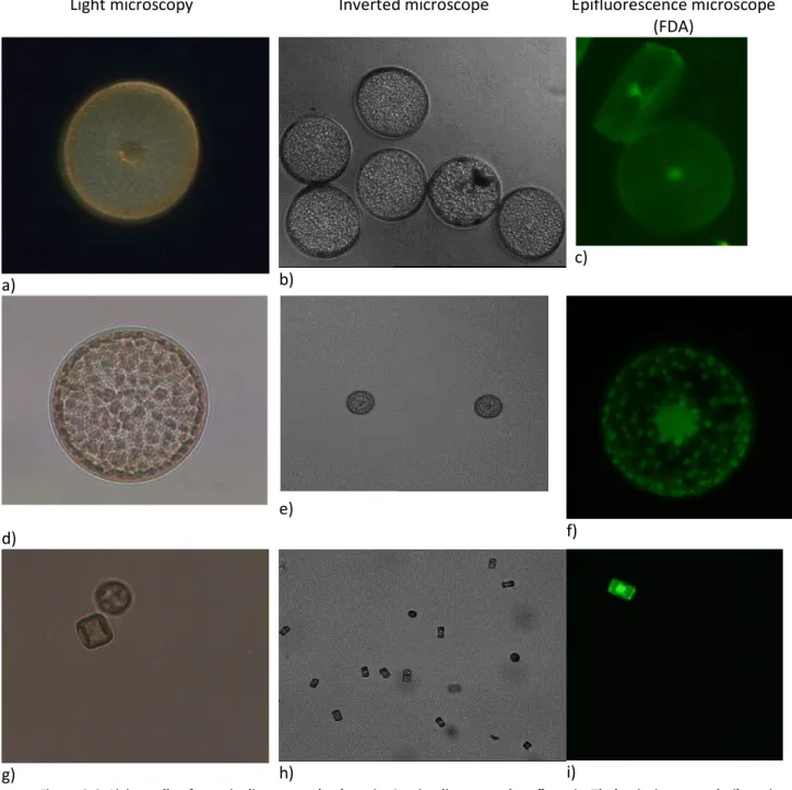
To study the Seminavis robusta growth, the Coulter Counter and the inverted microscope were used. Figure 2.7 describes the growth curve of all Seminavis strains using the Coulter. The growth curve is very similar for all diatoms, being the only significant difference the concentration. The same can be observed in the Seminavis growth curve using the inverted microscope (Figure 2.8). The exponential phase is very short (90 hours) and there is not a lag phase.
Figure 2.7. All Seminavis robusta growth curve using Coulter.
Seminavis robusta showed one particular feature during its growth: during the stationary
phase, by approximately 150 hours, the diatoms spontaneously detach from the wall of the culture flasks. That explains the decrease of the cell number assessed by inverted microscopy, as the cells in different plans were difficult to count.
18
Figure 2.8. All Seminavis robusta growth curve using inverted microscope.
Figure 2.9 shows the differences in Seminavis robusta 84 A growth using the three different methods used (Coulter Counter, inverted microscope and fluorimetry). The observation under the inverted microscope was the method the most affected by errors, being the curves obtained by the fluorimetry and by the Coulter Counter equivalent. Nevertheless, the pattern of the growth curve, comprising the two phases – exponential phase and stationary phase – are consistently observed in the curves generated by the three methods. Seminavis robusta does not have a lag phase: the exponential phase ends circa the 90 hours but the culture only is in a true stationary phase at 140 hours. The coincidence in the curves that resulted by the fluorimetry and the Coulter Counter stands for the significance of the results obtained by both methods.
19
Figure 2.9. Seminavis robusta 84A growth curve using three different methods (Coulter, inverted microscopy and fluorimetry).
2.3.2.2 Centric diatoms
The same methods (Coulter, inverted microscope, fluorimetry) were used for the study of the growth of the centric diatoms, but the cell size and the concentration of the cultures prevented the simultaneous use of the three methods in all cultures.
In the case of Coscinodiscus, the Coulter Counter was not used because the cell size is bigger than the probe detection limit. All the same, the results of fluorimetry and inverted microscopy were similar (Figure 2.10). Unlike Seminavis robusta, Coscinodiscus has a prolonged lag phase (lasting 90 hours), the cell needing more time to reach the stationary phase and being the curve during the exponential phase not very sharp.
20
Figure 2.10. Coscinodiscus sp. growth curve using two different methods (inverted microscopy and fluorimetry).
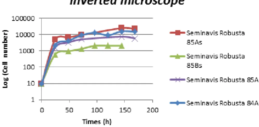
In the case of Thalassiosira, only the inverted microscope was used, since the culture was not sufficiently concentrated and neither the Coulter Counter nor the fluorimeter could be used. The use of centrifugation was considered to concentrate the samples and consequently to enable fluorimetry, but this approach had to be abandoned because huge volumes of culture would be needed. The growth curve (Figure 2.11) is similar to the one of Coscinodiscus. That is, the
Thalassiosira growth curve has an extended lag phase (with 90 hours) and a smooth exponential
21
Figure 2.11. Thallassiosira growth curve using inverted microscopy.
Cyclotella was the only freshwater diatom studied in this work. Here, as in Coscinodiscus, the
Coulter Counter could not be used as the size of Cyclotella is beneath the limit of detection. The curve (Figure 2.12) shows three well-defined phases (lag, exponential and stationary). Here, as it happened with Seminavis robust, the inverted microscope was error affected, because Cyclotella is very little and observation and counting in different plans of the microscope was limited.
22
2.4 Conclusions
The growth curves of all Semivavis robusta strains have the same shape, which was expected since they all belong to the same species. No morphological differences were observed among the strains with the exception of the cell size.
The three centric diatoms, Talassiosira, Coscinodiscus and the marine Cyclotella, were different in size and in chloroplasts distribution and their concentrations during the experiments were very low, which can be due to the high temperatures in the lab, around 30 °C.
All diatoms showed different growth curves patterns. All Seminavis strains belong to the same genus and they show the same behaviour. Talassiosira and Coscinodiscus were grown in the same conditions and have similar growth curves, even though they attend very different concentrations.
By the results, we can conclude that the three methods, the Coulter Counter, the inverted microscope and the fluorimetry were applicable methods for the study of diatom growth. However, these methods sometimes cannot be used due the cell size or population density. Furthermore, the inspection under the inverted microscope can result in significant errors, as the focusing of several focal plans cannot be efficiently accomplished rendering the results affected by errors.
3 Diatom characterisation
by MALDI-TOF ICMS
ABSTRACT. Diatom identification is sometimes intricate because it is based on the frustule morphology. To overcome the current difficulties, MALDI-TOF ICMS technique is proposed as a fast, inexpensive and reliable method. The use of MADI-TOF ICMS for diatoms identification is evaluated.
24
3.1 Introduction
The analysis and characterisation of macromolecules and their complexes which are the core of life, as well as the spectral typing of microbial cells for their taxonomic classification/identification and authentication using spectral analysis by Matrix Assisted Laser Desorption Ionisation – Time Of Flight Mass Spectrometry (MALDI-TOF MS) are both modern approaches for the life sciences and biotechnology studies (Shaah and Gharbia, 2010). MALDI-TOF MS emerged in the late 1980s as a sound technique to investigate the mass spectrometry of molecular high-mass of organic compounds through a soft ionisation of the molecules resulting in minimum fragmentation (Tanaka et al. 1998).
The MALDI-TOF MS technique has contributed to increasing scientific knowledge about the microorganisms and is now used as a reliable tool for rapid tests in hospitals and health centres. In this case the interest of the art in question is the analysis of the intact cell. The spectrum generated is analysed as fingerprint and the technique is called MALDI-TOF IC (Intact Cell) MS. In the MALDI-TOF ICMS technique the microbiological sample is covered with a UV-absorbing organic compound called matrix leading to a crystallised mixture. Then the crystallised sample is placed in a vacuum system that is targeted and irradiated with a pulsed light from a laser (e.g. N2, Nd:YAG or other) . The
charged matrix molecules and/or clusters transfer protons onto the sample molecules (e.g. peptide or proteins) in the expanding plume. The generated ions are accelerated into the TOF analyser, in which ions are separated according to their "time-of-flight" which is a function of molecular mass to charge. The TOF analyser determines the molecular mass to charge (m/z) ratio of ions by measuring velocities from accelerating ions to defined kinetic energies after calibration of the instrument with molecules of known molecular mass (Figure 3.1). In MALDI-TOF ICMS technique the linear mode set covers a huge mass range capable to get the appropriate fingerprint for each kind of microorganism (e.g. 2 - 20 kDa) (Santos et al. 2010).
25
Figure 3.1. Schematic representation of MALDI-TOF MS operation: After molecule ionisation the separation of ions occurs into the flight tube based on their molecular masses (Santos et al. 2010).
The success of diatoms in biotechnological and nanotechnological application primarily depends on the correct diatom identification and characterisation. Novel identification methods need to be fast, inexpensive and reliable (Santos et al. 2010; Shah, 2005). MALDI-TOF ICMS is a powerful technique with all of these adjectives. This technique has been proposed as an identification method for bacteria (Shaah and Gharbia, 2010), filamentous fungi (Santos et al. 2010), yeasts (Santos et al. 2011) and virus (Franco et al. 2010; McAlpin et al. 2010).
MALDI-TOF MS technique has been used for diatoms characterisation through the analyses of lipids (Vieler et al. 2007), chlorophylls (Suzuki et al. 2009), silaffins (Sumeper et al. 2007) and polyamines associated with silica (Sumper et al. 2006; Knott et al. 2007). In the works presented above, authors used a mass range between 100 and 3 000 Da. However, the remarkable reproducibility of the MALDI-TOF ICMS technique for the most common microorganisms (e.g. bacteria, filamentous fungi and yeasts) is based on the measurement of constantly expressed and highly abundant proteins. The usually observable molecular mass range is between 2 000 and 20 000 Da, where important proteins and other macromolecules appear, which is an advantage because these can be easily used as biomarkers (Santos et al. 2010).
26
In this work, seven isolates of diatoms belonging to four different genera namely, Seminavis;
Coscinodiscus; Thalassiosira and Cyclotella, were analysed by MALDI-TOF ICMS. All isolates were
obtained from the culture collection of the Laboratory of Protistology and Aquatic Ecology of University of Ghent (Ghent, Belgium). The molecular mass range between 2 000 and 20 000 Da was used as an attempt to generate a specific fingerprint for each diatom isolate.
3.2 Materials and methods
3.2.1 Growth conditions
All 7 diatoms isolates analysed in this study were obtained from the Laboratory of Protistology and Aquatic Ecology culture collection (PAE, http://www.pae.ugent.be/collection.html).
Escherichia coli strain DH5α was obtained from the Micoteca da Universidade do Minho (MUM,
www.micoteca.deb.uminho.pt). All cultures were maintained in falcon tubes and preserved in a dark room at 4.0 ±1.0 °C. Tubes were opened and strains sub-cultured according to the instructions issued by PAE. Homogenous inocula of diatoms cells were grown and maintained on f/2 medium.
Escherichia coli cells were grown and maintained on Luria-Bertani agar medium (LB: 10 g∙l 1
Bacto-tryptone, 5 g∙l-1 Bacto-yeast extract, 10 g∙l-1 NaCl).
3.2.2
MALDI-TOF AnalysisBenthic diatoms were initially detached from the culture flask through mechanical action, with a cell scraper (Cat # 5560500 - 23 cm - Orange Scientific). Centric diatoms were cultivated in 250 mL Elermeyers flasks. All the cultures were centrifuged at 3000 g for 10 minutes using a centrifuge 2 HERAEUS MEGAFUGE 1.0R. The pellet was collected, placed in an Eppendorf and centrifuged once again for 4 minutes using a micro-centrifuge Sigma 112 – B. Braun Botech international. The pellet obtained was transferred into an Eppendorf containing 30 µL acetonitrile aqueous solution (60 % acetonitrile in 40 % ultra-pure water), and vortexed for 1 minute. Half a microliter of the green suspension obtained above was placed on the MALDI sample plate. When the liquid phase was almost evaporated, 0.5 L DHB matrix solution (75 mg/mL 2,5-dihydroxybenzoic acid [DHB] in ethanol/water/acetonitrile [1:1:1; v/v/v] with 0.03% trifluoroacetic acid [TFA]) was added and mixed gently. Finally, all samples were air dried at room temperature and analysed in quadruplicate.
27
The MALDI-TOF ICMS analyses were performed in the Platform of Structural Analysis of the Centre of Biological Engineering of University of Minho on an MALDI-TOF Axima LNR system (KratosAnalytical, Shimadzu, Manchester, UK).The instrument was equipped with a nitrogen laser (337 nm), where the laser intensity was set above the threshold for ion production. Escherichia coli DH5α strain with known mass values of ribosomal proteins was used as an external calibrant. The mass range from 2 000 to 20 000 Da was recorded using the linear mode with a delay of 104 ns and an acceleration voltage of + 20 kV. Final spectra were generated by summing 20 laser shots accumulated per profile and 50 profiles produced per sample, leading to 1 000 laser shots per summed spectrum. The resulting peak lists were exported to the SARAMIS™ software package (Spectral Archiving and Microbial Identification System, AnagnosTec, Germany, www.anagnostec.eu) where the final identifications were achieved. This software uses a point system based on peak list with mass signals weighted according to their specificity. The similarity between individual spectra is expressed as the relative or absolute number of matching mass signals after subjecting the data to a single link agglomerative clustering algorithm. Microbial identifications by the SARAMIS™ package are based on the presence or absence of each peak in the spectra.
3.3 Results and discussion
The genetic and proteomic information are not available on the literature for the diatoms species studied in this work. The mass range from 2 000 to 20 000 Da was chosen taking into consideration the proteomic information available for bacteria (Shaah and Gharbia, 2010), filamentous fungi (Santos et al. 2010) and yeasts (Santos et al. 2011). However, through the results obtained preliminary in this work for all diatoms isolates it was possible to observe that the chemical compounds present on each diatom spectral fingerprint presented variations as a function of the isolate age. Moreover, for the same culture duplicate or triplicate MALDI-TOF ICMS samples presented different mass spectra. Figure 3.2 shows the duplicate spectra obtained from the same culture for Seminavis robusta strain 85A at 5 days old. The observation of these spectra leads to the conclusion that the majority of the compounds evaluated within the mass range from 2 000 to 20 000 Da are not ribosomal proteins (Appendices B).
28
Figure 3.2.Duplicates MALDI-TOF ICMS mass spectra for Seminavis robusta strain 85A at 5 days old.
In order to understand the change in molecular masses, the mass analyses of mature diatoms was performed at the stationary phase. It was developed according to the growth curve (Chapter 2) for each diatom species (Seminavis robusta strains 84A, 85A, 85As, and 85Bs, Cyclotella
meneghiniana, Coscinodiscus sp. and Thalassiosia sp.). All of the mass spectra obtained in these
analyses show similar behaviour as observed for Seminavis robusta strain 85A mass spectra at 5 days old described above (Figure 3.2). Considering both the growth curve obtained by classical techniques (Chapter 2) and the mass fingerprint by MALDI-TOF ICSM discussed above incongruent results are observed. The growth curves by classical techniques indicate that the stationary phase was 6 days old for all Seminavis robusta and 7 days old for Cyclotella meneghiniana, Thalassiosia sp. and
Coscinodiscus sp.. On the other hand, MALDI-TOF ICMS spectra performed at and/or after these
diatoms ages indicate to a potential different growth phase inside the same diatoms cultures.
Since the mass spectra change with the diatoms age the spectral change versus the diatom age was assessed. For each diatom strains a specific time course of the spectrum versus time was assessed based on the follow schedule: 7, 9, 13, 14, 18 and 30 days old. However, results indicate that because of the in situ bio-compounds extraction quality for some of the diatoms isolates the mass spectra where not generated for this entire schedule. Moreover, all the diatoms evaluated in this study presented a specific age where the mass spectrum becomes reproducible. For all
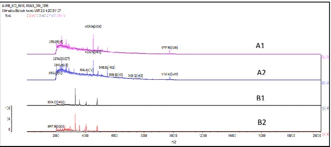
Seminavis robusta and Cyclotella meneghiniana this time was 9 days old (Figure 3.3) and for all Thalassiosia sp. and Coscinodiscus sp. it was 13 days old (Figure 3.4).
29
Figure 3.3. Reproducible MALDI-TOF ICMS mass spectra for (A1-2) Cyclotella meneghiniana and (B1-2) Seminavis robusta strains 84A at 9 days old.
Figure 3.4. Reproducible MALDI-TOF ICMS mass spectra for (A1-2) Thalassiosia sp. and (B1-2) Coscinodiscus sp. at 13 days old.
Furthermore, there was no compounds stability over time. Spectral data obtained for Coscinodiscus sp. at 13, 18 and 30 days old culture (Figure 3.5) is an example of instability over time for this species.
A1 A2 B2 B1 A1 A2 B1 B2
30
Figure 3.5. MALDI-TOF ICMS mass spectra for Coscinodiscus sp. at 13, 18 and 30 days old from top to bottom.
Using the first reproducible spectral data (Appendices B) including all peaks obtained from 2 000 to 20 000 Da a cluster analysis of the MALDI-TOF ICMS mass spectral data was assembled. This statistic analysis based on the mass spectral signatures allowed the grouping of all isolates into clusters according to their species designation (Figure 3.6). The dendrogram presents two main clusters with the two different pairs Thalassiosia sp./Cyclotella meneghiniana and Seminavis
robusta/Coscinodiscus sp. grouped at a threshold of about 30 % similarity. The pairs Thalassiosira
sp./Cyclotella meneghiniana was distinct at a threshold of about 44 %. Additionally, the pair
Seminavis robusta/Coscinodiscus sp. can be identified at a threshold of 38 %. Overall, the clustering
shows the isolates grouped altogether according to their species. Furthermore, Seminavis robusta strains 84A, 85A, 85As and 85Bs grouped altogether without a clear spectral differentiation for each strain.
31
Figure 3.6. MALDI-TOF ICMS spectra-based dendrogram of the diatoms isolates studied in this work.
3.4 Conclusions
MALDI-TOF ICMS, when the microbial age is known, appears to be a powerful highthroughput mass spectra method to discriminate diatoms genera. This technique may be alternative to classical and molecular biology methods that are time consuming and expensive approaches. The MALDI-TOF MS equipment is straightforward to operate and cost-effective on an individual sample basis. The best growth time for the diatom identification by MALDI-TOF ICMS was 9 days old for Seminavis robusta and Cyclotella meneghiniana and 13 days old for Thalassiosia sp. and
Coscinodiscus sp.. All of the diatoms evaluated in this study present a specific age where the mass
spectrum becomes reproducible. However, the reproducibility is not accompanied by the compounds
Benthic diatom (Marine) Centric diatom (Marine) Centric diatom (Freshwater)
32
stability. Based on the chemical variability of the molecular mass of some of the compounds evaluated within the mass range from 2 000 to 20 000 Da it is possible to conclude that the majority of the compounds presented on these fingerprints are not ribosomal proteins. The isolation and analysis of the diatom ribosomal proteins by MALDI-TOF MS could lead to a better understanding on this subject, inclusively the percentage of these proteins on the mass range evaluated in this work.
So, it is possible conclude that the MALDI-TOF ICMS have powerful to physiological characterisation of diatom cultures.
33
4 Immobilisation of diatoms
for long-term preservation
ABSTRACT. In every collection there is a need to preserve the genomic, the morphological and the physiological features of the specimens. In the case of long-term preservation of the marine centric diatom Coscinodiscus sp., the strategy of conserving its cultures in obscurity is the most commonly used, but this method can only be used for, at most, six months. In the present work, new methods of preservation were studied - immobilisation in gelatine and pectin beads - to try to improve the preservation time. Immobilized cells were stored for 4 weeks in absolute darkness at 4 °C, in gelatine beads. The immobilised cells were found dead when their viability was assessed.
34
4.1 Introduction
Cryopreservation consists in freezing the tissues (microorganisms, among others) at the temperature of liquid nitrogen (- 196 °C). This method is widely used for suspending the metabolic activity and keeping the organisms characteristics, that is, the genomic, the morphological and the physiological stability of preserved cells. The most commonly employed cryopotectants for diatoms are dimethyl sulphoxide (DMSO) and methanol. However this method is not suitable for the preservation of the diatom frustules. Indeed, as frustules are made of silica, they become very fragile at very low temperatures and tend to break up (Tzovenis et al. 2004; Gwo et al. 2005; Mitbavkar and Anil, 2006).
Obscurity is another method used to preserve diatoms. It consists in keeping the diatoms harvested during the exponential phase at 4 °C in the dark, for a period between 4 and 5 months. Diatoms were preserved between 4 and 5 months since the cells grow slowly (as diatoms are photosynthetic autotrophic microorganisms, they are growing in unfavourable conditions) (Montainia, 1995; Gillard et al. 2008).
There is another method of long-term preservation: the immobilisation. Cell Immobilisation is defined as a “natural or artificial method, to prevent cells from moving independently from its original location to all parts of an aqueous phase in a system” (de-Bashen and Bashan, 2010) with preservation of a required catalytic activity. In this work, the immobilisation of diatoms was done through different polymers described below.
Gelatine is an interesting candidate to immobilise diatoms for long-term preservation because it is a non-toxic, inexpensive and non-immunogenic material; nevertheless, it is biodegradable (Vandelli et al. 2004; Ratanavaporn et al. 2006; Huang et al. 2008).
Gelatine is a protein that it is derived from collagen by hydrolysis and denaturation (results of breaking of the collagen triple helix). These will cause an alteration in the collagen molecule, but not in their chemical composition.
To get the gelatine from denatured collagen, two basic processes are used: thermal treatment and hydrolytic degradation of covalent bonds. Thermal treatment (40 °C) occurs in the
35
presence of water (destroys both hydrogen and electrostatic interactions). Hydrolytic degradation of collagen can occur under acidic or basic conditions, leading to the formation of gelatine type A (acid pig skin) or type B (limed ossein), type C (limed bovine and cattle hide) and type AB (acid ossein) (Bosch and Gielens, 2003; Habraken et al. 2008). After hydrolysis and denaturation, gelatine chains undergo a gradual conformational change, known as the coil-to-helix transition. During this process, there is an increase in viscosity. Gelatine can be made into roll film and drug capsules which can also be used as a biomaterial in biomedical applications, such as in drug delivery systems (Zhang et al. 2006). Crosslink of gelatine is possible if aldehydes such as formaldehyde and glutaraldehyde are used. The crosslinked gelatine can form toxic products between the gelatine and the crosslinker (Vandelli et al. 2004).
Another candidate for the immobilisation is pectin. Pectin is a ubiquitous component (anionic polysaccharides) of the plant cell walls (Sila et al. 2009, Sørensen et al. 2009). Chemically, pectin is predominantly a linear polymer of mainly α-(1-4)-linked D-galacturonic acid (Sriamornsak et al. 2008, Sørensen et al. 2009, Souza et al. 2009). Pectin like gelatine can be used in drug delivery systems. This is because pectin can form gels by cross-linking with calcium ions. Intermolecular cross-links are formed between the divalent calcium ions and the negatively charged carboxyl groups of the pectin molecules, called an ‘egg-box’ conformations with interstices in which the calcium ions may pack and be coordinated (Sriamornsak et al. 2008).
4.2 Materials and methods
4.2.1 Cell cultures and culture conditions
The cell culture and culture condition are the same as described in Chapter 2.
4.2.2 Immobilisation 4.2.2.1 Gelatine
The gelatine solution (type A) is made at a concentration of 20 % (w/v) (5 g of gelatine was dissolved in 25 mL f/2 medium) in f/2 medium (Habraken et al. 2008). After this, the autoclave is used (15 min, 121 °C) as the gelatine solution needs to stay sterile. The gelatine solution is cooled, until its temperature reaches 30 °C after which it is placed in a hotplate. This is when the diatoms (25
36
mL of the culture) are mixed in the gelatine solution (800 rpm). The resulting solution is cooled for 30 min at room temperature. The solution (gelatine + diatom) was added drop-wise into paraffin oil (ice bath), while the mixture was mechanically stirred at 10 rpm to form an emulsion (Vandelli et al. 2004; Huang et al. 2008). The formed gelatine particles were filtered washed with f/2 medium and placed in petri dishes (Huang et al. 2008). These gelatine particles were kept at 4 °C in cold room.
The same process was repeated but with mPBS.
4.2.2.2 Cross-linking of Gelatine
The procedure of cross-linking gelatine beads is the same procedure of gelatine beads preparation, with the exception that the 8 % glutaraldehyde is added directly to the paraffin.
4.2.2.3 Pectin
Pectin (6 % w/v) was dispersed in solution with 50 % f/2medium and 50 % distilled water with agitation. The solution needs to cool down (fifteen minutes). After cooling, the pectin beads were obtained by dripping in an aqueous solution of 10 % (w/v) CaCl2 in low agitation. The formed
beads stayed in the solution for 30 minutes, then were separated and washed with f/2 medium, screen-filtered and dried at room temperature for 24 hours (Sriamornsak et al. 2008).
4.2.3 Viability
For viability determination the FDA was used (as described in Chapter 2).
4.3 Results and discussion
The stock culture was preserved by the obscurity method because it is very simple, easy and quick to carry on: a concentrated culture was placed at 4 ± 1 °C in the dark. With this method, it is not necessary to add anything to the culture, being this viable for 4 or 5 months and no contamination occurred.
Commonly referred in literature, the cryopreservation was not used because, as mentioned above, the low temperature involved can break diatoms frustule (causing their death) and the
37
cryopotectants are toxic for the cells. With this method it is necessary to be more careful and spend more time with sample preparation; however the cells may keep on viable for one year.
In the beads preparation, distilled water was normally used as a solvent, however in this work has been used f/2 medium and mPBS, since the diatoms (marine creatures) need salts to prevent the osmotic shock.
4.3.1 Gelatine
During this work, it was observed that it is very difficult to get diatoms concentrations higher than 20 % (w/v) (close to the solution saturation). Using higher gelatine concentration, the resulting beads were more perfect and consistent, as was expected. Figure 4.1 shows the difference in beads structure with different concentrations of gelatine. This experience was done with and without diatoms, to ensure that the problems did not arise from diatom enzymes, for instance.
a) b)
Figure 4.1. a) Gelatine beads in a concentration of 20 % (w/v); b) Gelatine beads in a concentration of 10 % (w/v).
In Figure 4.2, it is possible to observe that deterioration of the beads structure occurs over time. After 31 days, beads were completely solubilised. It seems that gelatine is not stable in the long-term (Wei et al. 2007), being this method difficult to use for long periods of time. The gelatine beads had, as solvent, the f/2 medium. The salt blocks some connection points and the gelatine structure is not compact enough to ensure long-term stability (Sarabia et al. 2000).
38
a) b) c)
Figure 4.2. a) Gelatine beads at time zero; b) Gelatine beads in 15 days after its production; c) Gelatine beads 31 days after its production.
A possible way to eliminate some of the salts effect is to make a medium with less salt (e.g. mPBS). In mPBS and with the same concentration of gelatine, beads had a better consistence (Figure 4.3). This is due to the fact that there is less salt available to occupy the connection points. As expected, the gelatine beads in mPBS did not dissolve over one month.
a) b) c)
Figure4.3 A – Gelatine beads in a concentration of 10 % (w/v) in f/2 medium; B - Gelatine beads in a concentration of 10 % (w/v) in ⁄ .of f/2 medium + ⁄ of mPBS; C – Gelatine beads in a concentration of 10 % (w/v) in mPBS.
After recovering the diatoms, it was necessary to assess the diatom viability, by using FDA. Unexpectedly, all cells were dead (Figure 4.4)which may be explained by a) the initial cell concentration was low (77 cell/mL), b) the cells were destroyed when placed on the coverslide, c) the diatoms chosen (centric diatoms) were not appropriate, since all known studies about diatom immobilisation are carried on with pennate diatoms (Gaudin et al. 2006).