R E V I E W
Open Access
A review on the
Scorpaena plumieri
fish
venom and its bioactive compounds
Fabiana V. Campos
1, Thiago N. Menezes
1, Pedro F. Malacarne
1, Fábio L. S. Costa
2, Gustavo B. Naumann
1,3,
Helena L. Gomes
1and Suely G. Figueiredo
1*Abstract
The most poisonous fish species found along the Brazilian coast is the spotted scorpionfishScorpaena plumieri. Though hardly ever life-threatening to humans, envenomation byS. plumierican be quite hazardous, provoking extreme pain and imposing significant socioeconomic costs, as the victims may require days to weeks to recover from their injuries. In this review we will walk the reader through the biological features that distinguish this species as well as the current epidemiological knowledge related to the envenomation and its consequences. But above all, we will discuss the challenges involved in the biochemical characterization of theS. plumierivenom and its
compounds, focusing then on the successful isolation and pharmacological analysis of some of the bioactive molecules responsible for the effects observed upon envenomation as well as on experimental models. Despite the achievement of considerable progress, much remains to be done, particularly in relation to the non-proteinaceous components of the venom. Therefore, further studies are necessary in order to provide a more complete picture of the venom’s chemical composition and physiological effects. Given that fish venoms remain considerably less studied when compared to terrestrial venoms, the exploration of their full potential opens a myriad of possibilities for the development of new drug leads and tools for elucidating the complex physiological processes.
Keywords:Scorpionfish,Scorpaena plumierivenom, Inflammatory response, Proteolytic activity, Cardiovascular activity, Sp-GP, Plumieribetin, C-type lectins, Sp-CTx
Background
The immense pharmacological potential contained in the venoms of several species throughout the globe has been profoundly remarked upon and — in relation to terrestrial animals— considerably well explored. On the other hand, marine and aquatic animals remain relatively underrepresented in the literature [1–3]. A search in the UniProtKB databank reveals a large number of entries for scorpion, spider and snake protein toxins, while data on marine and aquatic animals—particularly fish— re-main rather scarce (Fig. 1). This discrepancy can be somewhat explained by the fact that fish do not seem to pose as large a threat from an epidemiological point of view [1]. Moreover, the extreme lability of the toxic components combined with the challenges involved in
extracting, isolating and storing the venom makes their study and exploration a task that only the most ten-acious researchers can perform [1, 4, 5]. Nonetheless, fish comprise more than half of all venomous vertebrates [6, 7], so much so that a phylogenetic analysis conducted by Smith and Wheeler in 2006 [6] suggests that up to 1,200 fishes in 12 clades should be assumed venomous. Thus, an effort towards a deeper understanding of fish venoms contributes not only to the discovery of new drug leads but also to a more efficient exploration of our biodiversity.
The Brazilian coast is home to a large variety of ven-omous fish species, the most poisonous being the spot-ted scorpionfish Scorpaena plumieri [8–11]. It is noteworthy that the Scorpaeniformes (families Scorpae-nidae and Synanceiidae) are the most venomous marine fishes in the world [11, 12].
S. plumieriBloch, 1789, commonly known in Brazil as mangangá,niquim-de-pedraormamangava[11], can be found along the Brazilian southeastern coast, as well as
* Correspondence:[email protected]
1Departamento de Ciências Fisiológicas, Centro de Ciências da Saúde,
Universidade Federal do Espírito Santo, Av. Marechal Campos 1468, 29040-090 Vitória, ES, Brazil
Full list of author information is available at the end of the article
off Florida, in the Gulf of Mexico, the Caribbean, the Bahamas and Bermudas. It usually dwells in shallow wa-ters and reefs, remaining motionless and disguised among rocks and plants [13]. This camouflaging capabil-ity is paramount in order to ambush prey and to mislead predators (Fig. 2a). Like other scorpionfishes, the repre-sentatives of this species are fairly large (up to 50 cm), with 12 dorsal, 2 pelvic and 3 anal short and thick fin spines (Fig. 2b) covered with mucous-rich integumentary sheath [14]. The identification of the specimens is made through the observation of white spots or blotches on a black background on the inner portion of the pectoral fins [15] (Fig. 2c).
The venom gland in scorpionfishes is not a well-defined structure, but consists of a group of secretory cells lying within the spines anterolateral grooves, with-out an excretory duct [11, 16]. Therefore, the venom ap-paratus in this species comprises the spines plus the integumentary sheath associated with them. Envenom-ation occurs through mechanical pressure on the spines, which tears the integumentary sheath to allow the re-lease of the venom along with the mucus present in the skin [17, 18]. This quite primitive venomous apparatus,
common among poisonous fishes, has evolved mostly for defensive purposes, which is consistent with its in-voluntary delivery mechanism [1, 2].
Humans can become victims ofS. plumieriwhen fisher-men, divers and bathers accidentally tread on or handle the fish and have their skin perforated by the spines [11]. Fig. 1Fish venoms in the literature. Comparison between the
number of entries (%) obtained through a search for sequences of bioactive proteins from spiders, scorpions, snakes and fish in the UniProtKB database. Entry terms: spider/scorpion/snake/fish: organism; toxins: keyword
a
b
c
Fig. 2The spotted scorpionfishScorpaena plumieri.aPicture of a specimen ofS. plumierihighlighting its camouflage capability.b
Erected dorsal spines covered in mucous skin form—along with the pelvic and anal fin spines—the venom apparatus ofS. plumieri.
The clinical manifestations of accidents include local and systemic effects. The first symptom is always excruciating pain, followed by edema, erythema, occasional skin necro-sis, adenopathy, nausea, vomiting, agitation, malaise, sweating, diarrhea, tachycardia and arrhythmias, culminat-ing, in some cases, in severe hypotension [11]. The treat-ment is symptomatic and usually consists of soaking the affected limb in hot water (45–50 °C) at least until the pain is relieved, though why such heat is effective remains under discussion [11].
Envenomation byS. plumieri, though hardly ever life-threatening to humans, imposes considerable socioeco-nomic costs, given that fishermen — the group most prone to accidents — may require days to weeks to re-cover from their injuries [11]. And even if accidents in-volving S. plumieri are — at least according to the official reports made to the Notifiable Diseases Informa-tion System (SINAN)—somewhat rare when compared to other venomous aquatic species found in Brazil, the potential severity of the injuries caused by these stings justifies the need for a thorough investigation of these cases [19].
The Laboratory of Protein Chemistry of the Federal University of Espírito Santo (UFES), Brazil, which has been studying the venom of S. plumierifor over a dec-ade, is responsible for the great majority of the literature on this topic. Considerable progress has been made in relation to the biochemical and pharmacological prop-erties of the crude venom extract [20–23] and a few bioactive molecules have been isolated and analysed [20, 24–28]. In this review, we will focus on the dis-cussion of the chemical and physio-pharmacological properties of S. plumieri venom along with those of the bioactive molecules isolated so far.
Extraction and chemical composition ofS. plumierivenom
Given that the venom gland in S. plumieri is not a well-defined structure, the collection of the venom in an uncontaminated form is technically difficult. Hence,S. plumieri venom studies have been conducted using the extract from its venomous apparatus. This venomous ex-tract (referred to as SpV) has been obtained according to the batch method [4] adapted by Carrijo et al. [20], in which an average-sized fish (15–20 cm) yields≈10–16 mg of total protein.
SpV is mucous-rich, which presents a considerable challenge to its study. Nevertheless, the major hindrance to elucidating the nature of the venom has been the in-stability of its active components, which could be par-tially due to the presence of endogenous proteolytic enzymes [20, 24].
The protein complexity of SpV is evident from a num-ber of different components found when the extract was
subjected to two-dimensional SDS-PAGE. This analysis revealed about two hundred protein spots (6 to 120 kDa) with a predominance of anionic proteins [29]. A similar molecular weight range has been described for the protein components of other fish venoms [30–32].
In addition to the protein constituents, some other active compounds, such as biogenic amines have been described in fish venoms [33–37]. However, these components — which also present important implica-tions for venom activity — have yet to be explored in S. plumieri venomous extract.
Biological activities ofS. plumierivenom extract (Spv)
Studies conducted on SpV have shown the enormous di-versity and complexity of its biological activities. SpV was found to perform lethal, hemolytic, cardiovascular, inflammatory, integrin-binding-inhibitory and proteo-lytic activities [20, 22–24, 27, 29]. This spectrum of activities — observed in experimental animals — resem-bles those of other fish venoms previously described [1, 2]. The first study focusing on biological properties of SpV was reported by Carrijo et al. [20]. Intravenous injection of SpV in mice induced loss of muscular coordination, paraly-sis, urination, hypersalivation, convulsions and respiratory failure, followed by death. The LD50 was estimated to be
0.28 mg/kg, a value comparable to those reported for venoms of other scorpaeniform fish [4, 38, 39]. The venom also displays dose-dependent hemolytic activity on rabbit erythrocytes [20]. Furthermore, as SpV lacks phospholipase A2 activity—much like other fish venoms—the hemolysis can be explained by pore formation activity [25].
As previously mentioned, the first and most notable effect of the envenomation is clinically characterized by intense edema, erythema and excruciating pain, which are generally associated with an inflammatory response [11]. Experiments conducted using the mice paw test have shown that the injection of SpV into the footpad induces intense edema that is time- and dose-dependent [29]. In contrast, a pronounced nociceptive response reaches a plateau at low doses (≥15 μg/paw) [29]. This inflammatory response is characterized by a release of pivotal pro-inflammatory mediators (TNF, IL-6 and MCP-1) that may be associated with histopathological changes observed in paw tissue, distinguished by cellular infiltration of mainly neutrophils followed by mono-nuclear cells after 12 h [23]. SpV-induced edema was found to be significantly reduced by previous administra-tion of a serine-protease inhibitor (aprotinin) or a bradyki-nin B2 receptor antagonist (icatibant), while pretreatment with a non-selective COX inhibitor (diclofenac sodium) and a H1 receptor antagonist (promethazine) had less
In addition to the local inflammatory response, a sys-temic reaction is triggered after SpV injection in the footpad or peritoneal cavity of mice, leading to endothe-lial barrier dysfunction, microvascular hyperpermeability and sustained inflammatory response, culminating in al-veolar edema and neutrophilic inflammation. Alal-veolar macrophages (AM) and neutrophils act as a source of matrix metalloproteinases that together play a key role in the cascade of events leading to lung injury. These findings also confirm a central role for macrophage and neutrophils in the pathogenesis of venom-induced lung injury and also the importance of AMs in the resolution of this SpV-triggered process [21].
These inflammatory responses may be due to the ac-tivity of proteases, hyaluronidases and integrin-inhibiting factors that could affect the extracellular matrix (ECM). And indeed, enzymatic activities are prominently de-scribed in the literature on fish — and terrestrial — venoms [40–43]. These enzymes initiate reactions that can contribute to local and systemic effects by acting as
“spreading factors”, either increasing tissue permeability and facilitating the spread of other constituents of the venom or causing direct tissue damage to the prey [44]. Furthermore, these enzymes are also involved in the post-translational processing of the many toxins in the venom [45].
SpV was shown to hydrolyse casein and gelatin [20]. These proteolytic activities were also reported in the venoms of the fishPotamotrygon falkneriand Thalasso-phyne maculosa, respectively [31, 32]. Akin to most fish venoms, SpV is devoid of any phospholipase activity, al-beit phospholipase C activity has been detected in the Scatophagus argusvenom [46].
Due to their pivotal role, integrins—which are recep-tors of the ECM — are targets for several naturally oc-curring toxins. There are several literature reports of these molecules in snake venoms, including desintegrins [47] and C-type lectins [48–50]. On the other hand, only recently have these molecules been reported in fish venoms. A cell-free binding assay showed that SpV inhibited the binding of integrins α1β1,α2β1,α3β1 and
α7β1 to their respective ligands: collagen IV, collagen I, laminin-332 and laminin-111 [27].
Among all the effects caused by fish venoms, cardio-vascular activity has been the main subject of research in the field [1, 2]. Clinical reports have shown that the symptoms of S. plumieri envenomation include respira-tory distress and tachycardia [11]. Similarly, it was ob-served in animal models that SpV increases mean arterial pressure (MAP) in a dose-dependent manner. However, biphasic responses— characterized by an initial increase followed by a pronounced fall of the MAP—are achieved using higher doses (338μg/kg), leading to the death of the animal after a few minutes [22]. This phenomenon was
also observed in other fish venoms, such asP. volitans,S. horridaandS. guttata. The high-pressure phase has been associated with adrenoceptors while the hypotensive phase seems to involve muscarinic receptors and/or nitric oxide synthesis [51, 52].
In isolated hearts, SpV produces dose-dependent and transient positive ventricular chronotropic, inotropic and lusitropic effects. These responses are attenuated by a non-selective β-adrenergic antagonist, evidencing that the venom compounds could act — at least in part — directly through the presence of some adrenergic agonist in the venom and/or indirectly via the release of en-dogenous stores of norepinephrine from the sympathetic varicosities in the heart [22].
Besides the activity on the cardiac muscle, SpV also produces vascular effects. SpV induces a dose-dependent increase in perfusion pressure (CPP) on the coronary bed, and a biphasic effect on intact and pre-contracted rat aortic rings— characterized by an initial and transi-ent relaxing phase followed by a sustained contractile phase [22, 24]. It is noteworthy that variations in vascu-lar responses induced by the same fish venom have been observed in studies applying different experimental models [1].
The unravelling of the precise action mechanism behind all the biological activities attributed to venoms depends on the isolation of the substances responsible for each one of these activities.
An initial fractionation procedure applying gel filtration chromatography yielded five fractions from SpV [20]. This approach succeeded in separating the cardiovascular activ-ity from the integrin inhibitory activactiv-ity, though not from the hemolytic or inflammatory activities. In addition, this procedure also revealed a hemagglutinating fraction (Fig. 3). While the proteolytic and lectin-related biological activities were shown to be highly stable, a great deal of in-stability was shown by the hemolytic, cardiovascular and inflammatory activities [20, 24].
Finally, despite all the difficulties surrounding the puri-fication of active proteins from fish venoms our group has isolated four proteins from SpV. In the following section we will discuss the biochemical, physiological and pharmacological features of these proteins.
Bioactive proteins isolated from Spv
Scorpaena plumierigelatinolytic protease (Sp-GP)
optimal pH value for its activity was found to be within the range of 7–8 [20]. Although many fish venoms were found to perform proteolytic activity, the only other iso-lated fish venom proteases comprise a group of five toxins termed natterins (5.9–41.4 kDa) found in the venom of the toadfish Thalassophryne nattereri. These proteases cleave human kininogen and degrade type I and type IV collagen in vitro. The latter leads to direct induction of necrosis, stimulating an inflammatory re-sponse, which, in turn, correlates with the edema-inducing effects of the toxin [53, 54].
Lectins
Extracts from vegetable or animal sources — venoms, for instance — have the ability to induce the agglutin-ation of hemocytes and to disrupt cell-ECM interactions [48, 55]. These abilities are related to the activity of mol-ecules with carbohydrate-binding properties: the lectins.
Two lectins — (i) plumieribetin, a lectin homologous to monocot mannose-binding B-type lectin and (ii) a group of five isolectins (Sp-CL 1–5) homologous to fish C-type lectins — were purified fromS. plumieri venom [27, 28].
Plumieribetin was purified with a high degree of homogeneity by gel filtration chromatography — from both SpV (Fig. 3) and skin mucus — as a 14 kDa band in SDS-PAGE. Analytical gel filtration on a calibrated size exclusion column provided several peaks, most of which contained this same protein in different oligo-meric states (mainly as a tetramer). Cross-linkage studies
confirmed the oligomeric nature of this integrin-inhibiting factor. Plumieribetin is characterized by an abundance of anti-parallel beta strands, just as the afore-mentioned B-type lectins. The primary structure of plu-mieribetin is highly similar to those of homologous proteins isolated from other fishes, namelyPlatycephalus indicus(71.5%), the green puffer fishTetraodon nigrovir-idis (63.7%) and the Japanese pufferfish T. rubripes (56.8%) [27].
Plumieribetin binds toα1β1 integrin irrespective of N-glycosilation — indicating direct protein-protein inter-action — supressingα1β1 integrin binding to basement membrane collagen IV. It could not fully detach hepato-carcinoma HepG2 cells or primary arterial smooth muscle cells from collagen IV fragment CB3. It did, however, attenuate cell-collagen contacts and cell spreading, changing the actin cytoskeleton after blocking the compensatingα2β1 integrin as well [27].
In addition to the hemagglutinating fraction (FV) (Fig. 3), five main absorbance peaks were detected by re-verse phase high performance liquid chromatography (RP-HPLC) (RP1, 2, 3, 4 and 5). Mass spectrometry ana-lysis of these fractions on matrix-assisted laser desorp-tion/ionization— time of flight (MALDI-TOF) revealed a high degree of homogeneity with m/z signals and mo-lecular masses of 16.981, 16.982, 16.975, 16.841 and 16.842 kDa. The amino acid sequence of RP4 revealed homology (24–32% of identity) with various fish C-type lectins. Finally, the presence of the glycan moiety galactose-β(1→4)-N-acetylglucosamine was also re-vealed in the FV structure [28].
The similar chemical characteristics exhibited by RP fractions (elution in RP-HPLC and MALDI-TOF)— to-gether with the similarities found among amino acid se-quences— strongly suggest that RP1-5 are C-type lectin isoforms (isolectins) [28].
Scorpaena plumiericytolytictoxin (SP-Ctx)
Considerable evidence suggests that the cardiovascular, inflammatory and cytolytic effects attributed to Scorpae-nidae fish venoms are due to the action of a single labile
“lethal protein factor”[1, 5, 56].
A cytolysin denominated Sp-CTx — a glycoprotein with two subunits (of≈65 kDa each) — was purified from the venom of S. plumieri [24]. Next, an improved purification approach was established, which reduced the time and the number of chromatography steps needed to obtain the pure toxin [25]. Due to the lability of Sp-CTx, such a time reduction is crucial to the suc-cess of its isolation and functional characterization.
Orbitrap-MS analyses revealed thirty-seven Sp-CTx in-ternal amino acid sequences after proteolytic fragmenta-tion with trypsin. Through the protein database NCBInr, 29 tryptic peptide fragments were found to have identity Fig. 3Elution profile of the gel filtration fractionation of the
with other oligomeric cytolysins (SNTX, neoVTX, Pvtoxin or/and Patoxin, Fig. 4) from fishes belonging to the families Scorpaenidae and Synanceiidae [25]. The evolutionary implications of this similarity reinforces the idea of a close relationship between scorpionfish, lionfish and stonefish, already suggested based on phylogeny studies [6].
Like other fish cytolysins, Sp-CTx has shown hemolytic activity in rabbit erythrocytes attenuated by osmotic protectants (polyethylene glycol polymers) and molecules larger than 6 nm in diameter. This strongly suggested that Sp-CTx might be a pore-forming protein, since it lacks phospholipase A2 activity [25]. Further-more, previous reports have shown that the hemolytic effect induced by SNTX was fully prevented by osmotic protectants of adequate size while uncharged molecules of smaller size failed to avert cell lysis [57]. More re-cently, the pore formation mechanism was directly visu-alized through transmission electron microscopy of SNTX [58].
Despite its hemolytic effect, the focus of Sp-CTx re-search has been on its cardiovascular activities. In vivo and in vitro (isolated hearts) studies revealed that Sp-CTx reproduces the effects induced by SpV. In isolated papillary muscle, Sp-CTx produces a positive inotropic effect attenuated by propranolol and the catecholamine releasing agent tyramine, while increasing L-type Ca2+ current density in isolated ventricular cardiomyocytes. These results show that Sp-CTx induces cardiovascular
disorders through an increase of sarcolemmal calcium influx, partially due to the release of endogenous nor-adrenaline [26]. In addition, Sp-CTx reproduced the SpV-induced effect on aortic rings, although the relax-ation phase is less marked in this case. This relaxant ef-fect is abolished after endothelial denudation, suggesting that the release of endothelium-derived relaxing factors is involved in this response [24].
Besides the cytolytic and cardiovascular effects dis-played by cytolysins isolated from fish venoms, other pharmacological effects such as edematogenic and noci-ceptive activities have been reported [1]. As to Sp-CTx, a thorough investigation of its role in the inflammatory effect induced by SpV remains to be done.
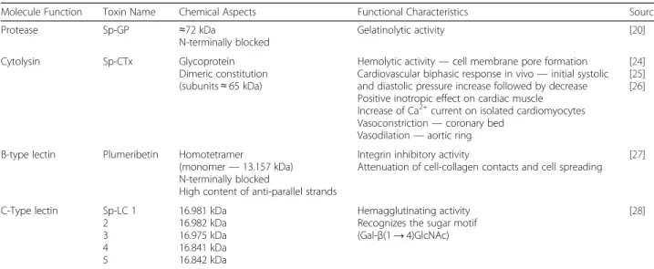
A summary of the bioactive proteins isolated from SpV is presented below (Table 1), along with their chem-ical and functional features.
Molecular genetics ofS. plumierivenom
The difficulties surrounding the study of fish venoms also affect their characterization at the molecular level. To date few reports have been published regarding the analysis of fish venoms from a genetic point of view [59–62]. Transcriptomic approaches performed on the venom glands of the stingray Neotrygon kuhlii [60] and the toadfish Thalassophryne nattereri [59] revealed a considerable number of proteins that are related to the pharmacological activity of these venoms — e.g. galec-tins [60] and C-type lecgalec-tins [59] —as well as some that
are novel to fish venoms. A preliminary analysis of expressed sequence tags (EST) obtained through a cDNA library from S. plumieri venom revealed that about 30% of the sequences had no similarities with pre-viously described ones, suggesting the presence of un-known genes of potential relevance in the venom gland. In addition, the screening of the library with antibodies against a lectin fraction from S. plumieri venom has shown that lectin-like genes account for 12% of all tran-scripts, a finding confirmed by extensivein silicoanalysis [61]. These constitute the very first steps towards the unraveling of the molecular diversity contained in fish venoms.
Neutralization ofS. plumieritoxic activities
Although there is no antivenom available for the enve-noming byS. plumieri, the commercial antivenom raised against the venom of the stonefish Synanceia trachynis (SFAV) — a horseFab’2 preparation made by CSL in Melbourne, Australia [63]—evoked a cross-reactive im-mune response to SpV.
SFAV neutralizes all known clinical effects of serious S. trachynis envenomation [64], and is also efficient in neutralising the inflammatory and cardiovascular re-sponses as as well as the hemolytic activity induced byS. plumieri in mice [29], suggesting that the compounds responsible for these effects share similar biochemical and antigenic properties to those found in stonefish venom. This antivenom also neutralises some of the toxic effects of other stonefish (S. verrucosa), lionfish (Pterois volitans, P. lunulata, P. antennata and Dendro-chirus zebra) and soldierfish (Gymnapistes marmoratus) [51, 65, 66].
This is in accordance with the hypothesis that venom-ous fishes belonging to different genera or inhabiting
different regions may share venom compounds with similar antigenic properties [1].
Conclusions
In conclusion, despite all the progress made recently, many questions remain to be answered, not only with respect to the physio-pharmacological effects and the precise action mechanism of some of the components already described, but also as to the considerable num-ber of molecules still unexplored in the venom ofS. plu-mieri. The study and exploration of the full potential contained in fish venoms can contribute to a better un-derstanding of complex physiological processes — such as the very pain induced by the envenomation —and to the discovery of new drugs, not to mention the develop-ment of more effective ways to treat the injuries caused by these animals.
Abbreviations
AM:Alveolar macrophages; CPP: Coronary perfusion pressure;
ECM: Extracellular matrix; EST: Expressed sequence tags; MALDI-TOF: Matrix-assisted laser desorption/ionization–time of flight; MAP: Mean arterial pressure; RP-HPLC: Reverse phase high performance liquid chromatography; SINAN: Notifiable diseases information system; Sp-GP:Scorpaena plumieri
gelatinolytic protease; SpV:S. plumierivenom extract; UFES: Federal University of espírito santo
Acknowledgements
The authors would like to thank all the members of the Laboratory of Protein Chemistry for the useful insights. Thanks are also due to the Center for the Study of Venoms and Venomous Animals (CEVAP) of UNESP for enabling the publication of this paper (Edital Toxinologia CAPES no. 063/ 2010, Process no. 230.38.006285/2011-21, AUXPE Toxinologia 1219/2011).
Funding
This work was funded by a FAPES/CNPq grant to FVC (grant no. 012/2014), by CAPES grants to TNM, PFM and GBN, by a FAPES-Profix grant to HLG (grant no. 009/2014) and by an INCTTOX/CNPq grant (grant no. 573790/ 2008-6). It was also supported by the Edital Toxinologia CAPES no. 063/2010, process no. 230.038.007890/2011-10, AUXPE Toxinologia 1824/2011 (granted to SGF).
Table 1Toxins purified from SpV to date
Molecule Function Toxin Name Chemical Aspects Functional Characteristics Source
Protease Sp-GP ≈72 kDa
N-terminally blocked
Gelatinolytic activity [20]
Cytolysin Sp-CTx Glycoprotein
Dimeric constitution (subunits≈65 kDa)
Hemolytic activity—cell membrane pore formation Cardiovascular biphasic response in vivo—initial systolic and diastolic pressure increase followed by decrease Positive inotropic effect on cardiac muscle Increase of Ca2+current on isolated cardiomyocytes Vasoconstriction—coronary bed
Vasodilation—aortic ring
[24] [25] [26]
B-type lectin Plumeribetin Homotetramer
(monomer—13.157 kDa) N-terminally blocked
High content of anti-parallel strands
Integrin inhibitory activity
Attenuation of cell-collagen contacts and cell spreading [27]
C-Type lectin Sp-LC 1 2 3 4 5
16.981 kDa 16.982 kDa 16.975 kDa 16.841 kDa 16.842 kDa
Hemagglutinating activity Recognizes the sugar motif (Gal-β(1→4)GlcNAc)
Authors’contributions
All authors contributed equally to the preparation of this manuscript. All authors read and approved the final manuscript.
Competing interests
The authors declare that they have no competing interests.
Ethics approval and consent to participate
Not applicable.
Author details
1Departamento de Ciências Fisiológicas, Centro de Ciências da Saúde,
Universidade Federal do Espírito Santo, Av. Marechal Campos 1468, 29040-090 Vitória, ES, Brazil.2Departamento de Bioquímica e Imunologia, Instituto de Ciências Fisiológicas, Universidade Federal de Minas Gerais, Belo Horizonte, MG, Brazil.3Diretoria do Centro de Pesquisa e Desenvolvimento, Fundação Ezequiel Dias, Belo Horizonte, MG, Brazil.
Received: 27 July 2016 Accepted: 30 November 2016
References
1. Church JE, Hodgson WC. The pharmacological activity of fish venoms. Toxicon. 2002;40(8):1083–93.
2. Ziegman R, Alewood P. Bioactive components in fish venoms. Toxins. 2015; 7(5):1497–531.
3. Lopes-Ferreira M, Grund LZ, Lima C.Thalassophryne nattererifish venom: from the envenoming to the understanding of the immune system. J Venom Anim Toxins incl Trop Dis. 2014;20:35. doi:10.1186/1678-9199-20-35. 4. Schaeffer Jr RC, Carlson RW, Russell FE. Some chemical properties of the
venom of the scorpionfishScorpaena guttata. Toxicon. 1971;9(1):69–78. 5. Figueiredo SG, Andrich F, Lima C, Lopes-Ferreira M, Haddad Jr V. Venomous
fish: a brief overview. In: de Lima ME, Pimenta AMC, Martin-Eauclaire MF, Zingali R, Rochat H, editors. Animal Toxins: State of the Art. Perspectives on Health and Biotechnology. Belo Horizonte: UFMG; 2009.
6. Smith WL, Wheeler WC. Venom evolution widespread in fishes: a phylogenetic road map for the bioprospecting of piscine venoms. J Hered. 2006;97(3):206–17.
7. Nelson JS. Fishes of the World. 3rd ed. New York: Wiley; 1984.
8. Haddad Jr V, Lupi O, Lonza JP, Tyring SK. Tropical dermatology: marine and aquatic dermatology. J Am Acad Dermatol. 2009;61(5):733–50.
9. Oliveira JS, Pires Junior OR, Morales RAV, Bloch Junior C, Schwartz CA, Freitas JC. Toxicity of puffer fish—two species (Lagocephalus laevigatus, Linaeus 1766 andSphoeroides spengleri, Bloch 1785) from the Southeastern Brazilian coast. J Venom Anim Toxins incl Trop Dis. 2003;9(1):76–88. 10. Simões EMS, Mendes TMA, Adão A, Haddad Jr V. Poisoning after ingestion
of pufferfish in Brazil: report of 11 cases. J Venom Anim Toxins incl Trop Dis. 2014;20:54. doi:10.1186/1678-9199-20-54.
11. Haddad Jr V, Martins IA, Makyama HM. Injuries caused by scorpionfishes (Scorpaena plumieriBloch, 1789 andScorpaena brasiliensisCuvier, 1829) in the Southwestern Atlantic Ocean (Brazilian coast): epidemiologic, clinic and therapeutic aspects of 23 stings in humans. Toxicon. 2003;42(1):79–83. 12. Gwee MC, Gopalakrishnakone P, Yuen R, Khoo HE, Low KS. A review of
stonefish venoms and toxins. Pharmacol Ther. 1994;64(3):509–28. 13. Humann P. Reef fish identification: Florida, Caribbean Bahamas. Deloach N,
editor. Florida: New World Publications; 1994.
14. Moyle PB, Cech Jr JJ. Fishes: an introduction to ichthyology. 3rd ed. USA: Prentice-Hall; 1996.
15. Bloch 1789 apud Eschmeyer WN. A systematic review of the scorpionfishes of the Atlantic Ocean (Pisces: Scorpaenidae). California: Academy of Sciences; 1969.
16. Russel FE. Marine toxins and venomous and poisonous marine animals. In: Russel FS, editor. Advances in Marine Biology, vol. 2. London: Academic; 1965. 17. Halstead BW. Injurious effects from the sting of the scorpionfish,Scorpaena
guttatawith report of a case. Calif Med. 1951;74(5):395–6.
18. Roche ET, Halstead BW. Fish Bulletin of the Department of Fish and Game of State of California. 1972;156:1–49. http://content.cdlib.org/view?docId= kt8z09n9rg&brand=calisphere&doc.view=entire_text .
19. Reckziegel GC, Dourado FS, Neto DG, Haddad Jr V. Injuries caused by aquatic animals in Brazil: an analysis of the data present in the information system for notifiable diseases. Rev Soc Bras Med Trop. 2015;48(4):460–7.
20. Carrijo LC, Andrich F, de Lima ME, Cordeiro MN, Richardson M, Figueiredo SG. Biological properties of the venom from the scorpionfish (Scorpaena plumieri) and purification of a gelatinolytic protease. Toxicon. 2005;45(7):843–50. 21. Boletini-Santos D, Komegae EN, Figueiredo SG, Haddad Jr V, Lopes- Ferreira
M, Lima C. Systemic response induced byScorpaena plumierifish venom initiates acute lung injury in mice. Toxicon. 2008;51(4):585–96. 22. Gomes HL, Andrich F, Mauad H, Sampaio KN, de Lima ME, Figueiredo SG,
et al. Cardiovascular effects of scorpionfish (Scorpaena plumieri) venom. Toxicon. 2010;55(2–3):580–9.
23. Menezes TN, Carnielli JB, Gomes HL, Pereira FE, Lemos EM, Bissoli NS, et al. Local inflammatory response induced by scorpionfishScorpaena plumieri
venom in mice. Toxicon. 2012;60(1):4–11.
24. Andrich F, Carnielli JB, Cassoli JS, Lautner RQ, Santos RA, Pimenta AM, et al. A potente vasoactive cytolysin isolated fromScorpaena plumieriscorpionfish venom. Toxicon. 2010;56(4):487–96.
25. Gomes HL, Andrich F, Fortes-Dias CL, Perales J, Teixeira-Ferreira A, Vassallo DV, et al. Molecular and biochemical characterization of a cytolysin from the
Scorpaena plumieri(scorpionfish) venom: evidence of pore formation on erythrocyte cell membrane. Toxicon. 2013;74:92–100.
26. Gomes HL, Menezes TN, Malacarne PF, Roman-Campos D, Gondim AN, Cruz JS, et al. Cardiovascular effects of Sp-CTx, a cytolysin from the scorpionfish (Scorpaena plumieri) venom. Toxicon. 2016;118:141–8.
27. Evangelista KS, Andrich F, Figueiredo de Rezende F, Niland S, Cordeiro MN, Horlacher T, et al. Plumieribetin, a fish lectin homologous to mannose-binding B-type lectins, inhibits the collagen-binding alpha1beta1 integrin. J Biol Chem. 2009;284(50):34747–9.
28. Andrich F, Richardson M, Naumann GB, Cordeiro MN, Santos AV, Santos DM, et al. Identification of C-type isolectins in the venom of the scorpionfish
Scorpaena plumieri. Toxicon. 2015;95:67–71.
29. Gomes HL, Menezes TN, Carnielli JBT, Andrich F, Evangelista KS, Chávez-Olórtegui C, et al. Stonefish antivenom neutralises the inflammatory and cardiovascular effects induced by scorpionfishScorpaena plumieri
venom. Toxicon. 2011;57(7–8):992–9.
30. Hopkins BJ, Hodgson WC. Cardiovascular studies on venom from the soldierfish (Gymnapistes marmoratus). Toxicon. 1998;36(7):973–83. 31. Sosa-Rosales JI, Piran-Soares AA, Farsky SH, Takehara HA, Lima C,
Lopes-Ferreira M. Important biological activities induced byThalassophryne maculosafish venom. Toxicon. 2005;45(2):155–61.
32. Haddad Jr V, Neto DG, de Paula Neto JB, de Luna Marques FP, Barbaro KC. Freshwater stingrays: study of epidemiologic, clinic and therapeutic aspects based on 84 envenomings in humans and some enzymatic activities of the venom. Toxicon. 2004;43(3):287–94.
33. Garnier P, Grosclaude JM, Goudey-Perrière F, Gervat V, Gayral P, Jacquot C, et al. Presence of norepinephrine and other biogenic amines in stonefish venom. J Chromotogr B Biomed Appl. 1996;685(2):364–9.
34. Church JE, Hodgson WC. Dose-dependent cardiovascular and neuromuscular effects of stonefish (Synanceja trachynis) venom. Toxicon. 2000;38(3):391–407. 35. Hopkins BJ, Hodgson WC, Sutherland SK. An in vitro pharmacological
examination of venom from the soldierfishGymnapistes marmoratus. Toxicon. 1997;35(7):1101–11.
36. Cohen AS, Olek AJ. An extract of lionfish (Pterois volitans) spine tissue contains acetylcholine and a toxin that affects neuromuscular transmission. Toxicon. 1989;27(12):1367–76.
37. Rodrigues RJ. Pharmacology of south American freshwater stingray venom (Potamotrygon motoro). Trans N Y Acad Sci. 1972;34(8):677–86.
38. Khoo HE, Yuen R, Poh CH, Tan CH. Biological activities ofSynanceja horrida
(stonefish) venom. Nat Toxins. 1992;1(1):54–60.
39. Garnier P, Goudey-Perrière F, Breton P, Dewulf C, Petek F, Perrière C. Enzymatic properties of the stonefish (Synanceia verrucosaBloch and Schneider, 1801) venom and purification of a lethal, hypotensive and cytolytic factor. Toxicon. 1995;33(2):143–55.
40. Gutiérrez JM, Rucavado A. Snake venom metalloproteinases: their role in the pathogenesis of local tissue damage. Biochimie. 2000;82(9–10):841–50. 41. Calvete JJ, Moreno-Murciano MP, Theakston RD, Kisiel DG, Marcinkiewicz C. Snake
venom disintegrins: novel dimeric disintegrins and structural diversification by disulphide bond engineering. Biochem J. 2003;372(Pt 3):725–34.
42. Poh CH, Yuen R, Chung MC, Khoo HE. Purification and partial
characterization of hyaluronidase from stonefish (Synanceja horrida) venom. Comp Biochem Physiol B. 1992;101(1–2):159–63.
44. Faiz MA, Falkous G, Harris JB, Mantle D. Comparison of protease and related enzyme activities in snake venoms. Comp Biochem Physiol B Biochem Mol Biol. 1996;113(1):199–204.
45. Borges MH, Figueiredo SG, Leprevost FV, De Lima ME, Cordeiro Mdo N, Diniz MR, et al. Venomous extract protein profile of Brazilian tarantula
Grammostola iheringi: searching for potential biotechnological applications. J Proteomics. 2016;136:35–47.
46. Ghafari SM, Jamili S, Bagheri KP, Ardakani EM, Fatemi MR, Shahbazzadeh F, et al. The first report on some toxic effects of green scat,Scatophagus argus
an Iranian Persian Gulf venomous fish. Toxicon. 2013;66:82–7.
47. McLane MA, Sanchez EE, Wong A, Paquette-Straub C, Perez JC. Disintegrins. Curr Drug Targets Cardiovasc Haematol Disord. 2004;4(4):327–55. 48. Eble JA, Niland S, Dennes A, Schmidt-Hederich A, Bruckner P, Brunner G.
Rhodocetin antagonizes stromal tumor invasion in vitro and other alpha2beta1 integrin-mediated cell functions. Matrix Biol. 2002;21(7):547–58. 49. Pilorget A, Conesa M, Sarray S, Michaud-Levesque J, Daoud S, Kim KS, et al.
Lebectin, aMacrovipera lebetinavenom-derived C-type lectin, inhibts angiogenesis both in vitro and in vivo. J Cell Physiol. 2007;211(2):307–15. 50. Sarray S, Delamarre E, Marvaldi J, El Ayeb M, Marrakchi N, Luis J. Lebectin and lebecetin, two C-type lectins from snake venom, inhibit alpha5beta1 and alphaV-containing integrins. Matrix Biol. 2007;26(4):306–13.
51. Church JE, Hodgson WC. Adrenergic and cholinergic activity contributes to the cardiovascular effects of lionfish (Pterois volitans) venom. Toxicon. 2002; 40(6):1083–93.
52. Carlson RW, Schaeffer Jr RC, La Grance RG, Roberts CM, Russel FE. Some pharmacological properties of the venom of the scorpionfishScorpaena guttata. Toxicon. 1971;9:379–91.
53. Magalhães GS, Lopes-Ferreira M, Junqueira-de-Azevedo IL, Spencer PJ, Araújo MS, Portaro FC, et al. Natterins, a new class of proteins with kininogenase activity characterized fromThalassophryne nattererifish venom. Biochimie. 2005;87(8):687–99.
54. Komegae EN, Ramos AD, Oliveira AK, Serrano SM, Lopes-Ferreira M, Lima C. Insights into the local pathogenesis induced by fish toxins: role of natterins and nattectin in the disruption of cell-cell and cell-extracellular matrix interactions and modulation of cell migration. Toxicon. 2011;58(6–7):509–17. 55. Kilpatrick DC. Animal lectins: a historical introduction and overview. Biochim
Biophys Acta. 2002;1572(2–3):187–97.
56. Khoo HE. Bioactive proteins from stonefish venom. Clin Exp Pharmacol Physiol. 2002;29(9):802–6.
57. Chen D, Kini RM, Yuen R, Khoo HE. Haemolytic activity of stonustoxin from stonefish (Synanceja horrida) venom: pore formation and the role of cationic amino acid residues. Biochem J. 1997;325(Pt 3):685–91.
58. Ellisdon AM, Reboul CF, Panjikar S, Huynh K, Oellig CA, Winter KL, et al. Stonefish toxin defines an ancient branch of the perforin-like superfamily. Proc Natl Acad Sci U S A. 2015;112(50):15360–5.
59. Magalhães GS, Junqueira-de-Azevedo IL, Lopes-Ferreira M, Lorenzini DM, Ho PL, Moura-da-Silva AM. Transcriptome analysis of expressed sequence tags from the venom glands of the fishThalassophryne nattereri. Biochimie. 2006; 88(6):693–9.
60. Baumann K, Casewell NR, Ali SA, Jackson TN, Vetter I, Dobson JS, et al. A ray of venom: combined proteomic and transcriptomic investigation of fish venom composition using barb tissue from the blue-spotted stingray (Neotrygon kuhlii). J Proteomics. 2014;109:188–98.
61. Costa FLS, de Lima ME, Pimenta AC, Figueiredo SG, Kalapothakis E, Salas CE. Expressed sequence tags in venomous tissue ofScorpaena plumieri
(Scorpaeniformes: Scorpaenidae). Neotrop Ichthyol. 2014;12(4):871–8. 62. Xie B, Li X, Lin Z, Ruan Z, Wang M, Liu J, et al. Prediction of toxin genes
from Chinese yellow catfish based on transcriptomic and proteomic sequencing. Int J Mol Sci. 2016;17(4):E556.
63. White J. Antivenom Handbook. Melbourne: CSL Ltd; 1995. http://catalogue. nla.gov.au/Record/1478936.
64. Church JE, Hodgson WC. Stonefish (Synanceia trachynis) antivenom: in vitro efficacy and clinical use. J Toxicol Toxin Rev. 2003;22(1):69–76.
65. Church JE, Hodgson WC. Stonefish (Synanceia spp.) antivenom neutralises the in vitro and in vivo cardiovascular activity of soldierfish (Gymnapistes marmoratus) venom. Toxicon. 2001;39(2–3):319–24.
66. Shiomi K, Hosaka M, Yamanaka H, Kikuchi T. Venoms from six species of marine fish: lethal and hemolytic activities and their neutralization by commercial stonefish antivenom. Mar Biol. 1989;103:285–89.
• We accept pre-submission inquiries
• Our selector tool helps you to find the most relevant journal
• We provide round the clock customer support
• Convenient online submission
• Thorough peer review
• Inclusion in PubMed and all major indexing services
• Maximum visibility for your research
Submit your manuscript at www.biomedcentral.com/submit