R E S E A R C H
Open Access
Sequence analysis of the cDNA encoding
for SpCTx: a lethal factor from scorpionfish
venom (
Scorpaena plumieri
)
Fábio L. S. Costa
1, Maria Elena De Lima
1, Suely G. Figueiredo
2, Rafaela S. Ferreira
1, Núbia S. Prates
1,
Tetsu Sakamoto
1and Carlos E. Salas
1*Abstract
Background:Lethal factors are multifunctional oligomeric proteins found in the venomous apparatus of Scorpaeniformes fish. These toxins elicit not only an array of biological responses in vitro but also cardiovascular disorders and strong hemolytic, nociceptive and edematogenic activities in vivo. This work describes the cloning and molecular identification of two toxin subunits, denominated Sp-CTx-αand Sp-CTx-β, from scorpionfish venom (Scorpaena plumieri).
Methods:The primary structures were deduced after cDNA amplification by PCR with primers from conserved sequences described in Scorpaeniformes toxins. Following DNA sequencing and bioinformatic analysis, the tridimensional structures of both subunits were modeled.
Results:The translated sequences (702 amino acids, each subunit) show homology with other lethal factors, while alignment between Sp-CTx-αand Sp-CTx-βshows 54% identity. The subunits lack N-terminal signal sequences and display masses of approximately 80 kDa each. Both Sp-CTx subunits display a B30.2/SPRY domain at the C-terminal region with typically conserved motifs as described in these toxins. Secondary structure prediction identified sixα-helices 18 residues long in bothαandβsubunits, some of them amphiphilic with their N-terminal flanked by many basic residues, creating a cationic site associated with the cytolytic activity of these toxins. Antimicrobial potential sites were identified in Sp-CTx and share some features with other peptides presenting variable and broad-spectrum activity. A phylogenetic tree built to represent these toxins supports the proximity between scorpionfish, lionfish and stonefish.
Conclusion:The study identified a putative toxin protein whose primary structure is similar to other fish toxins and with potential for production of antivenom against scorpionfish envenomation in Brazil. As a prelude to structure-function studies, we propose that the toxin is structurally related to pore-forming marine toxins.
Keywords:cDNA, Lethal factor,Scorpaena plumieri, Scorpionfish, Venom gland
Background
Scorpaeniformes from the families Scorpaenidae and Synanceiidae are the most venomous marine fishes known to date. Their venom apparatus encompasses dorsal, anal and pelvic fin spines associated with venom-containing tissues glands [1]. Occasional envenomation occurs by accidental poisoning by fish
spines. Clinical and pharmacological studies suggest that active components of fish venom exhibit cytolytic (hemolytic), inflammatory, neuromuscular and pro-nounced cardiovascular activities [2–5].
Scorpionfish members of the genus Scorpaenainhabit shallow waters of the tropical Atlantic Coast. Scorpaena plumieri, known in Brazil as “aniquim”, “mamangá” or “moréia-atí”, exhibits disguising coloration that predis-poses humans to poisoning along the Brazilian shore [6]. An array of symptoms including excruciating pain at the site of the puncture, edema and cardiovascular disorders are observed following envenoming [7].
* Correspondence:[email protected];[email protected]
1Departamento de Bioquímica e Imunologia, Instituto de Ciências Biológicas, Universidade Federal de Minas Gerais, Av. Antônio Carlos, 6627, Pampulha, Belo Horizonte, MG 31270-901, Brazil
Full list of author information is available at the end of the article
Many of the symptoms associated with injury caused by Scorpaeniformes are attributable to multifunctional proteins, described as “lethal factors” identified in the venom. Due to their strong hemolytic activity, these proteins have been designated as cytolytic toxins or “multifunctional cytolysins” (for a review, see [4, 8]). It was demonstrated that the hemolytic effect of these toxins is due to pore formation on the cell membrane of erythrocytes [9–12].
So far, cytolysins have been identified in the following groups: Pterois [13, 14], Scorpaenopsis, Sebastiscus and Sebastapistes[15] andScorpaena[16] from the Scorpae-nidae family, Hypodytes from the Tetraogidae family, Siganus fuscescens from the Siganidae family [17] and Inimicus [14] and Synanceia [18–20] from the family Synanceiidae.
The toxins are 148–160 kDa proteins composed by two homologous subunits, designated asαandβ, that remain
as-sociated via non-covalent interaction creating a dimeric structure. The domains MACPF/CDC (Membrane Attack Complex-Perforin/Cholesterol-Dependent Cytolysin), known for forming large, ring-shaped supramolecular oligomeric pore complexes on erythrocyte membranes, represent an an-cient pore-forming superfamily [10,19,20].
The cytolytic toxin (Sp-CTx) was purified from venom of the scorpionfishS. plumieri[11,16]. It displays vasor-elaxant activity and induces disorders in the cardiovas-cular system by an increase in sarcolemmal Ca+ 2, partially caused by release of endogenous noradrenaline [21, 22]. Sp-CTx is a dimeric glycoprotein (≈ 75 kDa/ subunit); its tryptic digestion yields peptide fragments whose Open Reading Frame (ORF) confirms its simila-rity to fish cytolysins [11,16].
A striking property shared by fish venoms is their abil-ity to induce hemolysis in vitro, arguing for a functional resemblance. The structural similarity between fish venoms was evident as most toxins were disabled upon re-action with horse-derived stonefish antivenom (SFAV) raised against crude venom of the stonefishSynanceia ver-rucosa (Commonwealth Serum Laboratories, Melbourne, Australia) [13, 14, 19, 20, 23–26]. The immune cross-reactivity among Scorpaeniformes toxins suggests that they share a common evolutionary ancestor. Based on these similarities, the design of DNA primers derived from the structure of stonefish toxin was instrumental for inferring the structure of S. verrucosa toxin [19, 27]. A similar strategy was applied to determine the primary structures of toxins from lionfish, waspfish and rabbitfish [14, 17], barchin scorpionfish, tassled scorpionfish and false kelpfish [15].
We previously described the production and partial characterization of a cDNA library from venomous tissue of S. plumieri, by using the random sequencing approach, and generated hundreds of partial sequences
[28]. This study aims to identify the coding sequences for S. plumieri toxin, and to verify the presence of determinants attributable to the protein that could be responsible for the pharmacological effects of this toxin. To find the mRNA encoding for the lethal factor in S. plumieri, we have used the library or the cDNAs source of this library and primers from conserved regions of the toxin to produce the in silico full amino-acid sequence of α- and β-subunits of Sp-CTx. We further analyzed
structural features of the hypothetic protein and the similarities with other fish venom toxins.
Methods
Biological specimens
Three live specimens of the scorpionfish S. plumieri (15–30 cm, length) were collected by a local fisherman off the Coast at Espírito Santo, Brazil and kept in an aquarium for a short duration prior to dissection. Fish-ing was authorized by the Instituto Brasileiro do Meio Ambiente e dos Recursos Naturais Renováveis–IBAMA (the Brazilian Public Agency for Environment Affairs). Glands tissue was dissected from the dorsal, pelvic and caudal ray-fin structures and kept in liquid N2 during homogenization in a grinder mill.
cDNA library construction
Total RNA was obtained from excised venom glands using the guanidinium isothiocyanate extraction proce-dure described by [29]. Poly(A)+ RNA was isolated by oligo(dT)-cellulose chromatography (mRNA Isolation Kit, Agilent Technologies, Inc. USA). Five μg of RNA
were transcribed into cDNA using the ZAP cDNA syn-thesis kit (ZAP-cDNA Gigapack III gold cloning kit, GE, USA).
RT-PCR procedure
A polymerase chain reaction was performed to amplify DNA from the excised bacteriophage library (~ 106pfus) or from the cDNA synthesized from 5 μg of total RNA
or 500 ng of mRNA chromatographically purified fol-lowing cDNA synthesis (GE Healthcare Life Sciences, USA), according to the manufacturer’s instructions.
DNA amplification was performed using Platinum®Taq DNA Polymerase (Invitrogen™, Life Technologies, Inc. USA) under the following conditions: pre-incubation at 94 °C for 5 min; 35 cycles consisting of denaturation at 94 °C for 30 s, annealing at 45–65 °C (Tm depending of the primer) for 30 s; extension at 72 °C for 1–2 min and final extension at 72 °C for 5 min. Amplified products were subcloned into pCR®8/GW/TOPO® TA Cloning with One Shot® TOP10 E. coli kit (Invitrogen™, Life Technologies, Inc.). The DNA of plasmid clones was isolated as described by Sambrook & Russell [30] and used for sequencing. Each PCR fragment encoding a putative region of the toxin was
cloned and the consensus sequence of at least 3–4 repli-cates assembled into the final sequence.
Primer design
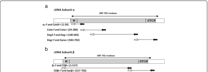
Initially, primers were designed based on conserved se-quences from toxins already described in other Scorpae-niform species. The nucleotide sequences of primers used in experiments are summarized in Table1. A total of twelve primers (eight for α-subunit and four for β-subunit) were employed to fully characterize the DNA
encoding both subunits (Fig.1). DNA primers Deg (for-ward and reverse) were used as described by Kiriake & Shiomi [13] to identify lionfish toxins while remaining primers were based on toxin sequences from stonefish [5,9,19,20].
Designations of the primers were based on reported DNA sequences corresponding to regions 60–83 (αT-f), 126–146 (Catα-f or Catαf-r), 498–517 (Deg2-f), 1790– 1809 (Deg-r or Degr-f) and (2151-2171) Synα-r from α-subunit and (52–71) βT-f, 1621–1638 (CDβ-r or CDβr-f), and 2139–2157 (Synβ-r) fromβ-subunit (Fig.1).
Comparative modeling
Comparative models of the Sp-CTx (α- andβ-subunits)
were constructed using the Automated Mode of SWISS-MODEL server [31]. The target sequences were used for identification of templates based on Blast and HHblits. The crystal structures of stonustoxin subunits
α (PDB ID:4WVM_A) and β (PDB ID:4WVM_B), at
3.1 Å resolution, were used for modeling of Sp-CTx sub-units. The alignment between target and template se-quences was conducted to generate 3D models. The stereochemical quality of the models was determined by Ramachandran plot assessment generated by RAMPAGE
[32]. The models were further evaluated through ProSA [33] and QMEAN statistical parameters [34]. We also calculated the RMSD values between the models and their corresponding template.
The HADDOCK 2.2 web server [35] was used for protein-protein docking of modeled structures. During the docking procedure, HADDOCK incorporated infor-mation about interacting residues at the interface of the protein complex. Therefore, before docking, contacts were identified with the InterProSurf web server [36], using template structures as an input to predict interac-ting residues.
Sequence and analysis of clones
Colonies grown overnight in ampicillin-supplemented medium at 37 °C were randomly selected. Plasmid DNA was isolated by the alkaline lysis method [30].
DNA sequences were obtained in the automated se-quencer 3.100 Genetic Analyzer System using BigDye™ Terminator v1.1, v3.1 Ready Reaction Mix (Applied Biosystems Inc., Foster City, CA, USA) in the presence of M13 forward primer or its reverse. Analysis of data was carried out using the software Phred for base calling and the quality score cutoff was set at 10 [37]. The nucleotide sequences from the vector, adaptors and Escherichia coli DNA were removed by the program VecScreen (http:// www.ncbi.nlm.nih.gov/tools/vecscreen).
Amino-acid sequences of toxin transcripts were de-duced via the program Open Reading Frame (ORF) Finder (https://www.ncbi.nlm.nih.gov/orffinder/). The isoelectric point (pI) and molecular mass (MM) from de-rived sequences were computed by the software Swiss-Prot/TrEMBL located in Expasy.
Table 1Nucleotide sequences of primers used for RT-PCR and cloning experiments
Primer identification Nucleotide sequence of primer Nucleotide positiona Subunitα
αT-f 5’-ATGTCTTCAGATTTGGTAATGCCT-3’ 60–83
Catα-f 5’-CGCAGAGAGAAACTGATCCCA-3’ 126–146
Catαf-r 5’-TGGGATCAGTTTCTCTCTGCG-3’ 126–146
Deg2-f 5’-GGGGCMAATGCYTTCTTTGT-3’ 498–517
Catα-r 5’-ATTGGCTCTCCTCTTCAGTTT-3’ 900–921
Degr-f 5’-ATTACTGGGAGGTGGAGTGG-3’ 1790-1809
Deg-r 5’-CCACTCYAMCTCCCAGTAAT-3’ 1790-1809
Synα-r 5’-TYAAAGTAATCTGASAGTTCC-3’ 2151-2171
Subunitβ
βTotal-f 5’-GTTGGAGTCATGCCTTCAGAC-3’ 52–71
CDβ-r 5’-GAGCTCACAGTCATACCA-3’ 1621-1638
CDβr-f 5’-TGGTATGACTGTGAGCTC-3’ 1621-1638
Synβ-r 5’-TAGTTTAATTTGACCATT-3’ 2139-2157
The amphiphilicity, α-helices, glycosylation sites and
peptide signal sequences in Sp-CTx were analyzed by the programs PSIPRED Protein Sequence Analysis Workbench (UCL Department of Computer Science), NETNGLYC (http://www.cbs.dtu.dk/services/NetNGlyc) and SignalP 4.0 [38], respectively. Cytolytic sites in
α-helices were predicted by designing a Helical Wheel as
described by Schiffer & Edmundson [39] and using the program (http://lbqp.unb.br/NetWheels) [40].
Phylogenetic analysis
Putative orthologues of Sp-CTx were identified by submit-ting derived protein sequences as queries to the BLASTP algorithm [41] on the NCBI webserver ( https://blas-t.ncbi.nlm.nih.gov/Blast.cgi) employing the non-redundant protein sequences (nr) database. From BLASTP retrieved protein accessions, we selected those accessions displaying a high similarity score with at least one of the query se-quences (coverage > 80%; identity > 50%) and pertaining to one of the species known to be venomous. Sequences were submitted to MUSCLE [42] and then to the Neighbor-Joining algorithm (bootstrap replicates: 500; substitution model: Maximum Composite Likelihood), both implemented in MEGA7 [43], for sequence align-ment and phylogenetic tree creation, respectively. For tree rooting analysis, we included the Stonustoxin subunit
β-like protein fromClupea harengus (accession number: XP_012674574.1) and considered it an outgroup.
Results
Cloning and sequencing of cDNAs encodingα- andβ -subunits of Sp-CTx
Initially, we designed the set of primers (Catαf-r) coding for the region containing many cationic residues
apparently involved in the hemolytic activity in Scorpae-niformes [44]. Using Catαprimers (Fig.1a) and cDNAS. plumieri as the template, a PCR fragment of approxi-mately 800 bp was amplified and cloned into pCR8/ GW/TOPO. The sequenced fragment contained an ORF encoding 265 amino-acid residues that aligned between positions 24–286 with α-subunits in Scorpaeniform
toxins found at the NCBI databank.
To characterize the N-terminal region, a reverse com-plement of Catα primer was designed and combined
with αT-f primer to produce an amplicon of 100 bp. After cloning and sequencing, this fragment generated an ORF of 24 residues corresponding to the N-terminal of the Sp-CTxα-subunit.
The C-terminal of Sp-CTx-αwas identified when
com-bining the complement of a Degr primer with Synα-r
primer to yield a 400 bp fragment (Fig.1a). After cloning and sequencing, a 126-amino-acid fragment was identi-fied and aligned to positions 583–584 of subunit-αfrom
fish toxins. In this fragment we identified three ter-mination codons (TAA) in frame, at the end of the sequence.
The identification of Sp-CTx-β followed PCR of the
excised library with primers βT-f and CDβ-r (Fig. 1b). After subcloning and sequencing, a 1545 bp PCR pro-duct yielded an ORF encoding a 515-amino-acid poly-peptide sharing 81% identity with β-subunit of Pterois. To determine the C-terminal portion of Sp-CTx-β, a
complement of CDβr-f primer was designed and
combined with Synβ-r primer in PCRs using a cDNA
template fromS. plumieri(Fig.1b). The resulting 600 bp fragment was cloned; and its sequence identified an ORF of 555 bp corresponding to 185 amino-acid residues located at C-terminals inβ-subunits.
a
b
Fig. 1Schematic cloning representation ofα- andβ-subunits of Sp-CTx. Forward and reverse primers are indicated by white and black arrows, respectively. The sequence of primers is shown in Table1. Amino-acid positions are relative to the primary structures of cDNAs from Synanceia. The positions of the arrows indicate the approximate size of the putative fragment. Initiation codon (M) and stop codon (STOP).a: Union of putative fragments inα-subunit was obtained by PCRs (αT-f and Catαf-r, Catα-f and Catα-r, Deg2-f and Deg-r, Degr-f and Synα-r).b: Union of putative fragments inβ-subunit (βT-f and CDβ-r were isolated from the cDNA library and CDβr-f and Synβ-r obtained by PCR)
Several primers were designed to attempt identifica-tion of the internal regions of Sp-CTx-αandβunder
dif-ferent PCR conditions (data not shown); one of them (Deg2-f, Deg-r) produced an amplicon of 1500 bp that was cloned and sequenced. Two related sequences were identified that aligned with internal regions of Sp-CTx-α
(1,365 bp - 455 residues) and Sp-CTx-β(1,104 bp - 368
residues). Assemblage of overlapping fragments pro-duced the entire sequence from Sp-CTx-α and
Sp-CTx-βas expected for Scorpaeniformes toxins.
Nucleotide sequence ofα- andβ-subunits of Sp-CTx Figure2ashows the assembled Sp-CTx-αsequence
con-taining 2192 bp. The 5′-untranslated region of this se-quence contains the initiation codon located at position 78, followed by an ORF encompassing 2106 bp encoding 702 amino-acid residues in frame with three stop codons in tandem, comprising the beginning of the poly A tail at the 3′-untranslated region. In this sequence the initial ATG (Met) is followed by two Ser, and the last two amino acids before the stop codons (TAA) are Leu.
In Sp-CTx-β, the initial ATG codon was found in
pos-ition 72, followed by an ORF containing 2106 bp (Fig. 2b). The initial coding ATG is followed by Pro and Ser; the 3′-terminal contains GGC-GAA (Gly-Glu) be-fore the single stop codon (TAA). However, the poly A tail was not identified in the 3′-untranslated region. No signal peptides were identified in the N-terminal regions of Sp-CTx-αor Sp-CTx-β.
The sequences of Sp-CTx subunits were deposited in the EMBL Nucleotide Sequence Database (DDBJ/EMBL/ GenBank nucleotide sequence databases) under the fol-lowing accession numbers: Seq1 MG053103/AVI44916 for the α-subunit and Seq2 MG53104/AVI44917 for the β-subunit ofS. plumieri.
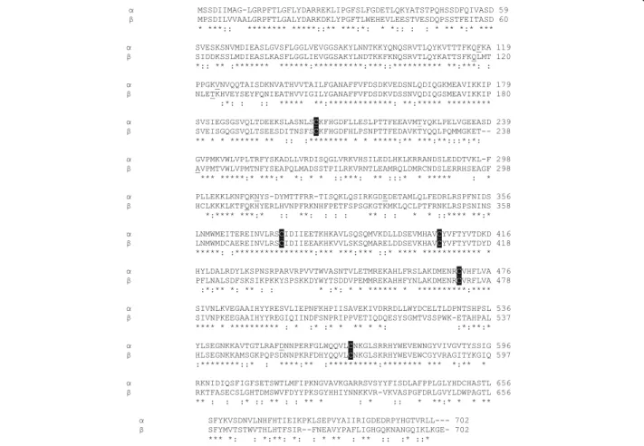
Amino-acid sequence ofα- andβ-subunits of Sp-CTx A comparison between deduced amino-acid sequences of Sp-CTxs α and βevidenced 54% identity confirming
their relatedness. Several insertions/deletions of one or two amino acids at various positions are detected in both subunits. Sp-CTx-α contain 7 cysteinyl residues
while 11 cysteinyl are found in Sp-CTx-β, five of which
(in positions 204, 374, 406, 470 and 568) are preserved in both subunits (Fig. 3). The deduced subunit-α has a
theoretical molecular mass of 79,801 kDa with pI 6.70, while subunit-βhas 80,126 kDa and pI 7.88.
Additional file 1 shows the deduced amino-acid se-quences of Sp-CTxs and their alignment with toxins from three species of scorpionfish (Sebastapistes strongia, Scor-paenopsis oxycephalaand Sebastiscus marmoratus), three species of lionfish (Pterois lunulata, Pterois volitans and Pterois antennata), two species of stonefish (Synanceia verrucosaand Syanceia horrida), one species of waspfish
(Hypodytes rubripinnis) and one species of devil stinger (Inimicus japonicus). The alignment shows that 176 resi-dues (24.5%), out of 717 amino acids (including gaps) are conserved in all toxins.
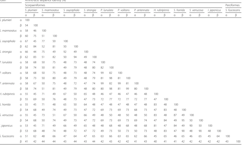
The amino-acid identities among these toxins are summarized in Table 2. It is shown that the identities between β subunits are somewhat stronger than for α-subunits. A strong identity was observed between the α-subunit inP. lunulata (99%) and theα-subunits from P. volitans and P. antennata. Overall, the S. plumieri toxin identities are stronger with those of scorpionfishes (Scorpaenopsis oxycephala, Sebastapistes strongia and Sebastiscus marmoratus), lionfishes (P. lunulata,P. voli-tans and P. antennata) followed by waspfish (H. rubri-pinnis), stonefish (S. verrucosa andS. horrida) and devil stinger (I. japonicus) toxins. The identities between Sp-CTx-αor -βsubunits and the corresponding
counter-parts listed in Table 2 show that Sp-CTx-β shares 84%
identity with toxin-β in scorpionfish S. oxicephala and 83% with S. strongia; meanwhile, the identity of Sp-CTx-α is 67% withα-subunit from S. oxicephala and 66% withα-subunit fromS. strongia. The identity between subunits from the same species ranks around 47–54%; the latter corresponds to the identity between subunitsαand βinS. plumieri. Meanwhile, within thePteroisgroup the identities betweenαandβsubunits attain 80–82%.
The PROSITE tool [45] revealed the presence of a B30.2/SPRY domain containing 197–198 residues at the C-terminal region on each subunit, although the amino-acid sequences within these domains are some-what variable.
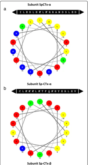
Predicted cytolytic domains
The cytolytic activity of many proteins is frequently re-lated to the presence of amphiphilicα-helices displaying
cationic sites (basic residues) flanked by hydrophobic surfaces that induce monomer aggregates able to form pores [44,46].
The prediction of secondary structures in Sp-CTxs (PSIPRED) posits the presence of five amphiphilic
α-helices with a minimum size of 20 amino-acid residues
(three in α- and two in β-subunit). Applying the “Edmunson Wheel” diagram, some predicted helixes ex-hibited cytolytic potential, as the hydrophobic portion is concentrated opposite to the hydrophilic side, revealing its amphiphilicity (Fig. 4). For instance, one amphiphilic
α-helix was predicted between Gln266 and Asp292 in Sp-CTx-α and the diagram design shows the final 18
a
b
Fig. 2(See legend on next page.)
to proteins displaying cytolytic activity. Another α-helix
with cytolytic potential was predicted between Cys300 and Val317 in Sp-CTx-β. The presence of amphiphilic residues Ser294 and His311 and the N-terminal flanking residues Lys302, Lys303, Lys304 and Lys306 support the cytolytic feature assigned to this domain (Fig.4b).
Comparative modeling
Using BLAST and Protein Data Bank tools, we found 55% and 68% sequence identity between α-subunit and β-subunit of Sp-CTx and venom homologues in SNTX
(S. horrida), considered sufficient to infer structural con-servation (Table2). The structure of the latter was deter-mined by X-ray crystallography at the resolution of
3.1 Å (PDB: 4WVMA and 4WVMB, chains α and β).
Despite its moderate resolution, SNTX was used as the template since it is the only structure available for this toxin in Scorpaeniformes. The automated mode of SWISS-MODEL was used for template identification, alignment and generation of the models. Using each subunit from Sp-CTx, a single model was built by the server followed by Ramachandran plot, ProSA and QMEAN analysis for model validation (Fig.5a).
Ramachadran Plot analysis of Sp-CTx model allocated 93.2–94.5% of amino-acid residues in favored regions, 4.2–5.8% in allowed regions and 1.0–1.3% in disallowed positions, confirming the stereochemical quality of the model. The ProSA server was used for evaluating
(See figure on previous page.)
Fig. 2Nucleotide and deduced amino-acid sequences of cDNAs encoding Sp-CTx-αaandβ-subunitb. Single-letter amino-acid notation is used. Underlined sequences refer to primers; boxed sequences were identical to peptide sequences isolated from tryptic digestion of purified Sp-CTx toxin [11]. Stop codons in frame are indicated by asterisks. The B30.2/SPRY domain is boxed. The nucleotide sequences forα- and
β-subunits fromS. plumierihave been deposited in the DDBJ/EMBL/GenBank nucleotide sequence databases under accession numbers 2,052,576 MG053103 and MG53104, respectively
Table 2A comparison of the amino-acid sequence identities between Sp-CTxα-βand other fish toxins Toxin Amino-acid sequence identity (%)
Scorpaeniformes Perciformes
S. plumieri S. marmoratus S. oxycephala S. strongia P. lunulata P. volitans P. antennata H. rubripinnis S. horrida S. verrucosa I. japonicus S. fuscescens
α β α β α β α β α β α β α β α β α β α β α β α β
S. plumieri α 100
β 54 100 S. marmoratus α 58 46 100
β 60 75 51 100 S. oxycephala α 67 45 77 50 100
β 62 84 52 81 50 100 S. strongia α 66 44 75 49 92 49 100
β 62 83 51 82 50 94 49 100 P. lunulata α 58 68 50 75 48 73 48 74 100
β 58 74 50 81 49 79 48 80 82 100 P. volitans α 58 68 50 75 48 73 48 74 99 82 100
β 58 73 50 80 49 79 48 79 81 98 81 100 P. antennata α 58 67 50 75 48 72 47 74 99 82 99 81 100
β 58 74 51 81 49 79 48 80 80 98 81 99 80 100 H. rubripinnis α 55 45 71 49 67 50 65 48 46 47 46 47 46 48 100
β 55 69 50 76 48 73 47 73 72 77 72 77 72 77 47 100
S. horrida α 55 45 71 48 65 50 64 48 47 48 47 48 47 48 83 48 100
β 54 68 49 74 49 73 47 72 69 73 69 73 68 73 47 83 48 100 S. verrucosa α 55 45 73 51 67 50 66 49 48 50 48 50 48 50 83 48 87 49 100
β 54 68 50 74 49 73 47 72 69 73 69 73 69 74 47 84 49 95 50 100
I. japonicus α 53 45 71 49 66 49 65 48 48 68 48 68 48 68 81 47 84 49 90 50 100
β 53 68 48 74 48 72 47 72 49 73 50 73 50 73 48 83 47 90 48 90 48 100
S. fuscescens α 51 62 48 66 47 64 47 65 63 66 63 65 62 66 45 65 46 65 46 65 45 64 100
β 41 42 44 44 43 44 43 44 42 43 42 42 41 43 40 41 41 42 42 42 42 42 43 100
Costa
et
al.
Journal
of
Venomous
Animals
and
Toxins
including
Tropical
Diseases
(2018) 24:24
Page
8
of
potential errors of the models. The overall quality for the Sp-CTx-α model, expressed as the z-score was − 11.71, while the z-score for the SNTX-αtemplate was− 9.82. The predicted z-score for the β-subunit was − 11.85, meanwhile the template z-score was −10.04. For both models (αandβ) the predicted z-scores for Sp-CTx
are within the range observed for experimentally deter-mined SNTX structures.
The QMEAN z-score for quality of Sp-CTx-α was − 3.23, and−2.57 for SNTX-α. The QMEAN score for Sp-CTx-βwas −2.87 and for the subunit βof the tem-plate was −2.05. Although the z-scores for Sp-CTxα-β are far from zero, they are within the range of values cal-culated for the respective template. According to QMEAN, the predicted differences between the models and the crystallographic structure are mainly due to changes of torsion angles exhibiting respective z-scores of 2.94 and−2.57 forα- andβ-subunit in Sp-CTx, while z-scores were−2.46 and−1.92 in α- and β-subunits from SNTX.
The modeled structures of SNTX and Sp-CTx were superimposed when RMSD in backbone atoms were 0.170 Å and 0.142 Å, forα- andβ-subunits, respectively
(Fig.5b). These low RMSD values highlight the extensive superposition between the model and the template with minimum deviation from backbone atoms. We then pre-dicted the structure of the heterodimer complex com-posed with the modeled subunits. For that purpose, interacting interface residues were predicted at the Inter-ProSurf web server and possible binding modes were calculated using HADDOCK. The protocol identified via rigid body docking, semi-flexible docking, and explicit solvent refinement 398 complex structures grouped into 5 clusters. According to HADDOCK protocol cluster 2 was the most reliable, encompassing 78 members and exhibiting a z-score of −1.2 (a more negative value is considered better, while remaining clusters had z-scores between−0.8 and 1.5). Each complex from cluster 2 was superposed with the crystallographic structure (PDB ID: 4WVM) and their respective RMSD calculated. The structure with the lowest RMSD (1.1 Å), calculated from the backbone atoms was selected for further analysis.
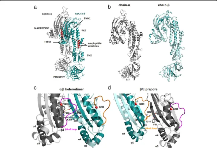
The 3D structure shows (Fig. 5a) that Sp-CTx-α and -βform a dimer containing a mixture ofα/βfolds com-prising four distinct domains: a MAPCPF/CDC domain, a focal adhesion-targeting (FAT) motif, thioredoxin (THX), and finally, the C-terminal domain containing PRYSPRY. A predicted secondary amphiphilic α-helix is
shown (red) in Fig.4within the FAT domain.
The interface between α- and β-subunits of Sp-CTx
has many features contained in the SNTX-α/β
heterodi-mer. Figure 5b reveals that both toxins present strong structural similarity within each heterodimer. A highly conserved loop was found in the interface between sub-units. In Sp-CTx, the β4-α6 binding site contains a
hydrophobic surface comprising TMH2, helix-α6 and
strand-β1, which is equivalent to MACPF and CDCs structures in SNTX, thus suggesting that this region is important for dimer formation, stability and oligomerization events (Fig. 5c, d). This analysis indi-cates that several noncovalent interactions stabilize the dimer interface in Sp-CTx.
a
b
Fig. 4Predicted amphiphilicα-helices in Sp-CTxα- andβ-subunits. Two potential amphiphilicα-helices were predicted by Helical Wheel Projections by Schiffer-Edmundson [39,40]. Residues are colored according to their chemical character as follows: acidic (blue), basic (red), uncharged polar (green) and nonpolar (yellow).aAmphiphilic
α-helix from Ile275 to Asp292 in Sp-CTx-αsubunit;bAmphiphilic
Phylogenetic study of Sp-CTx
The phylogenetic tree of Sp-CTx is shown in Fig.6. Ac-cordingly, toxins were grouped into three distinct clus-ters: i)Pteroissp. and Subunitsβgroup: PlTx-α, PaTx-α,
PvTx-α, PlTx-β, PvTx-β and PaTx-β from Pterois lunu-lata, Pterois antennata, Pterois volitans, Pterois lunu-lata, Pterois volitansandPterois antennatarespectively; Subunit β group: SmTx-β, Sp-CTx-β, SoTx-β, SsTx-β,
HrTx-β, IjTx-β, NeoVTX-βand SNTX-βfrom Sebastis-cus marmoratus,Scorpaena plumieri,Scorpaenopsis oxy-cephala, Sebastapistes strongia, Hypodytes rubripinnis, Inimicus japonicus, Synanceia verrucosa and Synanceia horrida, respectively; in addition SfTx-α is classified in
the same cluster despite its apparent differences with other members; ii) Subunits of the α group: Sp-CTx-α,
SoTx-α, SsTx-α, SmTx-α, HrTx-α, SNTX-α, NeoVTX-α
and IjTx-α from S. plumieri, Scorpaenopsis oxycephala, Sebastapistes strongia, Sebastiscus marmoratus, Hypo-dytes rubripinnis, Synanceia horrida, Synanceia verru-cosaand Inimicus japonicus, respectively; and iii) finally, β-subunit from Perciforme Siganus fuscescens included in a separate cluster. The phylogenetic analysis suggests that genes coding for subunits from all species belong to two different clusters (βandαclades) except forPterois, whose subunits are grouped together and branch out from the first clade.
Discussion
A pore-forming cytolysin from S. plumieri venom (Sp-CTx) that induces cardiovascular alterations and other pharmacological activities has been purified by [11,16]. Pharmacological effects similar to Sp-CTx have Fig. 5Three-dimensional modeled structure of Sp-CTx. The Sp-CTX modeled structures are shown in cartoon format.aModeled structure showing interactions between Sp-CTx subunits; Sp-CTx-α(gray) and Sp-CTx-β(blue). Identification of the N-terminal domains; MACPF/CDC, FAT, THX and PRYSPRY, the transmembraneα-helices TMH1 and TMH2, the amphiphilicα-helices (red) of Sp-CTx-αandβ-chains.bThe structure of Sp-CTx aligned with SNTX (Protein Data Bank ID code 4WVM) and schematic representation ofα-subunits (gray) on the left andβ-subunits (blue) on the right. Lighter tones depict the structure of SNTX.cHighlighted interface region within the heterodimer in the MACPF/CDC withβ-strands numbered according to their position in the centralβ-sheet. Theβ4-α6 loop is shown in pink, the conserved G208 (Sp-CTx-α) is shown as a red sphere. Hydrogen bonds between the residues F206 and K207 of strand-β4 (Sp-CTx-α) and T53, F54, E55 of strand-β1 (Sp-CTx-β) are displayed as yellow dashed lines.dThe interface region of prepore in MACPF/CDC. Theβ4-α6 loop is colored orange, the conserved G209 (Sp-CTx-β) is shown as a red sphere. Hydrogen bonds between residues T218 inβ4-α6 loop (Sp-CTx-β) and D52, T53 of strand-β1 (Sp-CTx-α) are shown as yellow dashed lines. Figures were generated using Pymol (v1.7.0.0), (http://www.pymol.org/; Delano Scientific LLC, South San Francisco, CA)
been attributed to other hemolytic factors from stonefish venoms [19, 20, 47, 48]. The cardiovascular effect in-duced by Sp-CTx is observable both in vitro and in vivo, and includes a vasorelaxant action that appears to in-volve the L-arginine-nitric oxide synthase pathway [16]. It has been suggested that the cardiovascular effect of Sp-CTx is caused by increased influx of sarcolemma Ca2 +affecting ventricular cardiomyocytes [22].
The structural features accounting for the pharma-cological properties of Sp-CTx are poorly defined mainly because of the limited amounts available in fish venom [4]. To gain insight into the venom protein composition, we initially produced a cDNA li-brary from S. plumieri to prospect by random EST the major gland components. While several lectins were identified in spine tissue, none of the readouts provided information on Sp-CTx [28].
Identification of Sp-CTx was then attempted using the library with two primers (βT-f and CDβ-r) whose se-quences were derived from Scorpaeniformes toxins. The sequenced fragment annealed to β toxins of three
families already identified as lethal factors and covering 74% of the β-subunit. Attempts to recover the missing
26% region in the library were unsuccessful. Instead, the missing C-terminal complement of Sp-CTx-βwas
identi-fied in the total cDNA fraction from S. plumieri with CDβr-f primers.
For Sp-CTx-αsubunit, the entire sequence was
identi-fied and assembled following amplification of four over-lapping segments from the same cDNA fraction (Fig. 1). The initiators for isolation of Sp-CTx-α were derived
prospects of lethal factors described in Scorpaeniformes. The deduced ORFs encode two polypeptides encompass-ing 702 amino-acids each and predicted mass of 80,153 kDa for Sp-CTx-αand 79,816 for Sp-CTx-β. The
predicted mass for Sp-CTx-α and -β subunits resemble
those of cytolysins identified in Scorpaeniformes venoms [13–15,19,20].
Gomes et al. [11], estimated the size of Sp-CTx complex to be 150 kDa based on non-reducing and denaturing electrophoretic evidence, in agreement with the figure deduced herein for Sp-CTx-α+
Sp-CTx-β. The authors also identified internal
pep-tides in Sp-CTx by Orbitrap-MS analysis of the tryp-sinized purified protein. Eight fragments totaling 79 residues (11.2%) were identified in Sp-CTx-α,
whereas twelve fragments totaling 116 residues (16.5%) were identified in Sp-CTx-β, matching the
sequences found herein, as shown in Fig. 2a, b (frag-ments highlighted in boxes). A search using SignalP 4.0 tool did not detect a signal peptide-like motifs in either Sp-CTx-α or β-subunit, similarly to other
Scorpaeniform toxins described to date [19, 49]. The absence of muscular tissue in venom glands indicates that mechanical pressure is required to release the venom through the spinal system [8]. An interesting feature in Scorpaeniformes toxins is the presence of a B30.2/SPRY domain in their C-terminal regions. This domain is also found in diverse protein fam-ilies, such as TRIM (Tripartite motif ), RBCC (RING-finger, B-box plus coiled-coil domain), BTN (butyrophilin) and SPSB (cytokine signaling box pro-tein) [50]. This highly variable domain possibly rec-ognizes a specific protein ligand [51]. The functional role of B30.2 and SPRY domains is unclear, although it is evolutionarily preserved. It displays three con-served motifs, containing LDP, WEVE and LDYE [50, 52]. The LDP motif is identifiable in Sp-CTx-α
at position 527–529, the WEVE motif is found both in Sp-CTx-α and -β at positions 578–581 and 579– 582, respectively, and the LDYE motif is absent in both subunits. On the other hand, the crystal struc-ture of SNTX reveals that the PRYSPRY domains in the heterodimeric toxin located distally to the N-terminal end are structurally similar to protein domains involved in innate immunity against micro-organism infection. The mediation of its action by protein-protein and protein-lipid interactions on the cell surface suggests a mechanism for toxicity in SNTX [10]. A comparative structural analysis be-tween Sp-CTx and SNTX crystals revealed the pres-ence of three shared domains: Membrane Attack Complex-Perforin/Cholesterol-Dependent Cytolysin (MACPF/CDC), focal adhesion-targeting (FAT) and thioredoxin (THX), [10].
MACPF/CDC proteins are perforins found in diverse organisms typically composing a ring-shaped supra-molecular oligomeric membrane pore complex, such as in pathogenic gram-positive bacteria and in the mam-malian complement immune system [53]. This domain interacts with FAT, which has a signaling function [54], and a region structurally similar to mitochondrial thior-edoxin (THX) fromSaccharomyces cerevisiae. However, the THX domain is not involved in redox reactions because it lacks a catalytic site [55].
Three-dimensional modeling was necessary because the alignment of primary sequences was insufficient to analyze the spatial orientation of Sp-CTx residues and their molecular interactions. By building the model for each subunit and obtaining the predicted heterodimer by docking, we were able to examine in detail the het-erodimer interface and to identify interactions that stabilize it.
The data led us to propose that Sp-CTx also belongs to the pore-forming MACPF/CDC superfamily, sharing a common four-strand folding and a highly twisted
β-sheet anchored to three small α-helix clusters, in
which two of these helical regions insert into the mem-brane (transmemmem-brane hairpins TMH1 and TMH2). Interestingly, the structural folding of these domains re-sembles the crystallographic structures of other proteins, such as those responsible for protein-cell interactions occurring during immunological recognition [52]. Previ-ous studies show that when pores are formed by CDCs, the monomers assemble into a prepore unit on the membrane surface and that the ensuing pore formation involves significant secondary and tertiary structural changes in TMH1 and TMH2 to penetrate the mem-brane as amphipathicβ-hairpins [56].
Similar to stonefish toxins, Sp-CTx displays 50% identity between its α- and β-subunits, while lionfish toxins are approximately 80% identical. It is unclear whether these variations in identities between subunits in stonefish and lionfish are related to species-specific functions. Because of this strong iden-tity [19], it was proposed that SNTX genes for α- and
β-subunits evolved separately from a common ances-tor after gene duplication.
A similarity search between Sp-CTx-α or Sp-CTx-β
and similar annotated sequences using the NCBI data-base and BLAST algorithm [41] revealed significant identity only with toxins from Scorpaeniformes. Five cysteinyl residues located at similar positions in described subunits appear to be involved in protein conformation through disulfide bridges. Ghadessy and cols. [19] identified, by titration of SNTX with DTNB, five free cysteines and ten cysteines involved in intra-chain disulfide bridges. However, in Sp-CTx these resi-dues did not interact in the heterodimer model.
Different from toxins in terrestrial animals displaying toxin isoforms encoded by more than two alleles, there is no information to indicate the number of copies in fish toxins. Chuang and Shiao, [15] suggested that gene duplication occurred in the mother species of Scorpae-niformes where they evolved intoαandβsubunits. The
authors identified an additional toxin duplication that can be found as a pseudogene in the lineage of lionfish.
Cationic residues like lysine and arginine and the hydrophobic amino acid tryptophan are essential for the cytolytic activity in toxins [44, 57]. The membrane-permeating ability of many peptides and proteins can be attributed to the presence of hydro-phobic segments or amphiphilic α-helices and β-sheets [44]. Chuang and Shiao, [15] reported 23
positively-charged amino acids and 6 conserved tryp-tophanyl residues in every Scorpaeniformes toxin de-scribed, a rule that is confirmed in Sp-CTx. Additional studies by site-directed mutagenesis would be useful for clarifying the role of these residues.
To investigate the evolutionary relationships among Scorpaeniformes toxins, a phylogenetic tree was con-structed and is displayed in Fig. 6. The classification of Sp-CTx agrees with previous evolutionary trees in-volving lethal factors [14, 15, 17]. In the diagram the amino-acid sequence of Sp-CTx is closest to those of scorpionfish and lionfish toxins followed by waspfish, stonefish and devil stinger toxins. Interestingly, the phylogenetic tree is consistent with the taxonomic classification based on the morphology of the venom glands described by Russell [58] and Halstead [1] who classified Scorpaeniformes into lionfish (Pterois) with shorter spines, scorpionfish (Scorpaena) with mode-rate spines and stonefish (Synanceia) with longer spines and highly developed tissue glands. The calcu-lated sequence identities are reflected in the phylo-genetic tree in which Siganus fuscescens toxin branches out from members of Scorpaeniformes, es-pecially for β-subunit [17].
Conclusion
In this study we identified the putative sequences coding for Sp-CTx, a lethal cytolysin from S. plumieri whose biochemical properties and pharmacological actions had been previously characterized. By comparative modeling with the SNTX structure, we identified potential deter-minants in Sp-CTx responsible for the cytolytic activity demonstrated in this toxin. The modeled Sp-CTxα-β
heterodimer fits appropriately with the structure of SNTX fromS. horridaidentified by crystallography, thus supporting the notion that these proteins share similar functions.
Additional file
Additional file 1:Alignment of the amino-acid sequences of Scor-paeniformes toxins.α-subunits of Sp-CTx, Sm-Tx, So-Tx, Ss-Tx, Pl-Tx, Pv-Tx, Pa-Tx, Hr-Tx, neoVTX, SNTX and Ij-Tx.β-subunits of Sp-CTx, Sm-Tx, So-Sm-Tx, Ss-Sm-Tx, Pl-Sm-Tx, Pv-Sm-Tx, Pa-Sm-Tx, Hr-Sm-Tx, neoVTX, SNTX and Ij-Tx. Single-letter amino-acid notation is used. Cysteine residues are highlighted in white on a black background. Identical residues are identified by a segment; (*) denotes conserved cationic residues; (\) denotes conserved tryptophan. Accession numbers (DDBJ/EMBL/Gen-Bank nucleotide sequence databases):α- andβ-subunits: Sm-Tx from
Sebastiscus marmoratustoxin, AIC84049 and AIC84050; So-Tx from
Scorpaenopsis oxycephalatoxin, AIC84047 and AIC84048; Ss-Tx from
Sebastapistes strongiatoxin, AIC84045 and AIC84046; Pl-Tx fromPterois lunulatatoxin, AB775453 and AB775454; Pv-Tx fromPterois volitans
toxin, AB623222 and AB623223; Pa-Tx fromPterois antennatatoxin, AB623220 and AB623221; Ij-Tx fromInimicus japonicustoxin, AB775455 and AB775456; Hr-Tx fromHypodytes rubripinnistoxin, AB775457 and AB775458; neoVTX fromSynanceia verrucosa, AB262392 and AB262393 and SNTX fromSynanceia horrida, U36237 and U32516. (DOCX 176 kb)
Abbreviation
Sp-CTx:Scorpaena plumieriCytolytic Toxin
Acknowledgements
The authors are grateful for the technical support offered by Dr. Evanguedes Kalapothakis during DNA sequencing protocols.
Funding
This work was supported by the National Institute of Science and Technology on Toxins (INCTTOX), the Coordination for the Improvement of Higher Education Personnel (CAPES), the State of Minas Gerais Research Foundation (FAPEMIG) and the National Council for Scientific and Technological Development (CNPq). RSF holds a CNPq Research Fellowship (Bolsa de Produtividade em Pesquisa) level 2. In addition, the publication of the present work was supported in part by CAPES through Programa Editoração CAPES (edital n. 13/2016, auxílio n. 0722/2017, processo n. 88881.142062/2017–01) and CNPq through Programa Editorial CNPq/CAPES (chamada n. 26/2017, processo n. 440954/2017–7).
Availability of data and materials
All data generated or analyzed during this research are included in this published article and its supplementary information files.
Authors’contributions
FLSC conducted and analyzed the experiments. MEL, CES, SGF and RSF analyzed the data and participated in the experimental design and the elaboration of the manuscript. NSP and TS participated in the modeling studies with the toxin. All authors read and approved the final manuscript.
Ethics approval and consent to participate
Fishing of the specimens used in the present study was authorized by the Brazilian Institute of Environment and Renewable Natural Resources (IBAMA).
Consent for publication
Not applicable.
Competing interests
The authors declare that they have no competing interests.
Publisher’s Note
Springer Nature remains neutral with regard to jurisdictional claims in published maps and institutional affiliations.
Author details
Belo Horizonte, MG 31270-901, Brazil.2Departamento de Ciências Fisiológicas, Universidade Federal do Espírito Santo, Vitória, ES, Brazil.
Received: 27 February 2018 Accepted: 3 August 2018
References
1. Halstead BW. Dangerous marine animals: that bites, sting, shock, are non-edible. In: Dangerous marine animals. 2nd ed. USA: Cornell Maritime Press-Maryland; 1980. p. 108–17.
2. Campos FV, Menezes TN, Malacarne PF, Costa FLS, Naumann GB, Gomes HL, et al. A review on theScorpaena plumierifish venom and its bioactive compounds. J Venom Anim Toxins incl Trop Dis. 2016;22:35.https://doi.org/ 10.1186/s40409-016-0090-7.
3. Church JE, Hodgson WC. The pharmacological activity of fish venoms. Toxicon. 2002;40(8):1083–93.
4. Figueiredo SG, Andrich F, Lima C, Lopes-Ferreira M, Haddad V Jr. Venomous fish: a brief overview. In: de Lima ME, Pimenta AMC, Martin-Eauclaire MF, Zingali R, Rochat H, editors. Animal Toxins: State of the Art. Perspectives in Health and Biotechnology. Belo Horizonte: UFMG; 2009.
5. Gwee MCE, Gopalakrishnakone P, Yuen R, Khoo HE, Low KSY. A review of stonefish venoms and toxins. Pharmacol Ther. 1994;64(3):509–28. 6. Carvalho-Filho A. Fishes: Brazilian Coast. Ed. Melro Ltda. 3rd ed. Brazil: Sao
Paulo: 1999, 320p.
7. Haddad V Jr, Martins IA, Makyama HM. Injuries caused by scorpionfishes (Scorpaena plumieriBloch, 1789 andScorpaena brasiliensisCuvier, 1829) in the southwestern Atlantic Ocean (Brazilian coast): epidemiologic, clinic and therapeutic aspects of 23 stings in humans. Toxicon. 2003;42(1):79–83. 8. Ziegman R, Alewood P. Bioactive components in fish venoms. Toxins. 2015;
7(5):1497–531.
9. Chen D, Kini RM, Yuen R, Khoo HE. Haemolytic activity of stonustoxin from stonefish (Synanceja horrida) venom: pore formation and the role of cationic amino acid residues. Biochem J. 1997;325(Pt 3):685–91.
10. Ellisdon AM, Reboul CF, Panjikar S, Huynh K, Oellig CA, Winter KL, et al. Stonefish toxin defines an ancient branch of the perforin-like superfamily. Proc Natl Acad Sci U S A. 2015;112(50):15360–5.
11. Gomes HL, Andrich F, Forte-Dias CL, Perales J, Teixeira-Ferreira A, Vassalo DV, et al. Molecular and biochemical characterization of a cytolysin from the
Scorpaena plumier(scorpionfish) venom: evidence of pore formation on erythrocyte cell membrane. Toxicon. 2013;74:92–100.
12. Ouanounou G, Malo M, Stinnakre J, Kreger AS, Molgo J. Trachynilysin, a neurosecretory protein isolated from stonefish (Synanceia trachynis) venom, forms nonselective pores in the membrane of NG108-15 cells. J Biol Chem. 2002;277(42):39119–27.
13. Kiriake A, Shiomi K. Some properties and cDNA cloning of proteinaceous toxins from two species of lionfish (Pterois antennataandPterois volitans). Toxicon. 2011;58(6–7):494–501.
14. Kiriake A, Suzuki Y, Nagashima Y, Shiomi K. Proteinaceous toxins from three species of scorpaeniform (lionfishPterois lunulata, devil stingerInimicus japonicusand waspfishHypodytes rubripinnis): close similarity in properties and primary structures to stonefish toxins. Toxicon. 2013;70:184–93. 15. Chuang PS, Shiao JC. Toxin gene determination and evolution in
scorpaenoid fish. Toxicon. 2014;88:21–33.
16. Andrich F, Carnielli JBT, Cassoli JS, Lautner RQ, Santos RAS, Pimenta AMC, et al. A potent vasoactive cytolysin isolated fromScorpaena plumieri
scorpionfish venom. Toxicon. 2010;56(4):487–96.
17. Kiriake A, Ishizaki S, Nagashima Y, Shiomi K. Occurrence of a stonefish toxin-like toxin in the venom of the rabbitfishSiganus fuscescens. Toxicon. 2017; 140:139–46.
18. Garnier P, Goudey-Perrière F, Breton P, Dewulf C, Petek F, Perrière C. Enzymatic properties of the stonefish (Synanceia verrucosaBloch and Schneider, 1801) venom and purification of a lethal, hypotensive and cytolytic factor. Toxicon. 1995;33(2):143–55.
19. Ghadessy FJ, Chen D, Kini RM, Chung MC, Jeyaseelan K, Khoo HE, et al. Stonustoxin is a novel lethal factor from stonefish (Synanceja horrida) venom. cDNA cloning and characterization. J Biol Chem. 1996;271(41): 25575–81.
20. Ueda A, Suzuki M, Honma T, Nagai H, Nagashima Y, Shiomi K. Purification, properties and cDNA cloning of neoverrucotoxin (neoVTX), a hemolytic lethal factor from the stonefishSynanceia verrucosavenom. Biochim Biophys Acta. 2006;1760(11):1713–22.
21. Gomes HL, Andrich F, Mauad H, Sampaio KN, De Lima ME, Figueiredo SG, et al. Cardiovascular effects of scorpionfish (Scorpaena plumieri) venom. Toxicon. 2010;55(2–3):580–9.
22. Gomes HL, Menezes TN, Malacarne PF, Roman-Campos D, Gondim AN, Cruz JS, et al. Cardiovascular effects of Sp-CTx, a cytolysin from the scorpionfish (Scorpaena plumieri) venom. Toxicon. 2016;118:141–8.
23. Shiomi K, Hosaka M, Yamanaka H, Kikuchi T. Venoms from six species of marine fish: lethal and hemolytic activities and their neutralization by commercial stonefish antivenom. Mar Biol. 1989;103(3):285–9. 24. Kreger AS. Detection of a cytolytic toxin in the venom of the stonefish
(Synanceia trachynis). Toxicon. 1991;29(6):733–43.
25. Church JE, Hodgson WC. Stonefish (Synanceiaspp.) antivenom neutralizes thein vitroandin vivocardiovascular activity of soldierfish (Gymnapistes marmoratus) venom. Toxicon. 2001;39(2–3):319–24.
26. Gomes HL, Menezes TN, Carnielli JB, Andrich F, Evangelista KS, Chávez-Olórtegui C, et al. Stonefish antivenom neutralises the inflammatory and cardiovascular effects induced by scorpionfishScorpaena plumierivenom. Toxicon. 2011;57(7–8):992–9.
27. Garnier P, Ducancel F, Ogawa T, Boulain JC, Goudey-Perrière F, Perrière C, et al. Complete amino-acid sequence of theβ-subunit of VTX from venom of the stonefish (Synanceia verrucosa) as identified from cDNA cloning experiments. Biochim Biophys Acta. 1997;1337(1):1–5.
28. Costa FLS, De Lima ME, Pimenta AC, Figueiredo SG, Kalapothakis E, Salas CE. Expressed sequence tags in venomous tissue ofScorpaena plumieri
(Scorpaeniformes: Scorpaenidae). Neotrop Ichthyol. 2014;12(4):871–8. 29. Chomczynski P, Sacchi N. Single-step method of RNA isolation by acid
guanidinium thiocyanate-phenol-chloroform extraction. Anal Biochem. 1987; 162(1):156–9.
30. Sambrook J, Russell DW. Molecular cloning: a laboratory manual. 3rd ed. New York: Cold Spring Harbor Laboratory Press; 2001.
31. Guex N, Peitsch MC, Schwede T. Automated comparative protein structure modeling with SWISS-MODEL and Swiss-PdbViewer: a historical perspective. Electrophoresis. 2009;30(S1):S162–73.
32. Lovell SC, Davis IW, Arendall WB 3rd, de Bakker PIW, Word JM, Prisant MG, et al. Structure validation by Calpha geometry: phi, psi and Cbeta deviation. Proteins. 2003;50(3):437–50.
33. Wiederstein M, Sippl MJ. ProSA-web: interactive web service for the recognition of errors in three-dimensional structures of proteins. Nucleic Acids Res. 2007;35:W407–10.
34. Benkert P, Tosatto SCE, Schomburg D. QMEAN: a comprehensive scoring function for model quality assessment. Proteins. 2008;71(1):261–77. 35. van Zundert GCP, Rodrigues JPGLM, Trellet M, Schmitz C, Kastritis PL, Karaca
E, et al. The HADDOCK2.2 web server: user-friendly integrative modeling of biomolecular complexes. J Mol Biol. 2016;428(4):720–5.
36. Negi SS, Schein CH, Oezguen N, Power TD, Braun W. InterProSurf: a web server for predicting interacting sites on protein surfaces. Bioinformatics. 2007;23(24):3397–9.
37. Ewing B, Hillier L, Wendl MC, Green P. Base-calling of automated sequencer traces using phred. I. Accuracy assessment. Genome Res. 1998;8(3):175–85. 38. Petersen TN, Brunak S, von Heijne G, Nielsen H. SignalP 4.0: discriminating signal peptides from transmembrane regions. Nat Methods. 2011;8(10):785–
6.
39. Schiffer M, Edmundson AB. Use of helical wheels to represent the structures of proteins and to identify segments with helical potential. Biophys J. 1967; 7(2):121–35.
40. Mól AR, Castro MS, Fontes W. NetWheels Tool: A web appication to create high quality peptide helical Wheel and net projections. 2006.http://lbqp. unb.br/NetWheels. Accessed 7 May 2018.
41. Altschul SF, Gish W, Miller W, Myers EW, Lipman DJ. Basic local alignment search tool. J Mol Biol. 1990;215(3):403–10.
42. Edgar RC. MUSCLE: multiple sequence alignment with high accuracy and high throughput. Nucleic Acids Res. 2004;32(5):1792–7.
43. Kumar S, Stecher G, Tamura K. MEGA7: molecular evolutionary genetics analysis version 7.0 for bigger datasets. Mol Biol Evol. 2016;33(7):1870–4. 44. Kini RM, Evans HJ. A common cytolytic region in myotoxins, hemolysins,
cardiotoxins and antibacterial peptides. Int J Pept Protein Res. 1989;34(4):277–86. 45. Hulo N, Bairoch A, Bulliard V, Cerutti L, De Castro E, Langendijk-Genevaux
PS, et al. The PROSITE database. Nucleic Acids Res. 2006;34:D227–30. 46. Pasupuleti M, Schmidtchen A, Malmsten M. Antimicrobial peptides: key
components of the innate immune system. Crit Rev Biotechnol. 2012; 32(2):143–71.
47. Colasante C, Meunier FA, Kreger AS, Molgó J. Selective depletion of clear synaptic vesicles and enhanced quantal transmitter release at frog motor nerve endings produced by trachynilysin, a protein toxin isolated from stonefish (Synanceia trachynis) venom. Eur J Neurosci. 1996;8(10):2149–56. 48. Poh CH, Yuen R, Khoo HE, Chung M, Gwee M, Gopalakrishnakone P.
Purification and partial characterization of stonustoxin (lethal factor) from
Synanceia horridavenom. Comp Biochem Physiol B. 1991;99(4):793–8. 49. von Heijne G. Transcending the impenetrable: how proteins come to terms
with membranes. Biochim Biophys Acta. 1988;947(2):307–33.
50. Perfetto L, Gherardini PF, Davey NE, Diella F, Helmer-Citterich M, Cesareni G. Exploring the diversity of SPRY/B30.2-mediated interactions. Trends Biochem Sci. 2013;38(1):38–46.
51. Woo JS, Imm JH, Min CK, Kim KJ, Cha SS, Oh BH. Structural and functional insights into the B30.2/SPRY domain. EMBO J. 2006;25(6):1353–63. 52. Henry J, Ribouchon MT, Offer C, Pontarotti P. B30.2-like domain proteins: a
growing family. Biochem Biophys Res Commun. 1997;235(1):162–5. 53. Hadders MA, Beringer DX, Gros P. Structure of C8alpha-MACPF reveals
mechanism of membrane attack in complement immune defense. Science. 2007;317(5844):1552–4.
54. Hayashi I, Vuori K, Liddington RC. The focal adhesion targeting (FAT) region of focal adhesion kinase is a four-helix bundle that binds paxillin. Nat Struct Biol. 2002;9(2):101–6.
55. Bao R, Zhang Y, Zhou CZ, Chen Y. Structural and mechanistic analyses of yeast mitochondrial thioredoxin Trx3 reveal putative function of its additional cysteine residues. Biochim Biophys Acta. 2009;1794(4):716–21. 56. Hotze EM, Tweten RK. Membrane assembly of the cholesterol-dependent
cytolysin pore complex. Biochim Biophys Acta. 2012;1818(4):1028–38. 57. Yew WS, Khoo HE. The role of tryptophan residues in the hemolytic activity
of stonustoxin, a lethal factor from stonefish (Synanceia horrida) venom. Biochimie. 2000;82:251–7.

![Fig. 6 Phylogenetic tree of Scorpaeniformes toxins. The tree was generated by the MUSCLE [42] and then analyzed with the Neighbor-Joining algorithm (bootstrap replicates: 500; substitution model: Maximum Composite Likelihood), both implemented in MEGA7 [43](https://thumb-eu.123doks.com/thumbv2/123dok_br/16029813.693971/11.892.88.806.133.616/phylogenetic-scorpaeniformes-generated-replicates-substitution-composite-likelihood-implemented.webp)
