R E S E A R C H
Open Access
Characterization of
Leiurus abdullahbayrami
(Scorpiones: Buthidae) venom: peptide profile,
cytotoxicity and antimicrobial activity
Efe Erde
ş
1,2, Tu
ğ
ba Somay Do
ğ
an
2,3,
İ
lhan Co
ş
ar
4, Tar
ı
k Dan
ış
man
4, Kadir Bo
ğ
aç Kunt
5, Tamay
Ş
eker
2,
Meral Yücel
2,6and Can Özen
1,2,7*Abstract
Background:Scorpion venoms are rich bioactive peptide libraries that offer promising molecules that may lead to the discovery and development of new drugs.Leiurus abdullahbayramiproduces one of the most potent venoms among Turkish scorpions that provokes severe symptoms in envenomated victims.
Methods:In the present study, the peptide profile of the venom was investigated by electrophoretic methods, size-exclusion and reversed-phase chromatography and mass spectroscopy. Cytotoxic and antimicrobial effects were evaluated on a breast cancer cell line (MCF-7) and various bacterial and fungal species.
Results:Proteins make up approximately half of the dry weight ofL. abdullahbayramicrude venom. Microfluidic capillary electrophoresis indicated the presence of 6 to 7 kDa peptides and proved to be a highly practical peptidomics tool with better resolution when compared to conventional polyacrylamide gel electrophoresis. Mass spectroscopy analysis helped us to identify 45 unique peptide masses between 1 to 7 kDa with a bimodal mass distribution peaking between molecular weights of 1 to 2 kDa (29%) and 3 to 4 kDa (31%).L. abdullahbayrami crude venom had a proliferative effect on MCF-7 cells, which may be explained by the high concentration of polyamines as well as potassium and calcium ions in the arachnid venoms. Antimicrobial effect was stronger on gram-negative bacteria.
Conclusions:This work represents the first peptidomic characterization ofL. abdullahbayramivenom. Considering the molecular weight-function relationship of previously identified venom peptides, future bioactivity studies may lead to the discovery of novel potassium and chloride ion channel inhibitors as well as new antimicrobial peptides fromL. abdullahbayramivenom.
Keywords:Scorpion venom, Toxin, Peptide,Leiurus abdullahbayrami, Microfluidic capillary, Electrophoresis, Peptidomics, Venomics, Cytotoxicity, Antimicrobial activity, Turkey
Background
Animal venoms compose a complex mixture of ions, small organic molecules, peptides and proteins, which evolved through millions of years of natural selection aiming at prey capture and defense mechanisms. A sig-nificant part of this rich mixture is composed of bio-active peptides. Due to their remarkable structural and
functional variety, these bioactive peptides offer almost limitless possibilities for the development of new thera-peutic agents [1,2].
Arachnid species including scorpions and spiders are of great interest for bioactive peptide research since the complex venom of these animals contain many peptide toxins [3]. Scorpion venoms are composed of inorganic salts, free amino acids, nucleotides, biogenic amines, peptides and proteins [4]. Peptide neurotoxins form most of their venom that may contain more than a hun-dred peptides [5]. So far, more than 600 scorpion pep-tides were described in the UniProt database [6]. These * Correspondence:[email protected]
1Department of Biotechnology, Graduate School of Natural and Applied
Sciences, METU, Ankara 06800, Turkey
2Molecular Biology and Biotechnology Research and Development Center,
Central Laboratory, METU, Ankara, Turkey
Full list of author information is available at the end of the article
toxins were functionally classified into four groups as Na+,
K+, Ca2+ and Cl− ion channel inhibitors [7]. During
400 million years of evolution, these toxins gained ex-cellent specificity and affinity for their targets, which makes them highly potent inhibitors [8].
According to latest records, Turkey has 23 species of scorpions in 11 genera. So far, venom characterization
of only three buthid species was carried out. A Na+
channelα-toxin Bu1 was discovered from the venom of
Buthacus macrocentrus [9]. Three K+channel inhibitors
MegKTx1, MegKTx2 and MegKTx3 were isolated from the venom ofMesobuthus gibbosus [10]. One of the two
scorpion species of Turkey that may cause lethal poison-ing in humans is Androctonus crassicauda. Biochemical
characterization of its venom is in progress and recently bioactive peptides Acra1, Acra2, Acra3 and Acra4 were isolated from the venom [11-13].
The Leiurus genus of Buthidae family includes five
species: L. quinquestriatus,L. jordanensis, L. savanicola, L. nasheri and L. abdullahbayrami. Leiurus quinques-triatus is the best characterized member of the genus
from which many well-known peptide toxins such as chlorotoxin and charybdotoxin were isolated [14,15].
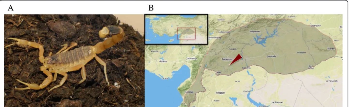
L. abdullahbayrami (Figure 1 – A) is a recently de-scribed species of Leiurus genus which was previously
identified as L. quinquestriatus. It is distributed around
southeastern Turkey, mainly western Euphrates basin and specimens were recorded in Adiyaman, Hatay, Kilis, Gaziantep, Sanliurfa, and Kahramanmaras provinces (Figure 1 –B) [16]. Electrophoretic profile of its venom indicated the strong expression of 4 and 6 kDa peptides. It was also shown that in vivo toxicity of its venom
(LD50) was 0.19 mg/kg on mice [17].
The aim of this study was to determine the peptide profile, cytotoxicity and antimicrobial effect of L. abdul-lahbayramivenom. Characterization studies included
pro-tein content determination, comparative electrophoretic
profiling and peptide mass determination. A combination of size-exclusion and reversed-phase chromatography (RPC) was used to focus on the peptide fraction of the crude venom. Bioactivity screening was carried out on a mammalian cell line (MCF-7) and a few selected bacterial and fungal species.
Methods
Specimen collection and venom milking
L. abdullahbayrami scorpions were collected from a
semi-arid steppe area of Sinanköy Village, Gaziantep Province, southeast of Turkey (37ο2
’16”N 37°35’58”E). Specimens were maintained in plastic boxes and fed mealworms twice a month. Specimens (six males and three females, adults) were milked by electrical stimula-tion (15 V) of the telson. Venom samples were collected in polypropylene tubes, diluted with double distilled water and centrifuged at 15,000 g for 15 minutes at 4°C. The supernatant was transferred to new tubes, lyophi-lized and stored at−80°C.
Protein content determination
Protein content of the crude venom was determined using Bio-Rad Quick Start™Bradford Protein Assay (USA).
Electrophoresis
Bio-Rad Mini-Protean® Tetra Cell system and microflui-dic capillary electrophoresis (MCE) with Agilent Protein 80 kit on an Agilent Bioanalyzer 2100 system (USA) were used. Tris-Glycine sodium dodecyl sulfate-polyacrylamide gel electrophoresis (SDS-PAGE) was performed using 4% stacking and 10% resolving gels under denaturing condi-tions at constant current (25 mA). For Tris-Tricine SDS-PAGE, 4% stacking, 10% spacer and 16% resolving gels with 6 M urea were used with 35 mA [18]. Protein bands were stained by silver staining; whereas 5μg of sam-ple was loaded per well for Tris-Glycine and Tris-Tricine
SDS-PAGE; and 0.1 μg of sample was loaded per well in MCE.
HPLC Fractionation
A Varian Prostar high-performance liquid chromatog-raphy (HPLC) system (USA) was used for venom
frac-tionation. First, 50 μL of crude venom (4 mg/mL)
dissolved in a running buffer (10% acetonitrile and 0.1% trifluoroacetic acid in deionized water) was applied to a Tosoh Bioscience TSKgel® G2000SW size exclusion column (Japan, 7.5 mm × 600 mm, 12.5 nm pore size). Total run time was 60 minutes with 0.5 mL/minute flow rate. Peptide fraction from size exclusion chromatog-raphy (SEC) run was collected, freeze dried and dis-solved in buffer A (0.1% trifluoroacetic acid in deionized water). Then, 50μL of resuspended sample (30 μg/mL) was apllied to a Vydac® (USA) 218TP54 C18 reversed-phase column (4.6 mm × 250 mm, 300 Å pore size). Pep-tide fractions were eluted at 0.7 mL/minute flow rate by a linear gradient of buffer A to 60% buffer B (0.1% tri-fluoroacetic acid in acetonitrile) over 90 minutes.
Mass spectroscopy
Agilent 1200 HPLC coupled LC/MS TOF 6530 mass spectroscopy system was used for the determination of molecular weight of venom peptides. Freeze dried peptide fraction from the SEC run was dissolved in buffer A (0.1% formic acid in deionized water) and 50μL resuspended sample (30μg/mL) was applied to an Agilent Technologies ZORBAX Eclipse XDB C18
col-umn (4.6 mm × 150 mm, 5μm). A linear gradient from
buffer A to 60% buffer B (0.1% formic acid in acetonitrile) at 0.7 mL/minute over 90 minutes was employed. Ionization was achieved with an electrospray ionization (ESI) module followed by mass detection by TOF detector that was operating at positive ion mode with 2000 V and 100–3200 m/z range. Data analysis was performed using Agilent MassHunter Workstation Qualitative Analysis software.
Cytotoxicity assay
MCF-7 human epithelial breast adenocarcinoma cell line was cultured in 4500 mg/L of glucose containing
Dulbecco’s modified Eagle’s medium (DMEM) with
10% FBS, 2 mM L-Glutamine and 100 units/mL of
penicillin-streptomycin at 37°C and 5% CO2. Then,
2 × 10³cells were seeded in 96-well microplate and
incu-bated for 24 hours before treatment. Crude venom (200μg/mL) and etoposide (60μM) were applied (24 and 48 hours) in serum-free medium to avoid interference of serum proteins. Cell viability was determined using an XTT Cell Viability kit from Cell Signaling Technology (USA). The experiments were conducted in triplicates and statistical significance of the observed difference
was determined using one-way ANOVA with Tukey’s
multiple comparison test of the GraphPad Prism soft-ware (USA).
Antimicrobial activity assay
Listeria monocytogenes, Escherichia coli, Enterobacter aerogenes,Pseudomonas aeruginosa,Candida kruseiand Candida albicans were obtained from the Laboratory
of Microbiology, Medical Faculty, Kırıkkale University.
Antibacterial activity of the venom was assessed by agar disc diffusion assay. Microorganisms were activated by inoculating a loop of the strain in the nutrient broth and incubated on rotary shaker. Then, 0.2 mL of inoculum (107-108 mL as per McFarland standard) was added to
the Mueller Hinton agar media. Subsequently, 40 μL of venom at a concentration of 20 mg/mL was applied on the disc (0.6 cm). After 18–24 ± 2 hours of incubation at 37 ± 0.1°C, microbial growth was determined by measur-ing the diameter of the inhibition zone. For antifungal activity investigation, yeasts (0.5-2.5 × 10⁶/mL) were
cul-tivated on Sabouraud 2%-dextrose agar. Venom solution was applied as mentioned above. After cultivation for 24–37 hours at 25 ± 2°C, the growth was determined by measuring the diameter of the inhibition zone. Standard antibiotic discs (RA5, TE30, AMC30 and E15) were used as positive controls. Phosphate buffered saline (PBS) soaked disks were used as negative control.
Ethics statement
This work required no approval by Middle East Technical University Local Ethical Committee for Animal Experi-ments based on the reason that invertebrate animals were used in the study. All precautions were taken to ensure no harm was inflicted on the scorpion specimens during venom milking.
Results
Protein content and electrophoretic profile
Protein content of L. abdullahbayrami venom was
de-termined by Bradford assay to be 54%. The venom of closely related member of the same genus,L. quinques-triatus, was previously determined to have 65% protein
in its composition [19].
40 kDa could not enter the 16% resolving part of the gel as expected. As a final electrophoretic approach, we also employed MCE and detected 6 and 7 kDa peptides in addition to multiple proteins peaks in 70–85 kDa range (Figure 2–C).
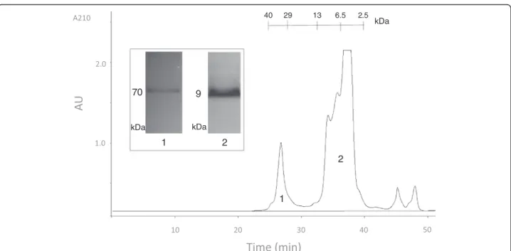
Size exclusion chromatography
In order to reduce venom complexity by eliminating small molecular compounds (<1 kDa) and high-molecular weight proteins (>10 kDa), we first applied the crude venom to size exclusion chromatography (SEC). As seen in Figure 3, two major fractions were obtained from this run: a rela-tively sharp protein peak with an estimated molecular weight of 36 kDa and a combination of multiple peptide peaks in approximately 4–10 kDa range. Surprisingly, the first SEC peak produced a single protein band of 70 kDa on the glycine SDS-PAGE. This significant molecular weight mismatch between SEC and PAGE experiments may be due to the highly compact globular structure of the protein causing retention in SEC matrix and delayed elu-tion from the column. SEC fracelu-tion 2, which will be re-ferred to as the peptide fraction from this point forward, was also analyzed by glycine SDS-PAGE and produced a single peptide band of 9 kDa.
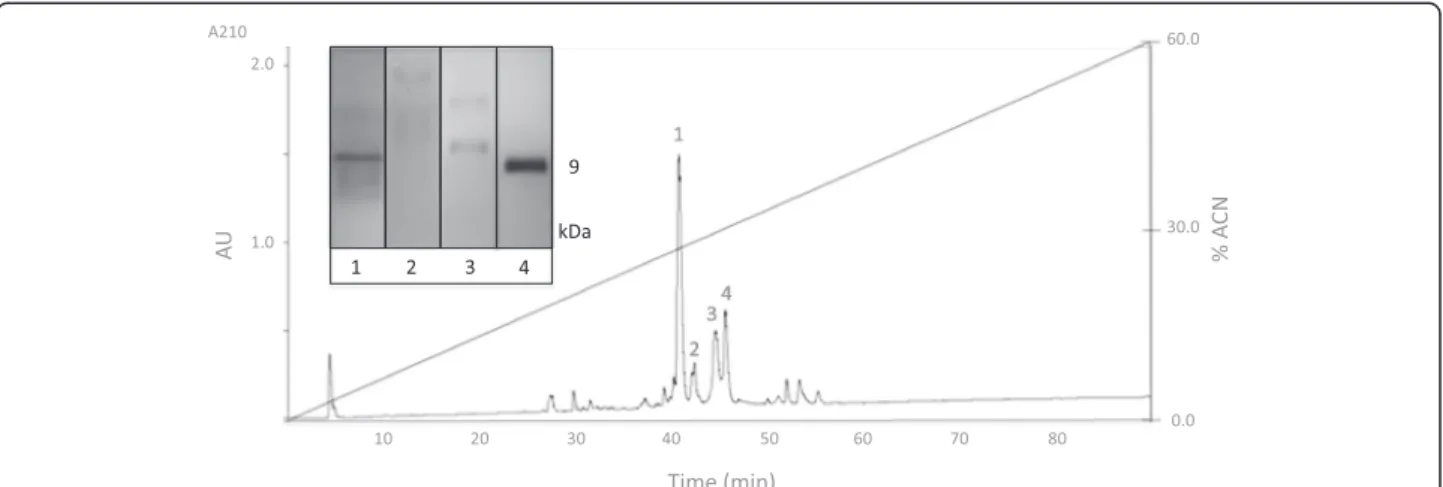
Reversed-phase chromatography
Separation of SEC fraction peptides based on their polarity was achieved by reversed-phase chromatog-raphy (RPC) (Figure 4). Since the venom complexity was reduced by a previous SEC run, a relatively clean
A B
C
Figure 2Electrophoretic profile ofL. abdullahbayramivenom. (A)Glycine SDS-PAGE;(B)Tricine SDS-PAGE and(C)MCE results. UM, LM and SP stand for upper marker, lower marker and system peak, respectively.
reversed-phase chromatogram was obtained. As shown in Figure 4, four major peaks were selected and loaded to glycine SDS-PAGE to confirm their peptide identity. As expected, a 9-kDa peptide band was observed in most samples.
Peptide mass determination
For molecular weight determination, the peptide fraction of the venom obtained from the SEC column was analyzed by liquid chromatography coupled to mass spectroscopy. Table 1 summarizes the deconvoluted molecular weights
of 45 peptides identified fromL. abdullahbayramivenom.
The mass range of the peptides was between 1032 and 6895 Da. Molecular weight distribution histogram shows a bimodal characteristic (Figure 5).
Cytotoxicity
Effect ofL. abdullahbayramicrude venom on cell
viabil-ity of MCF-7 human epithelial breast adenocarcinoma cell line was tested using XTT assay. As seen in Figure 6, venom (200μg/mL) had no cytotoxic effect on the cells following 24-hour and 48-hour treatments. On the other hand, venom treatment caused a significant increase in the metabolic activity after 48 hours. This increase strongly suggests a proliferative effect of the venom.
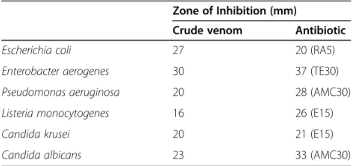
Antimicrobial activity
We also tested the antimicrobial activity of the venom on gram-negative (Escherichia coli, Enterobacter aero-genes and Pseudomonas aeruginosa) and gram-positive
(Listeria monocytogenes) bacteria as well as two fungal
species (Candida krusei and Candida albicans) using
agar disc diffusion assay. Zone of inhibition values (mm) after 24 hours of treatment are provided in Table 2. Antimicrobial effect of the venom seems to be stronger on the gram-negative bacteria.
Discussion
Microfluidic capillary electrophoresis offers significant resolution advantage
Peptidomics approach followed in this work has some differences when compared to commonly used crude venom characterization studies. In the literature, crude venom is typically separated by RPC that produces more peaks. Since our focus was on peptide components of the venom, we preferred to use SEC to separate the peptide fraction of the venom (1–10 kDa) from low- (<1 kDa) and Figure 4Reversed-phase chromatogram ofL. abdullahbayramivenom peptide fraction.The inset shows the glycine SDS-PAGE profile of the selected peaks.
Table 1 Retention times (RT) and deconvoluted molecular weights (MW) ofL. abdullahbayramivenom peptides determined by LC-ESI-TOF–45 molecular masses were
identified
RT (min) MW (Da) RT (min) MW (Da)
11.50 2961, 3000 29.14 4540, 6810
12.40 3024 30.00 5376
12.86 2948, 2988, 3183 33.32 3555, 3576, 3591,
3615, 3630
15.39 3198 33.39 1067
15.49 3234 34.65 1155
20.90 3768 36.87 1287
21.40 3772 36.95 1331, 1375
22.56 3996 37.67 1419
23.37 4056, 4092, 4168 38.20 1463
23.79 3996, 4000 39.71 1507
24.19 4036 43.05 1032
24.86 4056 64.08 1294, 1338
25.66 6780 64.15 1426
26.62 6805, 6820, 6840, 6855, 6895
high-molecular-weight components (>10 kDa) as sug-gested by Vassilevski et al. [20]. The peptide fraction
obtained by SEC was then loaded on a C18 RPC col-umn to separate peptides in the mix based on their polarity. This produced a clean HPLC chromatogram, which makes fraction collection and peptide bioactivity screening easier (Figure 4).
We also compared the performance of two electro-phoretic methods, namely PAGE and MCE. In terms of resolution, MCE has a clear advantage over PAGE. MCE peptide bands at 6 and 7 kDa have a perfect match to
MS determined peptide masses in 6–7 kDa range. Also, from a practical point of view, running an MCE experi-ment is significantly easier and shorter on Agilent 2100 MCE system. The major drawback of the platform was the lack of a suitable protein analysis kit for the detec-tion of peptides below 5 kDa. Due to this limitadetec-tion, we could not detect 1 to 5 kDa peptides, which were identi-fied by MS.
We detected a major 9-kDa band in tricine SDS-PAGE runs of SEC and RP-HPLC fractions. Since MS analysis does not indicate the presence of peptides with this mo-lecular weight, this must be due to the relatively poor resolution of our PAGE setup. Therefore what we deter-mined as 9 kDa on tricine SDS-PAGE must be 6–7 kDa peptides actually. Lack of 3–4 kDa peptide bands on our PAGE results was quite unexpected since 3.4 and 5 kDa bands can be clearly seen in the marker lanes Figure 5Molecular weight distribution ofL. abdullahbayramivenom peptides.
**
*
***
Figure 6Cytotoxicity assay ofL. abdullahbayramivenom on MCF-7 breast adenocarcinoma cell line.NC: negative control; Eto: etoposide (60μM ) positive control; CV: crude venom (200μg/mL).
Error bars represent SD (n = 3). Stars (*, **, ***) denote statistically significant differences atp≤0.05,p≤0.01 andp≤0.001, respectively.
Table 2 Antimicrobial activity ofL. abdullahbayrami
crude venom determined by agar disk diffusion assay
Zone of Inhibition (mm)
Crude venom Antibiotic
Escherichia coli 27 20 (RA5)
Enterobacter aerogenes 30 37 (TE30)
Pseudomonas aeruginosa 20 28 (AMC30)
Listeria monocytogenes 16 26 (E15)
Candida krusei 20 21 (E15)
Candida albicans 23 33 (AMC30)
(Additional file 1). Since Ozkan et al. [17] previously
showed the presence of 4-kDaL. abdullahbayrami
pep-tides on glycine SDS-PAGE, we suspect that dimer for-mation due to the intermolecular disulfide bridges may be responsible for the lack of the expected peptide band. On the other hand, increasing the concentration and type of the reducing agent and increased duration of reduction did not change the results. This leaves the sensitivity problem due to the low abundance of 3–4 kDa peptides in the venom as the best explanation for the observed outcome.
MostL. abdullahbayramivenom peptides are in the 1–4 kDa mass range
Molecular weight histogram provided in the excellent re-view by King and Hardy [6] indicates a bimodal mass distribution for the scorpion venom where the majority of the peptides fall in either 3.5 to 4.0 or 6.5 to 7.5 kDa ranges. Peptide mass profile ofL. abdullahbayrami
par-tially matches this general trend considering that one third of its peptides are in the 3–4 kDa range. However, instead of a major 6.5-7.5 kDa distribution, 30% of
L. abdullahbayramivenom peptides fall in the 1–2 kDa range. In the light of previous peptide bioactivity studies, 3–4 kDa peptides of the venom are expected to be potas-sium and chloride ion channel blockers while 1–2 kDa peptides may be non-disulfide-bridged peptides that typic-ally show antimicrobial activity [21].
L. abdullahbayramivenom shows proliferative effect L. abdullahbayrami crude venom did not show
cyto-toxic effect on MCF-7 cells at 200 μg/mL. Similarly, crude venom (250 μg/mL) of another Turkish scorpion,
Androctonus crassicauda, has also been shown to be not
toxic to various mammalian cells lines [12]. Interestingly,
L. abdullahbayrami venom has a clear proliferative
ef-fect on the cells especially after a 48-hour treatment. This may be due to several reasons. Arachnid venoms are rich in polyamines and ions, especially potassium and calcium [22]. These low-molecular-weight compo-nents and ions have been previously shown to induce cell proliferation [23]. In addition, amino acids and pro-teins of the crude venom might have provided a better growth medium for the MCF-7 cells considering the serum-free medium of the control cells. Sherifet al.[24]
has also reported a similar mitogenic effect of L. quin-questriatusvenom fractions on Vero and BGM cells and
concluded that increased calcium ion influx or interleu-kin and interleu-kinin-linked mechanisms might have stimulated the cell proliferation. Similarly, Heinen et al. [25] also
observed that Lonomia obliquacaterpillar venom led to
decrease in the production of nitric oxide and in-creased the viability of U138-MG and HT-29 cell lines. They also showed that activation of the cAMP signaling
pathway inhibited the effects of the venom, suggesting an interesting link between venom, nitric oxide and cAMP signaling [25].
Antimicrobial activity of the venom
Antimicrobial potency of the venom is comparable to pre-vious studies conducted on L. quinquestriatus [26]. We
identified 13 venom peptides with 1–2 kDa inL. abdullah-bayrami venom by mass spectroscopy and as discussed
earlier, these small peptides are typically categorized as non-disulfide-bridged peptides with antimicrobial activity.
Conclusions
This work is the first detailed bioactivity study that used
peptidomics methods for L. abdullahbayrami scorpion
venom. Protein content of the crude venom was deter-mined as 54%. We showed that MCE is a practical pepti-domics tool with high resolution for venom research. A total of 45 venom peptide masses were identified by mass spectroscopy in the present study, 60% of which are within 1–4 kDa range. L. abdullahbayrami crude venom has a proliferative effect on MCF-7 tumor cell line. Various venom components including potassium and calcium ions as well as polyamines, amino acids and proteins may play a role in this outcome. We also report antimicrobial effect of the venom, which is stronger on gram-negative bacteria.
Future studies on L. abdullahbayrami venom peptides
may lead to the discovery of novel potassium and chloride channel blockers as well as antimicrobial peptides.
Additional file
Additional file 1:Tricine SDS-PAGE profile of fraction number 3 and 4 of RP-HPLC (lanes 1 and 2) and protein ladder (lane 3).As seen from the protein ladder, tricine SDS-PAGE can resolve peptides in 3.4 to 10 kDa mass range.
Abbreviations
MCE:Microfluidic capillary electrophoresis; MW: Molecular weight; RPC: Reversed-phase chromatography; SDS-PAGE: Sodium dodecyl sulfate polyacrylamide gel electrophoresis; SEC: Size exclusion chromatography.
Competing interests
The authors declare that they have no competing interests.
Authors’contributions
EE drafted the manuscript and performed the electrophoretic analyses, cytotoxicity assays and data analysis. TSD conducted HPLC separation of the venom. KBK, TD and IC provided the specimens, identified species and extracted the venom. TD and IC also performed antimicrobial activity assays. TS supervised HPLC and mass spectrometry experiments and data analyses. MY contributed to experimental designs and data analyses. CO provided the idea, finalized the manuscript, gave supervision for experimental designs and data analyses. All authors read and approved the manuscript.
Acknowledgments
acknowledge Boy E. for his contributions to microfluidic capillary electrophoresis experiments. We also thank Ozen A., Muyan M., Igci N., Gerekci S., Durusu I., and Husnugil H. for critical review of the manuscript.
Author details
1Department of Biotechnology, Graduate School of Natural and Applied
Sciences, METU, Ankara 06800, Turkey.2Molecular Biology and Biotechnology Research and Development Center, Central Laboratory, METU, Ankara, Turkey. 3Department of Biology, Faculty of Science, Hecettepe University, Ankara,
Turkey.4Department of Biology, Faculty of Arts and Sciences, Kirikkale University, Kirikkale, Turkey.5Department of Biology, Faculty of Science, Anadolu University, Eskisehir, Turkey.6Department of Biological Sciences, Faculty of Arts and Sciences, METU, Ankara, Turkey.7Center of Excellence in Biomaterials and Tissue Engineering, METU, Ankara, Turkey.
Received: 23 June 2014 Accepted: 13 October 2014 Published: 3 November 2014
References
1. Lewis RJ, Garcia ML:Therapeutic potential of venom peptides.Nat Rev Drug Discov2003,2(10):790–802.
2. Escoubas P, Rash L:Tarantulas: eight-legged pharmacists and combinatorial chemists.Toxicon2004,43(5):555–574.
3. Possani LD, de la Vega RC R:Scorpion venom peptides.InHandbook of biologically active peptides. 1° edition.Edited by Kastin AJ. San Diego: Academic Press; 2006:339–354.
4. Quintero-Hernández V, Jiménez-Vargas JM, Gurrola GB, Valdivia HH, Possani LD:Scorpion venom components that affect ion-channels function.Toxicon2013,76:328–342.
5. Delepierre M, Prochnicka-Chalufour A, Boisbouvier J, Possani LD:Pi7, an orphan peptide from the scorpionPandinus imperator: a 1H-NMR analysis using a nano-NMR Probe.Biochemistry1999,38(51):16756–16765. 6. King GF, Hardy MC:Spider-venom peptides: structure, pharmacology, and
potential for control of insect pests.Annu Rev Entomol2013,58:475–496. 7. Srinivasan KN, Gopalakrishnakone P, Tan PT, Chew KC, Cheng B, Kini RM,
Koh JL, Seah SH, Brusic V:SCORPION, a molecular database of scorpion toxins.Toxicon2002,40(1):23–31.
8. Tan PTJ, Veeramani A, Srinivasan KN, Ranganathan S, Brusic V:SCORPION2: a database for structure-function analysis of scorpion toxins.Toxicon
2006,47(3):356–363.
9. Caliskan F, Quintero-Hernández V, Restano-Cassulini R, Batista CV, Zamudio FZ, Coronas FI, Possani LD:Turkish scorpionButhacus macrocentrus: general characterization of the venom and description of Bu1, a potent mammalian Na+-channelα-toxin.Toxicon2012,59(3):408–415.
10. Diego-García E, Peigneur S, Debaveye S, Gheldof E, Tytgat J, Caliskan F: Novel potassium channel blocker venom peptides fromMesobuthus gibbosus(Scorpiones: Buthidae).Toxicon2013,61:72–82.
11. Caliskan F, García BI, Coronas FI, Batista CV, Zamudio FZ, Possani LD: Characterization of venom components from the scorpion
Androctonus crassicaudaof Turkey: peptides and genes.Toxicon2006, 48(1):12–22.
12. Caliskan F, Ergene E, Sogut I, Hatipoglu I, Basalp A, Sivas H, Kanbak G: Biological assays on the effects of Acra3 peptide from Turkish scorpion
Androctonus crassicaudavenom on a mouse brain tumor cell line (BC3H1) and production of specific monoclonal antibodies.Toxicon2013, 76:350–361.
13. Caliskan F, Quintero-Hernández V, Restano-Cassulini R, Coronas-Valderrama FI, Corzo G, Possani LD:Molecular cloning and biochemical characterization of the first Na(+)-channelα-type toxin peptide (Acra4) fromAndroctonus crassicaudascorpion venom.Biochimie2013,95(6):1216–1222.
14. DeBin JA, Maggio JE, Strichartz GR:Purification and characterization of chlorotoxin, a chloride channel ligand from the venom of the scorpion. Am J Physiol1993,264(2 Pt 1):C361–C369.
15. Miller C, Moczydlowski E, Latorre R, Phillips M:Charybdotoxin, a protein inhibitor of single Ca2 +−activated K + channels from mammalian
skeletal muscle.Nature1985,313(6000):316–318.
16. Yağmur EA, Koç H, Kunt KB:Description of a new species ofLeiurus
Ehrenberg, 1828 (Scorpiones: Buthidae) from Southeastern Turkey. Euscorpius2009,85(85):1–20.
17. Ozkan O, Yagmur EA, Ark M:A newly described scorpion species,
Leiurus abdullahbayrami(Scorpion: Buthidae), and the lethal
potency andin vivoeffects of its venom.J Venom Anim Toxins Incl Trop Dis2011,17(4):414–421 [http://www.scielo.br/scielo.php? script=sci_arttext&pid=S1678-91992011000400008]
18. Schägger H:Tricine-SDS-PAGE.Nat Protoc2006,1(1):16–22.
19. Chicchi GG, Gimenez-Gallego G, Ber E, Garcia ML, Winquist R, Cascieri MA: Purification and characterization of a unique, potent inhibitor of apamin binding fromLeiurus quinquestriatus hebraeusvenom.J Biol Chem1988, 263(21):10192–10197.
20. Vassilevski AA, Kozlov SA, Egorov TA, Grishin EV:Purification and characterization of biologically active peptides from spider venoms. Methods Mol Biol2010,615:87–100.
21. Almaaytah A, Albalas Q:Scorpion venom peptides with no disulfide bridges: a review.Peptides2014,51:35–45.
22. Vassilevski AA, Kozlov SA, Grishin EV:Molecular diversity of spider venom. Biochemistry (Mosc)2009,74(13):1505–1534.
23. Weiger TM, Hermann A:Cell proliferation, potassium channels, polyamines and their interactions: a mini review.Amino Acids2014, 46(3):681–688.
24. Sherif N, Abu-Sinna G, El-Ghitanyl A:Effect ofLeiurus quinquestriatus
venom and venom fractions on cells culturedin vitro.Egyptian J Biol
2000,2:67–75.
25. Heinen TE, de Farias CB, Abujamra AL, Mendonça RZ, Roesler R, da Veiga AB: Effects ofLonomia obliquacaterpillar venom upon the proliferation and viability of cell lines.Cytotechnology2014,66(1):63–74.
26. Salama W, Geasa N:Investigation of the antimicrobial and hemolytic activity of venom of some Egyptian scorpion.J Microbiol Antimicrob2014, 6(1):21–28.
doi:10.1186/1678-9199-20-48
Cite this article as:Erdeşet al.:Characterization ofLeiurus abdullahbayrami(Scorpiones: Buthidae) venom: peptide profile, cytotoxicity and antimicrobial activity.Journal of Venomous Animals and Toxins including Tropical Diseases201420:48.
Submit your next manuscript to BioMed Central and take full advantage of:
• Convenient online submission
• Thorough peer review
• No space constraints or color figure charges
• Immediate publication on acceptance
• Inclusion in PubMed, CAS, Scopus and Google Scholar
• Research which is freely available for redistribution