(Annals of the Brazilian Academy of Sciences) ISSN 0001-3765
www.scielo.br/aabc
The extracellular matrix of the lung and its role in edema formation
PAOLO PELOSI1, PATRICIA R.M. ROCCO2, DANIELA NEGRINI3 and ALBERTO PASSI3 1Servizio di Anestesia B, Department of Ambient, Health and Safety, University of Insubria,
Ospedale di Circolo e Fondazione Macchi, viale Borri 57, 21100 Varese, Italy
2Laboratório de Investigação Pulmonar, Instituto de Biofísica Carlos Chagas Filho, Universidade Federal do Rio de Janeiro,
Centro de Ciências da Saúde, Ilha do Fundão, 21941-902 Rio de Janeiro, RJ, Brasil
3Department of Experimental, Biomedical and Clinical Sciences, University of Insubria,
Via HG Dunant 5, 21100 Varese Italy
Manuscript received on March 29, 2007; accepted for publication on April 24, 2007; presented byLUCIAMENDONÇAPREVIATO
ABSTRACT
The extracellular matrix is composed of a three-dimensional fiber mesh filled with different macromolecules such as: collagen (mainly type I and III), elastin, glycosaminoglycans, and proteoglycans. In the lung, the extracellular matrix has several functions which provide: 1) mechanical tensile and compressive strength and elasticity, 2) low mechanical tissue compliance contributing to the maintenance of normal interstitial fluid dynamics, 3) low resistive pathway for an effective gas exchange, d) control of cell behavior by the binding of growth factors, chemokines, cytokines and the interaction with cell-surface receptors, and e) tissue repair and remodeling. Fragmentation and disorganization of extracellular matrix components comprises the protective role of the extracellular matrix, leading to interstitial and eventually severe lung edema. Thus, once conditions of increased microvascular filtration are established, matrix remodeling proceeds fairly rapidly due to the activation of proteases. Conversely, a massive matrix deposition of collagen fiber decreases interstitial compliance and therefore makes the tissue safety factor stronger. As a result, changes in lung extracellular matrix significantly affect edema formation and distribution in the lung.
Key words:collagen, glycosaminoglycans, proteoglycan, interstitial pressure.
INTRODUCTION
The Extracellular Matrix (ECM) represents the three-dimensional scaffold of the alveolar wall, which is com-posed of a layer of epithelial and endothelial cells, their basement membrane and a thin layer of interstitial space lying between the capillary endothelium and the alveolar epithelium (West and Mathieu-Costello 1999).
In the segment where the epithelial and endothe-lial basement membranes are not fused, the interstitium is composed of cells, the fluid phase of the ECM and a macromolecular fibrous component. This fibrous mesh consists mainly of collagen types I and III, which provide
Correspondence to: Prof. Paolo Pelosi E-mail: [email protected]
the role of ECM components on the maintenance of in-terstitial fluid volume needs to be better clarified.
In this review, we will discuss the ECM organiza-tion and the relaorganiza-tionship between ECM and lung edema.
THE EXTRACELLULAR MATRIX ORGANIZATION
The ECM consists of fibrous proteins (collagen and elas-tin) and structural or adhesive proteins (fibronectin and laminin) embedded in a hydrated polysaccharide gel con-taining several glycosaminoglycans.
COLLAGEN
Collagen fibers constitute the main component of the ECM and are a fibrous protein that consists of threeα -chains, which form a rope-like triple helix, providing ten-sile strength to the ECM (Montes 1996). Despite their broad diversity in the connective tissue, types I, II, III (fibrillar) and IV, V, VI (non fibrillar or amorphous) rep-resent the main collagen fibers. Type III collagen fiber is more flexible and susceptible to breakdown, type I collagen is comprised of thicker more cross-linked fib-ril (Rocco et al. 2001, Santos et al. 2006), whereas type IV collagen fiber is located mainly in basement mem-brane. Their turnover is a dynamic process, necessary to the maintenance of the normal lung architecture (Rocco et al. 2003). The final collagen accumulation does not depend only on its synthesis, but also, on its degradation (Rocco et al. 2001, 2003). Consequently, the ECM is a dynamic structure, and equilibrium between synthesis and degradation of ECM components is required for the maintenance of its homeostasis (Santos et al. 2006).
ELASTIN
Elastic fibers represent another constituent of the ECM and comprise three components defined according to their amount of elastin and fibril orientation: 1) oxytalan fiber composed of a bundle of microfibrils, 2) elaunin fibers made up of microfibrils and a small amount of elastin, and 3) fully developed elastic fibers consisting of microfibrils and abundant elastin (Montes 1996). Many types of cells, including condroblasts, myofibroblasts, and smooth muscle cells synthesize these fibers. Due to their mechanical properties, elastic fibers provide recoil tension to restore the parenchyma to its previous configu-ration after stimulus for inspiconfigu-ration has ceased. In normal
alveolar septa, a subepithelial layer of elastic fibers com-posed mainly of fully developed elastic fibers, confers a great elasticity to the alveolar tissue in normal situa-tions (Mercer and Crapo 1990). Elastosis, as determined by septal inflammation and fiber fragmentation, leads to derangement of the alveolar wall architecture (Negri et al. 2000, Rocco et al. 2003). In response to increased tissue destruction, reactivation of elastin synthesis is ob-served, resulting in significant changes in the mechanical properties of the lung (Rocco et al. 2001).
GLYCOSAMINOGLYCANS AND PROTEOGLYCANS
In the connective tissue, proteoglycans (PGs) form a gelatinous and hydrated substance embedding the fib-rous proteins. Proteoglycans are constituted of a central protein bound to one or more polysaccharides, denomi-nated glycosaminoglycans (GAGs).
GLYCOSAMINOGLYCANS
GAGs are long, linear, and heterogeneous polysaccha-rides, which consist of repeating disaccharide units. There are two main types of GAGs: 1) Non-sulfated GAG (hyaluronic acid) and 2) Sulfated GAGs (heparan sulfate and heparin, chondroitin sulfate, dermatan sul-fate, and keratan sulfate). With the exception of hyalu-ronic acid, GAGs are usually covalently attached to a protein core, forming an overall structure referred to as proteoglycans (Souza-Fernandes et al. 2006).
Non sulfated glycosaminoglycan: hyaluronic acid
Hyaluronic acid is the most abundant and the largest non-sulfated GAG in the ECM. It is primarily synthe-sized by mesenchymal cells, being a necessary molecule for the assembly of a connective tissue matrix and an important stabilizing constituent of the loose connective tissue. It also participates in tissue hydration (Tammi et al. 2002), and is involved in several other functions, such as tissue repair (Li et al. 2000), and protection against infections and proteolytic granulocyte enzymes (Cantor et al. 2000).
Sulfated glycosaminoglycans
pro-teoglycans. The polyanionic nature of GAGs is the main determinant of the physical properties of PGs molecules, allowing them to resist compressive forces and to si-multaneously maintain tissue hydration. They are much smaller than HA, usually only 20 to 200 sugar residues long (Souza-Fernandes et al. 2006).
Within the lung parenchyma, the most abundant sul-fated GAG is heparan sulfate, a polysaccharide that is ex-pressed in virtually every cell in the body and comprises 50% to 90% of the total endothelial PGs (Whitelock and Iozzo 2005). Although it is initially produced in a cell-surface-bound form, it can also be shed as a soluble GAG. The mechanism of action of heparan sulfate includes a specific and non-covalent interaction with various pro-teins which affects the topographical destination, half-life, and bioactivity of the protein. Furthermore, hep-aran sulfate acts on morphogenesis, development, and organogenesis (Whitelock and Iozzo 2005). Heparan sulfate is also involved in a variety of biological pro-cesses including cell-matrix interactions and activation of chemokines, enzymes, and growth factors (Whitelock and Iozzo 2005).
Heparin is the most highly modified form of hep-aran sulfate. This GAG, which can be considered an oversulfated intracellular variant of the heparan sulfate, is commonly used in patients as an anticoagulant drug (Whitelock and Iozzo 2005). Heparin and heparan sul-fate are very closely related and may share many struc-tural and functional activities. The lung is a rich na-tive source of heparin. Heparin’s abundance may be ex-plained by the fact that the lung is rich in mast cells, which may be heparin’s sole original cell. Mast cell hep-arin resides in secretory granules, where most of the GAG chains are linked to a core protein (serglycin), forming macromolecular PGs much larger than commercial hep-arin. Very little heparin is incorporated into cell surface PG of epithelial and endothelial cells that are more likely to contain heparan sulfate, which in turn is under-sulfated compared with heparin. Some heparan sulfate chains of vascular endothelium contain short heparin-like se-quences. However, most native lung heparin is locked up in mast cells as large PGs. This does not necessarily mean that heparin’s physiological action occurs exclu-sively within cells, because stimulated mast cells secrete heparin outside the cell along with granule-associated
mediators, such as histamine, chymase, and tryptase (Ruoss et al. 1991).
PROTEOGLYCANS
In the lung, the main PGs families may be distinguished based on GAGs composition, molecular weight, and function: chondroitin sulfate-containing PG (CS-PG: Versican), heparan sulfate-containing PGs (HS-PGs: Perlecan and Glypican), chondroitin and heparan sulfate-containing PGs (CS-HS-PGs: Syndecan), and dermatan-sulfate-containing PGs (DS-PG: Decorin). They are lo-calized in different areas of the extracellular matrix: ver-sican in the pulmonary interstitium, perlecan in the vas-cular basement membrane, decorin in the interstitium and in the epithelial basement membrane linked with col-lagen fibrils, syndecan and glypican in the cell surface (Roberts et al. 1997).
Versican is a large molecule found around lung fi-broblasts and blood vessels in regions not occupied by collagen and elastin (Roberts et al. 1997). The precise function of versican is unclear but it is thought to be in-volved in tissue hydration in mature tissues. It may form aggregates with hyaluronic acid, fibronectin and various collagens, playing an important role in cell to matrix in-teraction. Versican has been shown to link with smooth muscle cells in the walls of airways and lung vessels, in-hibiting cell-matrix adhesion (Iozzo and Murdoch 1996), regulating mesenchymal cells differentiation and, stim-ulating matrix synthesis, favoring wound healing.
Perlecan is the largest PG in the lung and is a typical component of vascular basement membrane (Yurchenko and Schittny 1990), although it has also been identified within the ECM of some tissues, remote from basement membrane. Certainly its complex core protein provides the potential to interact with numerous proteins. In the basement membranes it provides a filtration barrier inter-acting with collagen IV limiting flow of macromolecules or cells between two tissue compartments. It also regu-lates the interaction of the basic fibroblast growth factors with their receptors and modulates tissue metabolism.
Decorin is the smallest dermatan-sulfate-containing PGs. The presence of decorin alters the kinetics of fibril formation and the diameter of the resulting fibril (Roberts et al. 1997) modulating tissue remodeling. Indeed, its name was derived from its surface decoration of collagen fibrils when viewed in the electron microscope.
These data indicate that the function of PGs and GAGs in the lung is not limited to the maintenance of the mechanical and fluid dynamic properties of the par-enchyma. These molecules are also important in tissue development and recovery after injury, interacting with inflammatory cells, proteases, and growth factors. Thus, the ECM transmits essential information to pulmonary cells, regulating their proliferation, differentiation, and organization. The structural integrity of the pulmonary interstitium largely depends on the balance between the regulation of synthesis and degradation of ECM components.
EXTRACELLULAR MATRIX METALLOPROTEASIS
Although many proteases are able to cleave ECM mo-lecules, the family of Zn++ matrix metalloproteinases
(MMPs) and their inhibitors are likely to be the normal physiologically relevant mediators of ECM degradation (Lanchou et al. 2003, Elkington and Friedland 2006, Greenlee et al. 2007). They can degrade many proteins including collagens, fibronectin, laminin, proteoglycans, entactins, and elastin. In particular, they are very impor-tant in: a) the breakdown of ECM and basement mem-brane, b) tissue remodeling and angiogenesis, and c) the restoration of functional connective tissue in the wound-healing process.
Gelatinases (MMP-2 and MMP-9) are the subgroup of MMPs most extensively studied in human intersti-tial lung diseases and in experimental models of lung fibrosis, probably due to the facility and great sensitivity of revealing gelatinolytic activity through gelatin zymo-grams. Both gelatinases contain fibronectin type II-like repeats within their catalytic domain, resulting in a higher binding affinity to gelatin and elastin. The MMP-2 is produced by fibroblasts, endothelial and epithelial cells, and macrophages. MMP-2 (gelatinase A) is known to be effective primarily against type IV collagen and other basement-membrane components although a weak abil-ity to degrade stromal collagens has been also reported.
On the contrary MMP-9 (gelatinase B) is not produced by resident cells, but under various forms of stimula-tion bronchial epithelial cells, ciliar cells, alveolar type II cells, fibroblasts, smooth muscle cells, and endothelial cells may produce MMP-9. Alveolar type I, neuroen-docrine, and goblet cells have not yet been shown to produce MMP-9. Leukocytes in the lung can also be a source of MMP-9. Macrophages, eosinophils, mast cells, lymphocytes, and dendritic cells all can produce MMP-9.
Because MMPs may cause significant host dam-age, their proteolytic activity is tightly regulated. Thus, MMPs are rarely stored, but require gene transcription before secretion, with the exception of neutrophil MMP-8 and -9. In fact, these are either secreted as pro-enzymes that require proteolytic cleavage or are activated intra-cellularly by pro-protein convertases such as furin. Spe-cific inhibitors of MMPs – the tissue inhibitors of met-alloproteinases (TIMPs) – which bind MMPs in a 1:1 manner are secreted to prevent enzymatic activity; as a result the balance between MMPs and TIMPs determines matrix turnover, where either an excess of MMPs or a deficit of TIMPs may result in excess ECM degradation (Elkington and Friedland 2006).
The majority of MMPs are not expressed in nor-mal healthy tissues, but are expressed in diseased tis-sues, that are inflamed or undergoing repair and remod-eling (Lanchou et al. 2003). MMP expression may be upregulated by exogenous stimuli, cytokines, and cell-cell contact. Conversely, cytokines such as interferon (IFN)-γ and interleukins (IL)-4 and -10 may down reg-ulate MMP expression. Both inflammatory and stromal cells can express MMPs, although the profile is both cell and stimulus specific.
THE EXTRACELLULAR MATRIX AND THE BASAL MEMBRANE
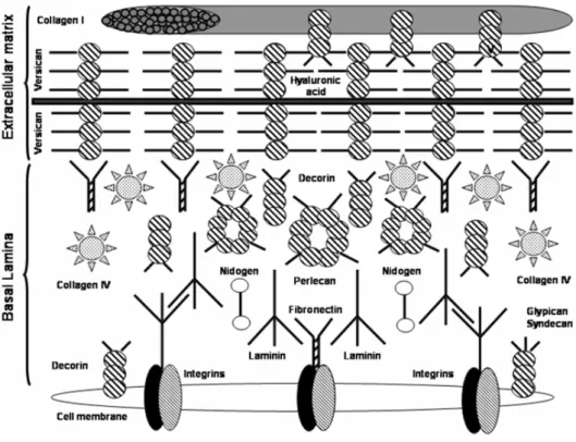
The basal lamina of pulmonary cells is extremely com-plex and it is composed of different molecules like la-minin, nidogen, and perlecan (Fig. 1) (Furuyama and Mochitate 2000).
basement membranes. The ‘sword’ tips can bind cell receptors, and the crosspieces allow laminin to bind to other laminin molecules. Other sites for nidogen and perlecan binding are also present in the molecule;
2) Nidogen, also known as entactin, is a dumbbell shaped 150 kDa sulfated glycoprotein, with three domains. Nidogen connects laminin to both the col-lagen layers and perlecan in the basal lamina;
3) Perlecan, which has a 400 kDa core protein with three heparan sulfate chains attached, is the pre-dominant proteoglycan in the basal lamina. It binds to collagen, laminin, nidogen, and other perlecan molecules (Nguyen et al. 2002).
On one side of the cell membrane, the basal lamina is linked to the cell by means of integrins (Tschumper-lin et al. 2004, Ingber 2006,), while on the other side it is liked to the ECM through links to collagen type IV. Integrins are adhesive membrane receptors that exist as heterodimers. They exhibit a “Velcro” effect: i.e. they have strength in numbers, but are individually easy to disrupt. They require Ca++or Mg++to bind, and their
job is to link the ECM to the cytoskeleton. Fibronectin reinforces the connections between the basal lamina with both the cell membrane and the other ECM components. It is likely that type IV collagen is chiefly responsible for the great tensile strength of the ECM and that the other molecules play important roles in consolidating the struc-ture of the ECM and its connections with overlying cells.
PULMONARY INTERSTITIAL FLUID DYNAMICS
The interstitial compartment of the lung is kept at a sub-atmospheric pressure in physiological conditions, with a low amount of extravascular water. In the lung, a rela-tively “dry” interstitial space allows a minimum thick-ness of the air-blood barrier to optimize gas diffusion. A rise in extravascular lung water may occur because of an increase in the pressure gradient across the microvas-cular barrier and/or by an increase in perm-porosity of the endothelial barrier.
The maintenance of the steady state interstitial fluid turnover results from the balance between several fac-tors, such as: 1) the transendothelial fluid and solutes
filtration, 2) the convective outflows into the lymphatic system, and 3) the mechanical and hydrophilic prop-erties of the solid elements of the ECM. The hydraulic pressure of the free liquid phase of the pulmonary inters-titium (Pi) reflects the dynamic situation that results
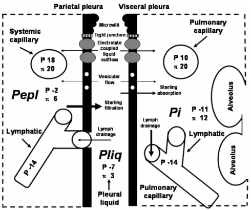
from the complex interaction between the factors stated above representing a key variable to understand the mechanisms which control lung fluid balance (Miseroc-chi et al. 1990, 1997, Negrini et al. 2003). The end-expiratory Pi is significantly lower than intra-pleural (Pli q)and extra-pleural(Pepl)liquid pressure,
indicat-ing that the lung parenchyma is relatively “dehydrated” compared to the other two tissue compartments (Fig. 2). ThePidistribution is not uniform within the lung
paren-chyma, decreasing by∼0.7 cmH2O/cm of lung height.
The gravity – dependent Pi distribution reflects: a) the
uneven mechanical stress developing in lung tissue at various lung heights, b) the inhomogeneous perfusion of the lung parenchyma, in analogy with that found in the pleural space, and c) the greater drainage of intersti-tial fluid into the lymphatic system in the lower regions. During inspiration, sustained by lowered pleural surface pressure, bothPli qandPi become more subatmospheric
(Miserocchi et al. 1990, Miserocchi and Negrini 1997, Negrini et al. 2003).
The fluid bulk flow (Jv) between the pulmonary
capillaries(c)and the surrounding interstitium(i)is de-scribed by the well-known Starling law:
Jv=Lp·S· [(Pc−Pi)−σ (πc−πi)] (1)
wherePeπare the hydraulic and colloidosmotic
pres-sures in the two compartments, Lp is the hydraulic
fil-tration coefficient of the pulmonary capillary endothelial layer,Sis the exchange surface area andσ is the reflec-tion coefficient of the endothelium for total proteins, a correction factor accounting for the protein to endothe-lial pore radii ratio. The factor in the square parenthe-sis gives the net pressure gradient across the membrane,
Pnet.
THE EXTRACELLULAR MATRIX AND LUNG EDEMA
Figure 3 shows examples of hydraulic(P)and
Fig. 1 – Links among the cell membrane, the basal lamina, and the extracellular matrix.
very negative in normal lungs, ensuring the maintenance of a net fluid filtration into the interstitial space, the early phase of interstitial edema is associated to an increase in
Piwith no significant change in interstitial fluid volume.
This phenomenon is caused by the low tissue compli-ance, provided by the tight structure of the fibrous ma-trix, and represents an important “tissue safety factor” to counteract further progression of pulmonary edema. With the worsening of the edema,Pi drops back to zero
and subsequently remains unchanged despite a marked increase in the wet-to-dry weight ratio of the lung. The worsening of edema occurs because of an increasingly high filtration rate, in spite of a progressive augment in
Pi and thus a reducedPnet. Hence, at least two
fac-tors interact to determine the development of pulmonary edema: 1) the loss of the tissue safety factor, which re-flects in an increase of tissue compliance and 2) a rise in microvascular permeability (Passi et al. 1998).
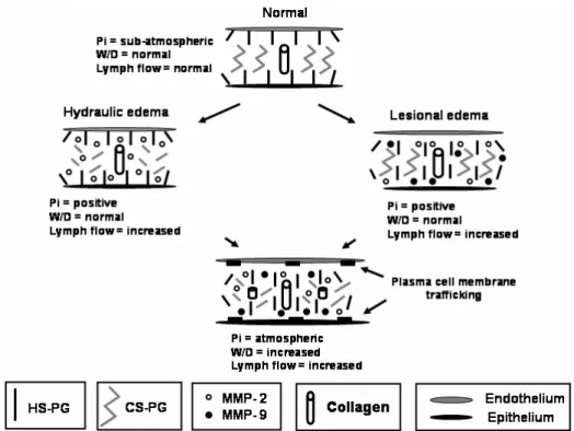
EFFECTS OF HYDRAULIC AND LESIONAL EDEMA ON ECM COMPONENTS
Interstitial lung edema led to some degree of disorgan-ization of the extracellular matrix, despite its strong
me-chanical resistance, particularly at the expense of pro-teoglycans (Negrini et al. 2003). These molecules are responsible for the structural integrity of pulmonary in-terstitium as they control fluid dynamics through their influence on microvascular permeability and tissue com-pliance. They also are involved in cell and cell-matrix interactions and in the cytokine network since they regulate the traffic of the molecules within the in-terstitial space and promote interactions (Roberts et al. 1997). To better understand the relationship between pulmonary interstitial pressure and the functional state of the matrix, hydraulic and lesional edema were devel-oped in anesthetized rabbits by intravenous injection of saline solution (0.5 ml·min-1·kg-1) (Miserocchi et al.
1993) and a single bolus of pancreatic elastase (200µg,
7 IU) (Negrini et al. 1998), respectively.
ex-Fig. 2 – Schematic representation of transpleural liquid flows (arrows) under physiological conditions. Mesothelial cells and adjoining interstitium, with embedded capillaries and lymphatics are shown. At the top, the mesothelial cell depicts the cell mechanisms involved in pleural liquid turnover [microvilli, vesicles, electrolyte transporters (grey circles)]. Arrow size is proportional to the corresponding flow magnitude. Hydraulic(P)and colloidosmotic(π ) pressures (cmH2O) are involved in the Starling balance. Pepl
(extrapleural interstitial pressure),Pli q(intrapleural pressure) andPi(lung interstitial pressure).
Fig. 3 – Hydraulic(P)and colloidosmotic(π )pressures of the microvasculature(cap)and surrounding interstitial space(i nt)under normal condition, during mild, and severe interstitial edema. Grey arrow represents transendothelial flow while black arrow represents trans-lymphatic flow. Hydraulic(P)and colloidosmotic(π )pressures are shown as cmH2O.
travascular water yielding tissue stress and weakness of PGs non-covalent bands with other matrix molecules or tissue metalloproteinases activation (Passi et al. 1999).
hep-aran sulfate proteoglycan lesion led to an augment in microvascular permeability (Negrini et al. 1998). Early in the course of hydraulic edema there was an increase in MMP-2, while in lesional edema both MMP-2 and MMP-9 were augmented (Passi et al. 1998).
Contrary to hydraulic edema, hypoxic pulmonary edema led to fragmentation of intermediate molecular weight heparin-sulfate PGs, such as perlecan of the base-ment membrane (Miserocchi et al. 2001b).
Recent data also suggest that the integrity of the heparan-sulfate proteoglycan components of the pulmo-nary ECM is required to maintain the three-dimensional architecture of the matrix itself, which in turn, ensures its mechanical response to increased fluid filtration (Ne-grini et al. 2006).
In an experimental model of fibrosis there was a marked reduction in the absolute volume of edema, in parallel with a reduction in lung volume, suggesting that fibrosis deeply affects the distribution of edema in the lung (Zwinkler et al. 1994).
EXTRACELLULAR MATRIX AND CELL PLASMA MEMBRANE
The response of epithelial cells to basement membrane deformation is affected by the extracellular matrix, plasma membrane, and cell cytoskeleton. Proteins that link the extracellular matrix with stress bearing cytos-keletal biopolymers play a pivotal role in mechanosens-ing and transduction (Geiger et al. 2001). They are as-sembled in so-called focal adhesion sites, plaques, or complexes. Focal adhesions are highly plastic structures and remodel in a force-dependent manner (Coppolino and Dedhar 2000). This allows adherent cells to probe the regional impedance of the surrounding scaffolding and, in turn, provides cues for directional control of locomotion and cell shape (Geiger et al. 2001). The most extensively studied adhesion receptors involved in mechanotransduction are the integrins. After ligand-induced activation, integrins transduce matrix-depend-ent intracellular biochemical signals. Other adhesion molecules such as cadherins are also increasingly recog-nized as playing important roles in mechanotransduction and as molecular targets of deformation induced impair-ments of the epithelial and endothelial barrier function (Cavanaugh et al. 2001).
The relationship between the ECM and plasma membrane has important implications in preventing lung injury. Cells experience plasma membrane stress failure when the matrix to which they adhere undergoes large deformations (Vlahakis and Hubmayr 2005). There are some adaptive responses of the lipid surface to deforming stress in alveolar epithelial cells:
1) the cell might unfold plasma membrane, thus pre-venting any increase in plasma membrane tension,
2) once unfolded, the plasma membrane stretches,
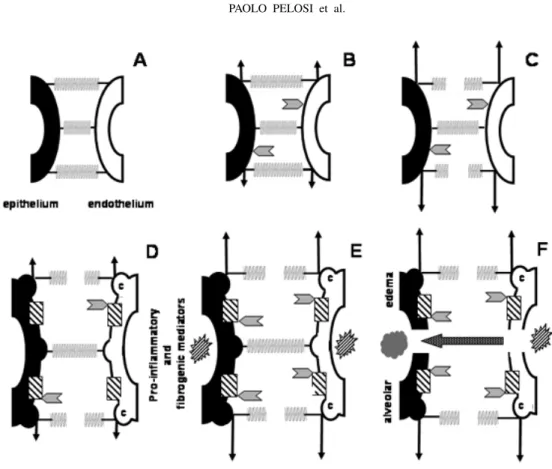
3) if these responses are overwhelmed, the plasma membrane might either undergo stress failure or further adapt its stress-bearing elements to prevent plasma membrane breaks (Fig. 5) (McNeil and Steinhardt 1997, Akinlaja and Sachs 1998, Liu et al. 1999, Tschumperlin et al. 2000).
Lipid trafficking to the cell surface is another adaptive mechanism in alveolar epithelial cells (Vlahakis et al. 2001) (Fig. 5). Several observations on normal and in-jured lungs raise interest in the molecule and pathway specificity of deformation triggered vesicular trafficking. When lungs suffer relatively mild forms of interstitial pulmonary edema, the lipid microdomains of lung cell surface membranes undergo a substantial reorganization (Palestini et al. 2003). The functional consequence of membrane remodeling, which is almost certainly accom-panied by changes in surface protein expression, remains to be explored.In vitro, such changes are associated with
changes in cell phenotype and by inference, changes in the cells susceptibility to mechanical injury. In endothe-lial cells, fibroblasts, and some epitheendothe-lial cells, sphin-golipids, cholesterol, and glycosylphosphatidylinisotol-anchored proteins appear to have a preferential associ-ation with 50- to 100-nm pits called caveolae (Razani et al. 2002) (Fig. 5). These structures play an impor-tant role in endocytosis processes and can be identified in type I alveolar epithelium cells (Gil 1983, Kasper et al. 1998). Caveolae are plasma membrane invaginations that may unfold when laterally stressed, and may be important not only for the mechanotransduction of de-formation-induced lipid trafficking, but also central for the maintenance of sublytic membrane tension.
phys-Fig. 4 – Changes in extracellular matrix during hydraulic and lesional edemas. Pi: interstitial pressure, W/D: wet-to-dry weight ratio, HS: Heparan sulfate, PG: Proteoglycan, CS: Chondroitin sulfate. MMP: Matrix Metalloproteasis. During hydraulic edema the prevalent lesion is the fragmentation of the CS, whilst in the lesional edema HS is damaged.
ical stimuli on endothelial cell surface. The increase in tissue forces at interstitial level is a consequence of in-terstitial fluid accumulation, particularly in the air-blood barrier as demonstrated by the increased thickness of the extracellular matrix. Interstitial edema causes a raise in interstitial fluid pressure from the physiological value of approximately –10 cmH2O up to∼5 cmH2O
(Mise-rocchi et al. 1993, 2001a). As capillary pressure remains essentially unchanged at∼10 cmH2O in mild interstitial
edema (Negrini 1995), the above condition should cause a substantial alteration in the transmural pressure. In fact, in physiological conditions, the transmural pressure is relatively high, averaging 20 cmH2O, with the
subatmo-spheric interstitial pressure pulling the basal side of the cell. Conversely, in edema the transmural pressure gra-dient is reduced to 5 cmH2O, and the positive interstitial
pressure pushes the basal side of the cell. Furthermore, parenchymal forces exerted at focal points where cell-matrix attachments occur are expected to increase as a consequence of swelling of the extracellular matrix.
Daffara and colleagues hypothesize a specific “sens-ing” function by lung cells resulting from a perturbation in cell-matrix interaction (Daffara et al. 2004). In this context, the cell-matrix interaction may differ according to the type of edema since differences were found in the sequential degradation of PGs family and in the interac-tion properties of PGs to some matrix components (Mis-erocchi et al. 2001a). Based on these considerations, Botto and coworkers described a differential response of lung endothelial cells to hydraulic and hypoxic pul-monary edema, inducing a specific cascade of cellular events from signaling-transduction to cellular functional attitude aimed at tissue repair (Botto et al. 2006).
CONCLUSIONS
perivas-Fig. 5 – Extracellular matrix (ECM) and cell response to increased lung interstitial pressure. Springs represent ECM components. Arrowheads: the intensity of interstitial pressure. Arrows: the intensity of stress on endothelium and epithelium. A: Normal pressure in the interstitium with no cell stress or ECM lesion. Panel B: Increased pressure in the interstitium, with limited cell stress since the ECM is intact. Panel C: During interstitial pressure increase, some components of ECM are ruptured, plasma membrane unfolded and stretched. Panel D: Adaptative cell response: the translocation of lipids from intracellular stores to the plasma membrane (dashed square). Note the presence of caveolae (c) in endothelial and epithelial cells. Panel E: Even though cells sensed the stress early, with further increase in interstitial pressure, pro-inflammatory and fibrogenic mediators were released. Panel F: Maintenance of increased stress associated with the effects of pro-inflammatory and fibrogenic mediators contribute to the fragmentation of extracellular matrix components and epithelial and endothelial cell rupture resulting in edema.
cular and interstitial sieve in relation to plasma proteins, thus modulating both interstitial protein concentration and trans-endothelial fluid filtration; b) a mechanical sup-port to lymphatic vessels sustaining and modulating their draining function, and c) a rigid three dimension low compliant scaffold opposing fluid accumulation into in-terstitial space. Fragmentation of proteoglycans induced by different stimuli such as: fluid overload, exposure to proteolytic or inflammatory agents, hypoxic or hyper-oxic gas mixtures, or increased tissue strain/stress, result in a progressive loosening of proteoglycans intermolec-ular connections with other ECM components. The loss
both the endothelial and the epithelial barrier and pos-sibly of the interstitial matrix.
ACKNOWLEDGMENTS
We would like to express our gratitude to Mr. Andre Benedito da Silva, and Mrs. Jaqueline Lima do Nasci-mento for their skillful technical assistance. Supported by: Programa de Apoio a Núcleos de Excelência – Fun-dação Carlos Chagas Filho de Amparo à Pesquisa no Es-tado do Rio de Janeiro (PRONEX-FAPERJ), Conselho Nacional de Desenvolvimento Científico e Tecnológico (CNPq), Fundação Carlos Chagas Filho de Amparo à Pesquisa no Estado do Rio de Janeiro State (FAPERJ).
RESUMO
A matriz extracelular é um aglomerado tridimensional de macromoléculas composta por: fibras colágenas (principal-mente, tipos I e III), elastina, glicosaminoglicanos e proteogli-canos. No pulmão, a matriz extracelular tem várias funções, tais como: 1) promover estressetensile elasticidade tecidual, 2) contribuir para a manutenção da dinâmica de fluidos no interstício, 3) propiciar efetiva troca gasosa, 4) controlar a função celular através de sua ligação com fatores de cresci-mento, quimiocinas, citocinas e interação com receptores de su-perfície, e 5) remodelamento e reparo tecidual. A fragmentação e a desorganização da matriz extracelular pode acarretar edema intersticial e, eventualmente, edema alveolar grave. Logo, quando há aumento da filtração microvascular ocorre rápido remodelamento da matriz por ativação de proteases. Destarte, a deposição de fibras colágenas reduz a complacência intersti-cial limitando o edema. Em conclusão, modificações na matriz extracelular podem afetar a formação e distribuição do edema no pulmão.
Palavras-chave:colágeno, glicosaminoglicanos, proteoglica-nos, pressão intersticial.
REFERENCES
AKINLAJAJ ANDSACHS F. 1998. The breakdown of cell membranes by electrical and mechanical stress. Biophys J 75: 247–254.
BOTTO L, BERETTA E, DAFFARA R, MISEROCCHI G
ANDPALESTINIP. 2006. Biochemical and morpholog-ical changes in endothelial cells in response to hypoxic interstitial edema. Respir Res 7: 7.
CANTOR JO, SHTEYNGART B, CERRETA JM, LIU M,
ARMAND G AND TURINO GM. 2000. The effect of hyaluronan on elastic fiber injury in vitro and elastase-induced airspace enlargement in vivo. Proc Soc Exp Biol Med 225: 65–71.
CAVANAUGHJRKJ, OSWARIJANDMARGULIESSS. 2001. Role of stretch on tight junction structure in alveolar ep-ithelial cells. Am J Respir Cell Mol Biol 25: 584–591. COPPOLINOMGANDDEDHARS. 2000. Bi-directional
sig-nal transduction by integrin receptors. Int J Biochem Cell Biol 32: 171–188.
DAFFARAR, BOTTOL, BERETTAE, CONFORTIE, FAINI A, PALESTINIPANDMISEROCCHIG. 2004. Endothe-lial cells as early sensors of pulmonary interstitial edema. J Appl Physiol 97: 1575–1583.
ELKINGTONPTANDFRIEDLANDJS. 2006. Matrix metal-loproteinases in destructive pulmonary pathology. Thorax 61: 259–266.
FURUYAMA A ANDMOCHITATE K. 2000. Assembly of the exogenous extracellular matrix during basement mem-brane formation by alveolar epithelial cells in vitro. J Cell Sci 113: 859–868.
GEIGER B, BERSHADSKY A, PANKOV R AND YAMADA
KM. 2001. Transmembrane crosstalk between extracellu-lar matrix-cytoskeleton crosstalk. Nat Rev Mol Cell Biol 2: 793–805.
GILJ. 1983. Number and distribution of plasmalemmal vesi-cles in the lung. Fed Proc 42: 2414–2418.
GREENLEEKJ, WERBZANDKHERADMANDF. 2007. Ma-trix metalloproteinases in lung: multiple, multifarious, and multifaceted. Physiol Rev 87: 69–98.
INGBERDE. 2006. Cellular mechanotransduction: putting all the pieces together again. FASEB J 20: 811–827. IOZZO RV ANDMURDOCH AD. 1996. Proteoglycans of
the extracellular environment. Clues from the gene and protein side offer novel perspective in molecular diversity and function. FASEB J 10: 598–614.
JOHNSON Z, PROUDFOOT AANDHANDEL T. 2005. In-teraction of chemokines and glycosaminoglycans: A new twist in the regulation of chemokine function with oppor-tunities for therapeutic interventions. Cytokine Growth Factor Rev 16: 625–636.
KASPER M, REIMANN T, HEMPEL U, WENZEL KW, BIERHAUSA, SCHUHD, DIMMERV, HAROSKEGAND MULLERM. 1998. Loss of caveolin expression in type I pneumocytes as an indicator of subcellular alterations dur-ing lung fibrogenesis. Histochem Cell Biol 109: 41–48. LANCHOUJ, CORBELMANDTANGUYM. 2003. Imbalance
between matrix metalloproteinases (MMP-9 and MMP-2) and tissue inhibitors of metalloproteinases (TIMP-1 and TIMP-2) in acute respiratory distress syndrome patients. Crit Care Med 31: 536–542.
LIY, RAHMANIANM, WIDSTROMC, LEPPERDINGERG, FROSTGIANDHELDINP. 2000. Irradiation induced ex-pression of hyaluronan (HA) synthase 2 and hyaluronidase 2 genes in rat lung tissue accompanies active turnover of HA and induction of types I and III collagen gene expres-sion. Am J Resp Cell Mol Biol 23: 411–418.
LIU M, TANSWELL AK ANDPOST M. 1999. Mechanical force-induced signal transduction in lung cells. Am J Phys-iol Lung Cell Mol PhysPhys-iol 277: L667–L683.
MCNEILPLANDSTEINHARDTRA. 1997. Loss, restoration, and maintenance of plasma membrane integrity. J Cell Biol 137: 1–4.
MERCERRRANDCRAPOJD. 1990. Spatial distribution of collagen and elastin fibres in the lungs. J Appl Physiol 69: 756–765.
MISEROCCHIGANDNEGRINID. 1997. Pleural space: pres-sures and fluid dynamics. In: CRYSTALRG, WESTJB, WEIBELERANDBARNESPJ (Eds), The Lung. 2nded., Philadelphia: Lippincott-Raven, p. 1217–1225.
MISEROCCHIG, NEGRINIDANDGONANOC. 1990. Direct measurements of interstitial pulmonary pressure in in situ lung with intact pleural space. J Appl Physiol 69: 2168– 2174.
MISEROCCHIG, NEGRINID, DELFABBROMAND VEN-TUROLID. 1993. Pulmonary interstitial pressure in intact in situ lung: The transition to interstitial edema. J Appl Physiol 74: 1171–1177.
MISEROCCHIG, NEGRINID, PASSIAANDDELUCAG. 2001a. Development of lung edema: interstitial fluid dy-namics and molecular structure. News Physiol Sci 16: 66–71.
MISEROCCHIG, PASSI A, NEGRINI D, DEL FABBRO M ANDDELUCAG. 2001b. Pulmonary interstitial pressure and tissue matrix structure in acute hypoxia. Am J Physiol Lung Cell Mol Physiol 280: L881–L887.
MONTES GS. 1996. Structural biology of the fibres of the collagenous and elastic systems. Cell Biol Int 20: 15–27.
NEGRI EM, MONTES GS, SALDIVA PHN AND
CAPPE-LOZZIVL. 2000. Architectural remodelling in acute and chronic interstitial lung disease: fibrosis or fibroelastosis Histopathology 37: 393–401.
NEGRINID. 1995. Pulmonary microvascular pressure profile during development of hydrostatic edema. Microcircula-tion 2: 173–180.
NEGRINID, PASSIA, DELUCAGANDMISEROCCHIG. 1998. Proteoglycan involvement during development of lesional pulmonary edema. Am J Physiol 274: L203– L211.
NEGRINID, PASSIA, DELUCAGANDMISEROCCHIG. 2003. Matrix proteoglycans in development of pulmonary edema. In: GARGHG, ROUGHLEYPJANDHALESCA (Eds), Proteoglycans in lung disease, New York: Marcel Dekker, p. 143–168.
NEGRINID, TENSTADO, PASSIAANDWIIGH. 2006. Dif-ferential degradation of matrix proteoglycans and edema development in rabbit lung. Am J Physiol Lung Cell Mol Physiol 290: L470–L477.
NGUYEN NM, BAIY, MOCHITATE KANDSENIOR RM.
2002. Laminin alpha-chain expression and basement membrane formation by MLE-15 respiratory epithelial cells. Am J Physiol Lung Cell Mol Physiol 282: L1004– L1011.
NOBLEPWANDJIANGD. 2006. Matrix regulation of lung injury, inflammation, and repair: the role of innate immu-nity. Proc Am Thorac Soc 3: 401–404.
PALESTINIP, CALVIC, CONFORTIE, DAFFARAR, BOTTO LANDMISEROCCHIG. 2003. Compositional changes in lipid microdomains of air-blood barrier plasma mem-branes in pulmonary interstitial edema. J Appl Physiol 95: 1446–1452.
PASSI A, NEGRINI D, ALBERTINI R, DE LUCA G AND MISEROCCHI G. 1998. Involvement of lung interstitial proteoglycans in development of hydraulic and elastase induced edema. Am J Physiol 275: L631–L635. PASSIA, NEGRINID, ALBERTINIR, MISEROCCHIGAND
DELUCAG. 1999. The sensitivity of versican from rabbit lung to gelatinase A (MMP-2) and B (MMP-9) and its involvement in the development of hydraulic lung edema. FEBS Lett 456: 93–96.
RAZANIB, WOODMANSEANDLISANTIMP. 2002. Cave-olae: from cell biology to animal physiology. Pharmacol Rev 54: 431–467.
ANDBARNESPJ (Eds), The Lung. 2nded., Philadelphia: Lippincott-Raven, p. 757–767.
ROCCO PRM, NEGRI EM, KURTZ PM, VASCONCELLOS
FP, SILVAGH, CAPELOZZIVL, ROMEROPVANDZIN WA. 2001. Lung tissue mechanics and extracellular ma-trix in acute lung injury. Am J Respir Crit Care Med 164: 1067–1071.
ROCCO PRM, SOUZA AB, FAFFE DS, PASSARO CP,
SANTOSFB, NEGRIEM, LIMAJGM, CONTADORRS, CAPELOZZIVLANDZINWA. 2003. Effect of corticos-teroid on lung parenchyma remodeling at an early phase of acute lung injury. Am J Respir Crit Care Med 168: 677–684.
RUOSSSJ, GOLDWMANDCAUGHEYGH. 1991. Mast cell exocytosis: evidence that granule proteoglycan processing is not coupled to degranulation. Biochem Biophys Res Commun 179: 140–146.
SANTOS FB, NAGATO LKS, BOECHEM NM, NEGRI
EM, GUIMARÃESA, CAPELOZZIVL, FAFFEDS, ZIN WAANDROCCOPRM. 2006. Time course of lung par-enchyma remodeling in pulmonary and extrapulmonary acute lung injury. J Appl Physiol 100: 98–106.
SOUZA FERNANDES AB, PELOSI P AND ROCCO PRM.
2006. Bench-to-bedside review: The role of glycosamino-glycans in respiratory disease. Critical Care 10: 237. TAMMI MI, DAY AJAND TURLEY EA. 2002.
Hyaluro-nan and homeostasis: a balancing act. J Biol Chem 277: 4581–4584.
TSCHUMPERLINDJ, OSWARIJANDMARGULIESSS. 2000. Deformation- induced injury of alveolar epithelial cells. Am J Respir Crit Care Med 162: 357–362.
TSCHUMPERLIN DJ ET AL. 2004. Mechanotransduction through growth-factor shedding into the extracellular space. Nature 429: 83–86.
TUMOVAS, WOODSAANDCOUCHMANJR. 2000. Hep-aran sulfate proteoglycans on the cell surface: versatile coordinators of cellular functions. Int J Biochem Cell Biol 32: 269–288.
VLAHAKISNEANDHUBMAYR RD. 2005. Cellular Stress Failure in Ventilator-injured lungs. Am J Respir Crit Care Med 171: 1328–1342.
VLAHAKISNE, SCHROEDERMA, PAGANOREAND
HUB-MAYRRD. 2001. Deformation-induced lipid trafficking in alveolar epithelial cells. Am J Physiol Lung Cell Mol Physiol 280: L938–L946.
WEST JBANDMATHIEU-COSTELLOO. 1999. Structure, strength, failure, and remodeling of the pulmonary blood-gas barrier. Annu Rev Physiol 61: 543–557.
WHITELOCKJMANDIOZZORV. 2005. Heparan sulfate: a complex polymer charged with biological activity. Chem Rev 105: 2745–2764.
YURCHENKOPDANDSCHITTNYJC. 1990. Molecular ar-chitecture of basement membrane. FASEB J 4: 1577– 1590.
ZHAOJ, SIMEPJ, BRINGAS JRP, GAULDIEJAND WAR-BURTON D. 1999. Adenovirus-mediated decorin gene transfer prevents TGF-beta-induced inhibition of lung morphogenesis. Am J Physiol Lung Mol Cell Physiol 277: L412–L422.