Shading affecting growth and leaf structure of Ocimum selloi
Bragantia, Campinas, v.69, n.2, p.349-359, 2010 349
EFFECTS OF COLOURED SHADE NETTING ON THE VEGETATIVE
DEVELOPMENT AND LEAF STRUCTURE OF
OCIMUM SELLOI
(
1)
LARISSA CORRÊA DO BOMFIM COSTA (2*), JOSÉ EDUARDO BRASIL PEREIRA PINTO (3), EVARISTO MAURO DE CASTRO (3), EDUARDO ALVES (3),
SUZAN KELLY VILELA BERTOLUCCI (3), LOUISE FERREIRA ROSAL (3)
ABSTRACT
The objective of this work was to investigate the effects of shading on the growth and development of the medicinal plant Ocimum selloi after 90 days. Seedlings were maintained under three different light treatments, namely, full sunlight vs coloured shade treatments provided by red and blue ChromatiNet® 50% coloured netting. After 90 days of cultivation, height, stem diameter, petiole length, total leaf area (TLA), specific leaf area (SLA), leaf area ratio (LAR) and leaf weight ratio (LWR) of each plant hare measured. Microscopic examination of leaf sections were conducted in order to determine the leaf blade thicknesses, together with the size and density of the stomata, the number and area of chloroplasts, and chloroplast starch grains present in the palisade parenchyma cells. Although coloured shade-grown plants were taller in comparison with those grown in full sunlight, total dry biomass together with root, stem and leaf tissue biomasses were lower. Plants exhibited phenotypic plasticity, as demonstrated by the different TLA, SLA, LAR and LWR values that were recorded under the various light treatments. Stomatal density and leaf thickness was increased in plants maintained in full sunlight owing to the expansion of the abaxial epidermis and the spongy parenchyma. Chloroplasts were more numerous and larger in plants grown under shading, whilst the accumulation of chloroplastic starch grains was greater in plants grown under red shading or in full sunlight.
Key words: light quality and intensity; plastic coloured netting; plant anatomy; leaf morphology and structure.
RESUMO
EFEITO DO SOMBREAMENTO COM TELAS COLORIDAS NO DESENVOLVIMENTO VEGETATIVO E ESTRUTURA FOLIAR DE Ocimum selloi
O objetivo deste trabalho foi verificar o efeito do sombreamento de Ocimum selloi com telas coloridas do tipo Cromatinet® (50%) vermelha, azul e a condição natural de pleno sol (sem tela) sobre as características biométricas, fisiológicas, anatômicas e ultraestruturais da planta após 90 dias. As plantas nas telas coloridas tiveram maior crescimento em altura, mas o maior acúmulo de biomassa seca total e particionada e relação raiz: parte aérea (R:PA) ocorreram na condição de pleno sol. A espécie proporcionou plasticidade fenotípica, verificada pelos ajustes das variáveis área foliar total (AFT), área foliar específica (AFE), razão de área foliar (RAF) e razão de massa foliar (RPF) em função dos tratamentos. Ocorreu maior espessamento foliar na condição de pleno sol pelo somatório do maior espessamento da epiderme abaxial e do parênquima esponjoso. A densidade estomática foi maior nas plantas a pleno sol. O número de cloroplastos e seu tamanho foi maior nas telas coloridas, enquanto o maior acúmulo de amido ocorreu a pleno sol e tela vermelha.
Palavras-chave: planta medicinal, telas coloridas, qualidade de luz, ultraestrutura foliar.
(1) Receipt for publication in August 27, 2008 and accept in December 12, 2009.
(2) Departamento de Ciências Biológicas, Universidade Estadual de Santa Cruz, 45662-900 Ilhéus (BA) Brazil. E-mail:
[email protected] (*) Autora correspondente.
1. INTRODUCTION
Plants react to changes that occur in the spectrum of electromagnetic radiation to which they are exposed through alterations in morphology and physiological functions that result in adaptation to different environmental conditions (KASPERBBAUER and HAMILTON, 1984). Such alterations are mediated by pigments, known as phytochromes, which have absorption peaks in the red and blue/ultraviolet regions of the spectrum (LI et al., 2000). These photoreceptors are able to detect variations in light composition and induce photomorphogenetic responses, either in vivo or in vitro (KIM et al., 2004; MA C E D O et al., 2004) that influence growth and development (LE E et al., 1997; LI et al., 2000), morphology (STUEFER and HUBER, 1998), leaf and stem anatomy (SCHUERGER et al., 1997; LEE et al., 2000), distribution of photosynthetic products (KASPERBBAUER, 1987; BROWN et al., 1995), photosynthetic efficiency (KA S P E R B B A U E R and PE A S L E E, 1973) and chemical composition (MACEDO et al., 2004).
Specific leaf area (SLA) values depend on the quality of light and vary amongst species. Increases in SLA and reductions in the thickness of the leaf blade under shading conditions are common alterations that confer functional advantages to plants grown in environments with low light intensity (BUISSON and LEE, 1993). Thus it is important to know the anatomical and ultra-structural leaf changes that may be somehow related with biomass production.
However, whilst the effects of light quality on plants are well known, the response of different species to light management is variable (KIM et al., 2004), and it is clearly important to treat plants with the correct type of light filters (MCMAHON and KELLY, 1995), especially medicinal plants with economical interests.
A common light quality management is the supplementation with artificial light sources (BROWN et al., 1995) through the use of coloured plastic films (OYAERT et al., 1999), spectral filters (RAJAPAKSE et al., 1992) and reflective coloured mulches (LOUGHRIN and KASPERBBAUER, 2001) to induce physiological responses in plants. Cromatinet® coloured netting modifies the spectrum of the incident radiation in the visible region and enriches the relative content of scattered light such that transmittance of light by the blue netting is in the 400-540 nm region, whilst that of the red netting is in the 590-760 nm region (OREN-SHAMIR et al., 2001). Although the red:far red ratio (R:FR), which is the main regulatory factor leading to a phytochrome response, is not greatly modified by the use of such nets, the blue:red ratio (B:R) is enhanced by blue netting and reduced by red netting (SHAHAK et al.,
2004). Coloured shade netting not only exhibit special optical properties that allow the control of light, but also have the advantage of influencing the microclimate to which the plant is exposed to (OREN -SHAMIR et al., 2001) and offer physical protection against excessive radiation, insect pests and environmental changes (SHAHAK et al., 2004).
Ocimum selloi Benth. (Lamiaceae), known as “elixir-paregórico”, “alfavaquinha” and “atroveran”, is a perennial shrub, native from the southern and south-eastern regions of Brazil. Leaf infusions are widely used in popular medicine as anti-diarrhoeic, anti-spasmodic and anti-inflammatory agents, and the plant is also valued as an insect repellent. Various chemotypes of O. selloi have been reported in which the major component of the essential oil, namely methyl chavicol, is accompanied by either methyl eugenol (MARTINS, 1998) or trans-anethol (MORAES et al., 2002), which are commonly found in the leaves of the specie.
Based on previous positive results of coloured shade netting benefits to plants development, we investigated the hypothesis that the light quality of the red- and blue-shade spectrum, rather than the light intensity (full sunlight vs. shade conditions), is responsible for the major leaf biomass components of
O. selloi, related to morphological, physiological and anatomical information.
The objective of the present study was to determine the effects of coloured shade netting on the growth after 90 days and leaf anatomy and ultra-structure of O. selloi with the aim of optimising biomass yields of this important medicinal plant.
2. MATERIALS AND METHODS
The plantlets with 10 cm of high of O. selloi
Shading affecting growth and leaf structure of Ocimum selloi
Bragantia, Campinas, v.69, n.2, p.349-359, 2010 351
The biometric analyses of plants were performed 90 days (flower phase) after the experiment was set. Plant height, stem diameter and petiole length of leaves at the second node below the main inflorescence were determined.
Total leaf area (TLA; cm²) was measured using a Li-Cor LI–3100 leaf area meter, from which the specific leaf area (SLA; cm² g-1), leaf area ratio (LAR; cm² g-1), and leaf weight ratio (LWR; g g-1) were calculated according to BENINCASA (2003).
Following the determination of leaf growth parameters, the roots, stems, leaves and inflorescences of each plant were separated, packed into individual paper bags, and dried in a fan oven at 70 ºC to constant weight. The root and above ground biomass was determined and the ratio of root: shoot tissues (R:S) was calculated.
Leaves of ten plants grown under different light conditions were examined microscopically. Mature leaves located at the second node below the main inflorescence were excised and fixed with formalin-acetic acid-alcohol solution for 48 h, followed by 70% ethanol solution.
Paradermic sections of leaf samples obtained from the median region of the leaf blade were free-hand cut and stained with 1% safranin solution. Leaf sections were observed under an Olympus (São Paulo, Brazil) model BX41 light microscope equipped with a Cannon® (São Paulo, Brazil) PowerShot A510 digital camera.
Scanning electron microscopy (SEM) was used to evaluate the thickness of the adaxial and abaxial epidermis, thickness of the palisade and spongy parenchyma, thickness of the leaf blade (transversal sections) and the size and density of stomata.
Leaf sections were cut, fixed with modified Karnovsky solution [2.5% glutaraldehyde and 2.5% formaldehyde in 0.05 M sodium cacodylate buffer (pH 7.2) containing 0.001 M calcium chloride], washed with sodium cacodylate buffer, immersed in 30% glycerol and then in liquid nitrogen. The sections were then placed onto a pre-cooled metallic surface, parted using a surgical blade, fixed with 1% osmium tetroxide, dehydrated in an acetone gradient and dried with a Bal-Tec (Balzers, Liechtenstein) model CPD 030 critical point drier. Sections were then sputtered with gold using a Bal-Tec model SCD 050 evaporator. Microscopic observations and electron micrographs were made using a Nano Technology Systems (Carl Zeiss, Oberkochen, Germany) model Evo® 40 VP SEM at an accelerating voltage of 20 kV.
Transmission electron microscopy (TEM) was employed to determine the number and area of
chloroplasts and starch grains present in the palisade parenchyma cells. Sections were fixed with modified Karnovsky solution as described for SEM, washed with sodium cacodylate buffer and post-fixed with 1% osmium tetroxide. The sections were transferred to 0.5% uranyl acetate solution, dehydrated using a gradient of acetone, infiltrated with an increasing gradient of acetone/Spurr’s resin mixture and placed into moulds. Following polymerisation for 24 hours in an oven at 70 ºC, the resin blocks were trimmed and cut into ultra thin slices (< 100 nm) using a Reichert Jung (Heidelberg, Germany) microtome equipped with a diamond blade. Slices were contrasted with 2% uranyl acetate followed by 1%lead acetate for 3 min, and examined using a Zeiss EM 109 microscope.
Digital images were captured using AnalySIS image software (Soft Imaging System GmbH, Münster, Germany) and analysed with a WinCELL Pro software (Regent Instruments, (Sainte-Foy, Québec, Canada).
A totally randomised design was adopted for the investigation, which included three different treatments, seven pots (experimental units) and two plants per repetition. Data were analysed by analysis of variance (one-way ANOVA) and meanvalues were compared using the Tukey’s test. Differences were considered significant at P< 0.05.
3. RESULTS AND DISCUSSION
The average values of light intensity obtained during the study time were 1500 µmol m-2 s-1 (full sunlight), 650 µmol m-2 s-1 (under 50% blue shading) and 690 µmol m-2 s-1 (under 50% red shading). The plant height was not different between red- and blue-shadding; however, shade-grown plants were significant taller than those grown under direct full sunlight (Table 1). Moreover, the management of light intensity had no effect on the diameter of stems or the length of petioles. Since the stem is characterized by house the vascular tissue, stem diameter is correlated directly with the capacity for transporting water and carbohydrates (ST U E F E R and HU B E R, 1998), and indirectly with the capacity for storing these metabolites (LEE et al., 1997).
Table 1
. Effects of different light regimes on the linear growth and on the production of biomass in
Ocimum selloi plants ( 1) Treatments Height Diameter of Length Dry mass Root:shoot stems of petioles Total Roots Stems Leaves Inflorescences ratio cm ( 2 ) g Full sunlight
60.80 ± 1.5
B
1.36 ± 0.04
A
1.65 ± 0.05
A
153.03 ± 7.2
A
65.63 ± 5.9
A
54.84 ± 1.5
A
24.86 ± 0.6
A
7.76 ± 0.3
B
0.83 ± 0.11
A
Red shading
72.60 ± 1.8
A
1.31 ± 0.08
A
1.82 ± 0.11
A
98.78 ± 2.9
B
28.86 ± 2.3
B
39.84 ± 0.9
B
19.52 ± 0.4
B
10.63 ± 0.6
A
0.42 ± 0.03
B
Blue shading
70.20 ± 1.5
A
1.13 ± 0.07
A
1.83 ± 0.09
A
53.72 ± 4.3
C
11.16 ± 1.8
C
21.21 ± 1.5
C
11.24 ± 0.8
C
10.19 ± 0.7
A
0.25 ± 0.02
B
Coefficient of variation (%)
7.61 16.87 15.87 15.94 34.41 11.09 11.10 19.55 54.87 ( 1 )
Results are expressed as mean values ± standard error (
n
= 7). Within a column, mean values bearing different superscript letters were significantly different (Tukey’s test; p<0.05).
(
2 ) Measurements were performed at the root-stem transition zone (the crown).
In general, the vegetative growth of plants maintained under low light intensity is optimised in order to increase light interception and to facilitate photosynthetic processes (KA S P E R B B A U E R, 1987). Therefore, the overall improvement in the growth of O. selloi plants maintained under full sunlight compared with those under shaded conditions can be explained by the lower light intensity associated with the latter.
The striking reduction in total dry biomass observed in plants grown under blue shading (Table 1) may have resulted from a decrease in the rate of CO2 assimilation (OYAERT et al., 1999). Blue light, either alone or in combination with other types of visible radiation, is known to be a critical factor in the photomorphogenic response of plants (RAJAPAKSE et al., 1992; BROWN et al., 1995), and it has recently been reported (Kim et al., 2004) that there is a reduction in the net photosynthetic rate of plants submitted to blue and blue-far red light treatment.
Although absorbance of light by chlorophyll is maximal in the red portion of the spectrum, biomass production in O. selloi was not increased under red shading. This can be explained by the fact that normal plant growth and development can be sustained only by balancing amounts of complementary types of radiation such that plants grown under red shading require supplementation with blue radiation (BROWN et al., 1995).
As shown in Table 1, the root:shoot ratio (R:S) was higher in plants cultivated in full sunlight compared to that of plants cultivated under coloured shading, indicating that the distribution and accumulation of photosynthetic products in the roots is significantly enhanced by intense sunlight. Shading treatments diminished the storage of photosynthetic products in the roots, but the effects of red and blue shading on such accumulation were similar. Normally, environmental variations modify the R:S ratio by altering the distribution of photosynthetic products within the plant (FERREIRA et al., 2004). Although red and far red radiation may control the plant phytochromes and influence the partition between roots and aerial tissues under controlled conditions (KASPERBBAUER, 1987), the alteration in the R:S ratio observed in O. selloi could be attributed to the variation in light intensity alone.
Shading affecting growth and leaf structure of Ocimum selloi
Bragantia, Campinas, v.69, n.2, p.349-359, 2010 353
TLA decreased in the order red shading > full sunlight > blue shading, whilst both SLA and LAR decreased in the order blue shading > red shading > full sunlight. Regarding LWR, the effects of blue and red shading were similar, but the average values were significantly higher than that produced by full sunlight (Table 2).
Considering that leaf biomass was maximized when plants were maintained under full sunlight, it is likely that the increased TLA exhibited by plants maintained under red shading was due to the expansion of individual leaves, probably influenced by the smaller R:B (red:blue) light ratio. A similar finding has been reported for O. basilicum, specimens of which produced greater leaf area and higher fresh biomass yields when cultivated in soil covered with red plastic mulches
(LOUGHRIN and KASPERBBAUER, 2001). In plants maintained under blue shading, for which the B:R light ratio was the highest, leaf expansion was inhibited indicating that O. selloi is not tolerant towards this wavelength. According to LI et al. (2000), reduction in leaf size results in a reduction in photosynthetic area and consequently in dry biomass yield. The inhibitory effect of blue shading on the vegetative growth of O. selloi has been reported by some authors (OREN-SHAMIR et al., 2001; SHAHAK et al., 2004), whilst reductions in TLA have been observed for
Chrysanthemum (OYAERT et al., 1999) and Capsicum (LI et al., 2000) spp. grown under photoselective films and copper sulphate filters. Although variations in leaf area are often associated with variations in the levels of sunlight (GONÇALVES, 2001), in O. selloi this parameter was strongly influenced by the quality of light.
Table 2. Effects of different light regimes on leaf development in Ocimum selloi plants(1)
Treatments TLA (2) SLA (2) LAR (2) LWR (2)
cm² cm².g-1 g g-1
Full sunlight 3592.45 ± 101.9B 154.59 ± 2.4C 23.81 ± 0.9C 0.16 ± 0.005B Red shading 4064.96 ± 155.2A 217.25 ± 3.3B 41.35 ± 1.7B 0.20 ± 0.006A Blue shading 2401.13 ± 117.6C 233.63 ± 2.8A 48.69 ± 1.9A 0.21 ± 0.004A
Coefficient of variation (%) 11.97 4.49 13.19 9.42
(1)Results are expressed as mean values ± standard error (n = 7). Within a column, mean values bearing different superscript letters were significantly different (Tukey’s test; p<0.05).
(2) TLA - total leaf area; SLA - specif leaf area; LAR - leaf area ratio; LWR – leaf weight ratio.
The SLA is inversely related to the thickness of the leaf blade, and alterations in its value reflect changes in leaf structure. Whilst SLA values depend on the quality of light and vary amongst species, increases in SLA and reductions in the thickness of the leaf blade under shading conditions are common alterations that confer functional advantages to plants grown in environments with low light intensity (BUISSON and LEE, 1993). This was demonstrated to be the case for O. selloi plants maintained under red or blue shading, the leaves of which were significantly thinner than those of plants cultivated in full sunlight. For blue-grown plants, however, the larger SLA value was compensated by a reduced TLA resulting in a smaller leaf biomass. Species of Chrysanthemum and
Capsicum maintained under photoselective plastic also exhibited reduced SLA values (LI et al., 2000), whilst in Hopea spp. the quality of light was reported to have no effect on SLA (LEE et al., 1997),
LAR reflects the magnitude of the photosynthetic area (BENINCASA, 2003). The increased LAR values obtained for O. selloi grown under blue shading indicate that these plants required a larger leaf area for the production of one gram of dry matter than those grown under red shading or in full sunlight. According to
BENINCASA (2003), alterations in LAR values may be understood, within certain limits, as the capacity of a species to adapt to different light conditions.
O. selloi plants grown under shading presented larger LWR values than those grown under full sunlight, demonstrating that in the former situation some of the photosynthetic products were retained in the leaves, whilst in the latter these products were more efficiently exported to other tissues. In fact, the capacity to export photo-assimilates from leaves to other parts may be a genetic characteristic that can be influenced by environmental changes, including the spectral quality of light (BENINCASA, 2003).
Shading affecting growth and leaf structure of
Ocimum selloi
Bragantia, Campinas, v.69, n.2, p.349-359, 2010
355
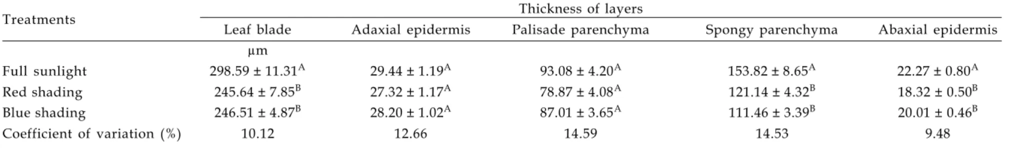
Table 3. Effects of different light regimes on the structure of Ocimum selloi leaves(1)
Treatments Thickness of layers
Leaf blade Adaxial epidermis Palisade parenchyma Spongy parenchyma Abaxial epidermis
µm
Full sunlight 298.59 ± 11.31A 29.44 ± 1.19A 93.08 ± 4.20A 153.82 ± 8.65A 22.27 ± 0.80A
Red shading 245.64 ± 7.85B 27.32 ± 1.17A 78.87 ± 4.08A 121.14 ± 4.32B 18.32 ± 0.50B
Blue shading 246.51 ± 4.87B 28.20 ± 1.02A 87.01 ± 3.65A 111.46 ± 3.39B 20.01 ± 0.46B
Coefficient of variation (%) 10.12 12.66 14.59 14.53 9.48
(1)Results are expressed as mean values ± standard error (n = 10). Within a column, mean values bearing different superscript letters were significantly different (Tukey’s test;
(p<0.05).
Table 4. Effects of different light regimes on the size and density of stomata in Ocimum selloi leaves(1)
Size of stomata
Density of stomata
Treatments Abaxial epidermis Adaxial epidermis
Abaxial epidermis Adaxial epidermis
Polar diameter Equatorial diameter Polar diameter Equatorial diameter
µm number per mm2
Full sunlight 28.13 ± 0.49A 17.22 ± 0.5B 29.98 ± 0.27A 17.99 ± 0.26B 283.84 ± 18.22A 123.02 ± 6.35A
Red shading (2) 28.87 ± 0.63A 19.14 ± 0.48A 30.56 ± 0.56A 17.84 ± 0.28B 232.38 ± 6.72B 105.33 ± 5.28AB
Blue shading 28.47 ± 0.42A 18.71 ± 0.4AB 31.08 ± 0.37A 19.53 ± 0.19A 201.82 ± 11.21B 99.71 ± 4.97B
Coefficient of variation (%) 8.02 10.86 6.14 5.88 17.10 16.10
(1)Results are expressed as mean values ± standard error (n = 10). Within a column, mean values bearing different superscript letters were significantly different (Tukey test; (p<0.05).
Shading affecting growth and leaf structure of Ocimum selloi
Bragantia, Campinas, v.69, n.2, p.349-359, 2010 357
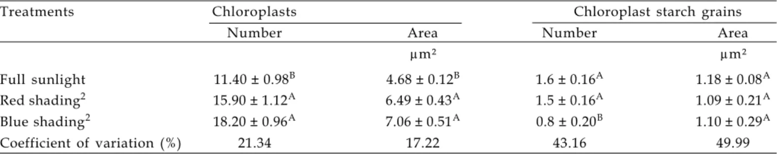
Table 5. Effects of different light regimes on chloroplasts and starch grains in Ocimum selloi palisade parenchyma cells(1)
Treatments Chloroplasts Chloroplast starch grains
Number Area Number Area
µm² µm²
Full sunlight 11.40 ± 0.98B 4.68 ± 0.12B 1.6 ± 0.16A 1.18 ± 0.08A Red shading2 15.90 ± 1.12A 6.49 ± 0.43A 1.5 ± 0.16A 1.09 ± 0.21A Blue shading2 18.20 ± 0.96A 7.06 ± 0.51A 0.8 ± 0.20B 1.10 ± 0.29A
Coefficient of variation (%) 21.34 17.22 43.16 49.99
(1)Results are expressed as mean values ± standard error (n = 10). Within a column, mean values bearing different superscript letters were significantly different (Tukey test; p<0.05).
(2) Red and blue shading was provided by 50% ChromatiNet® coloured plastic netting.
The thicknesses of the adaxial epidermis and the palisade parenchyma were unaltered by shading, but plants exposed to full sunlight showed an increase in thickness of the abaxial epidermis and spongy parenchyma resulting in the formation of a thicker leaf blade (Figure 2 a, c, e).
For plants maintained under shading conditions, a reduction in leaf thickness appears to be a common phenomenon that may be attributed to diverse factors including an increase of the R:FR (red:far red) light ratio (KASPERBBAUER and PEASLEE, 1973) and a decrease in light intensity (BUISSON and LEE, 1993). Since there were no variations in the R:FR light ratio in the experiments performed with O. selloi, the increased leaf thickness observed in plants grown in sunlight resulted exclusively from the variation in light intensity. These results corroborate those found by LEE et al. (2000), who reported that changes in the quality of light did not significantly affect the thickness of the leaf blade or alter the structure of the mesophyll layer.
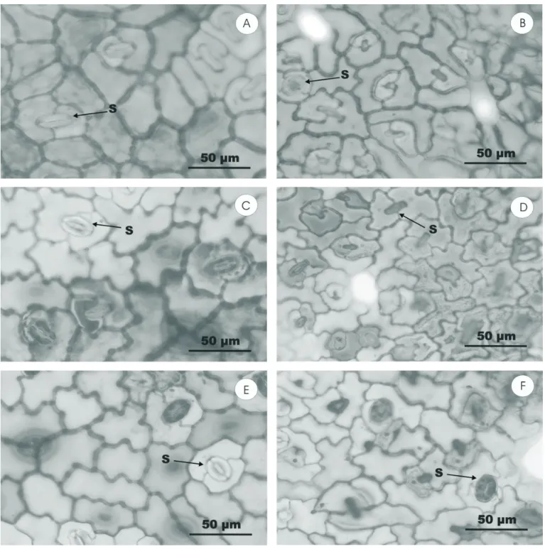
SEM examination revealed that the epidermal layers of O. selloi leaves contained diacytic stomata of equal size and positioned at the same level as other epidermal cells (Table 4). Although stomata were observed in both the lower and upper epidermis, their density was greater on the abaxial surface (hypo-amphistomatal leaves; Fig. 1). Moreover, the density of stomata was significantly greater in leaves of plants grown in full sunlight (Table 4), a finding that is in agreement with that of BUISSON and LEE (1993), who reported a reduction in stomatal density in plants maintained under neutral and filtered shading compared with those cultivated in full sunlight, and of LEE et al (1997), who found a higher stomatal density in plants of Hopea odorata maintained under strong light intensity. In the case of O. selloi, the polar diameter of the stomata in both epidermal layers was
not influenced by light treatment whereas the equatorial diameter varied according to the quality of the light such that red shading increased the diameter of abaxial stomata whilst blue shading increased the diameter of adaxial stomata.
SEM examination of transverse leaf sections revealed a dorsiventral structure and a uniseriate epidermis. The mesophyll consisted of one layer of palisade parenchyma cells and four to five layers of spongy parenchyma cells (MARTINS, 1998; GONÇALVES, 2001). Additionally, cells of the adaxial epidermis were observed to be larger than those of the abaxial layer. Variations in the shape of epidermal cell wallsfollowing exposure of
Mikania glomerata to different photoperiod treatments have been previously reported (CASTRO et al., 2005).
Chloroplasts were generally observed near to the cell walls (Figure 2a, d, f). The number and size of chloroplasts located in cells of the palisade parenchyma was significantly larger in plants grown under red and blue shading compared with plants maintained under full sunlight (Table 5). These results can be explained by the fact that the leaves of shaded plants are thinner and contain larger pigment-rich chloroplasts.
4. CONCLUSION
Various modifications in the anatomy and morphology of O. selloi, including plant height, total and organ-specific (roots, stems, leaves and inflorescences) dry biomass, R:AP ratio, LWR, leaf thickness, stomatal density and size of chloroplasts, resulted from the alteration of light intensity. However, TLA, SLA, LAR, and the number of chloroplasts and starch grains were influenced by the quality of the incident radiation (R:B light ratio).
ACKNOWLEDGMENTS
The authors are grateful to Polysack Indústrias (Leme, SP, Brazil) for the coloured shade netting donation, to Fundação de Amparo à Pesquisa de Minas Gerais for financial support and to the Conselho Nacional de Desenvolvimento Científico e Tecnológico (CNPq) for one scientific productivity fellowship.
REFERENCES
BENINCASA, M.M.P. Análise de crescimento de plantas. 2nd
Ed. FUNEP: Jaboticabal, 2003. 41p.
BROWN, C.S.; SCHUERGER, A.C.; SAGER, J.C. Growth and photomorphogenesis of pepper plants under red light-emitting diodes with supplemental blue or far-red lighting. J. American Society of Horticultural Science, v. 120, p. 808-813, 1995.
BUISSON, D.; LEE, D.W. The developmental responses of papaya leaves to simulated canopy shade. American Journal of Botany, v.80, p. 947-952, 1993.
CASTRO, E.M.; PINTO, J.E.B.P.; MELO, H.C.; SOARES, A.M.; ALVARENGA, A.A.; LIMA JÚNIOR, E.C. Aspectos anatômicos e fisiológicos de plantas de guaco submetidas a diferentes fotoperíodos. Horticultura Brasileira, v.23, p.846-850, 2005.
FERREIRA, M.M.; MOTA, M.B.; PINTO, J.E.P.B.; CASTRO, E.M. Crescimento e alocação e alocação de biomassa de planas de vinca (Catharanthus roseus (L.) G. Don) em função da adubação orgânica e época de colheita. Revista Brasileira de Plantas Medicinais, v.6, p.72-76, 2004.
GONÇALVES, L.A. Ontogenia dos tricomas glandulares e influência da radiação solar no desenvolvimento e no teor de óleo essencial de Ocimum selloi Benth. (Lamiaceae). Viçosa. 2001. 95p. (MSc Dissertation). Universidade Federal de Viçosa.
KASPERBAUER, M.J. Far-red light reflection from green leaves and effects on phytochrome-mediated assimilate partitioning under field conditions. Plant Physiology, v.8, p.350-354, 1987.
KASPERBAUER, M.J.; HAMILTON, J.L. Chloroplast structure and starch grain accumulation in leaves that received different red and far-red levels during development. Plant Physiology, v.74, p.967-970, 1984.
KASPERBAUER, M.J.; PEASLEE, D.E. Morphology and photosynthetic efficiency of tobacco leaves that received end-of-day red or far red light during development. Plant Physiology, v.52, p.440-442, 1973.
KIM, S-J.; HAHN, E-J.; HEO, J.; PAEK, K-Y. Effects of LEDs on net photosynthetic rate, growth and leaf stomata of chrysanthemum plantlets in vitro. Scientia Horticulturae, v.101, p.143-151, 2004.
LARCHER, W. Ecofisiologia Vegetal. RIMA: São Carlos, 2000. 319p.
LEE, D.W.; OBERBAUER, S.F.; KRISHNAPILAY, B.; MANSOR, M.; MOHAMAD, H.; YAP, S.K. Effects of irradiance and spectral quality on seedling development of two Southeast Asian Hopea species. Oecologia, v.110, p.1-9, 1997.
LEE, D.W.; OBERBAUER, S.F.; JOHNSON, P.; KRISHNAPILAY, B.; MANSOR, M.; MOHAMAD, H.; Yap, S.K. Effects of irradiance and spectral quality on leaf structure and function in seedlings of two Southeast Asian Hopea (Dipterocarpaceae) species. American Journal of Botany, v.87, p.447-455, 2000.
LEONG, T.; GOODCHILD, D.J.; ANDERSON, J.M. Effect of light quality on the composition, function and structure of photosynthetic thylakoid membranes of Asplenium australicum (Sm.) Hook. Plant Physiology, v.78, p.561-567, 1985.
LI, S.; RAJAPAKSE, N.C.; YOUNG, R.E.; OI, R. Growth responses of chrysanthemum and bell pepper transplants to photoselective plastic films. Scientia Horticulturae, v.84, p.215-225, 2000.
LOUGHRIN, J.H.; KASPERBAUER, M.J. Light reflected from colored mulches affects aroma and phenol content of sweet basil (Ocimum basilicum L.) leaves. Journal of Agriculture and Food Chemistry, v.49, p.331-1335, 2001.
MACEDO, A.F.; LAGE, C.L.; ESQUIBEL, M.A.; SOUZA, M.M.; SILVA, K.L.; NIERO, R.; CECHINEL-FILHO, V. Preliminary phytochemical and pharmacological studies on plantlets of Alternanthera brasiliana cultured under different spectral quality of lights. Acta Farmaceutica Bonaerense, v.23, p.515-519, 2004.
MARTINS, E.R. Estudos em Ocimum selloi Benth: isoenzimas, morfologia e óleo essencial. In: MING, L.C.; SCHEFFER, M.C.; CORRÊA JÚNIOR, C.; BARROS, I.B.I.; MATTOS, J.K.A. (Eds.) Plantas medicinais e aromáticas condimentares:avanços na pesquisa agronômica, v. 2. Botucatu: UNESP, 1998. p.97-126.
MCMAHON, M.J.; KELLY, J.W. Anatomy and pigments of chrysanthemum leaves developed under spectrally selective filters. Scientia Horticulturae, v.64, p.203-209, 1995.
Shading affecting growth and leaf structure of Ocimum selloi
Bragantia, Campinas, v.69, n.2, p.349-359, 2010 359
OREN-SHAMIR, M.; GUSSAKOVSKY, E.E.; SPIEGEL, E.; NISSIM-LEVI, A.; RATNER, K.; GILLER, Y.E.; SHAHAK, Y. Colored shade nets can improve the yield and quality of green decorative branches of Pittosporum variegatum. Journal of Horticultural Science and Biotechnology, v.76, p.353-361, 2001.
OYAERT, E.; VOLCKAERT, P.C.; DEBERGH, P.C. Growth of chrysanthemum under coloured plastic films with different light qualities and quantities. Scientia Horticulturae, v.79, p.195-205, 1999.
RAJAPAKSE, N.C.; POLLOCK, R.K.; MCMAHON, M.J.; KELLY, J.W.; YOUNG, R.E. Interpretation of light quality measurements and plant response in spectral filter research. HortScience, v.27, p.1208-1211, 1992.
SCHUERGER, A.C.; BROWN, C.S.; STRYJEWSKI, E.C. Anatomic features of pepper plants (Capsicum annuum L.) grown under red light-emitting diodes supplemented with blue or far-red light. Annals of Botany, v.79, p.273-282, 1997.
SHAHAK, Y.; GUSSAKOVSKY, E.E.; GAL, E.; GANELEVIN, R. Colornets: crop protection and light-quality manipulation in one technology. Acta Hort. 659, 143-151. 2004. Available at http://www.actahort.org/books/659/659_17.htm Accessed in 16th May 2006.