1. Department of Physical Medicine and Rehabilitation, Recep Tayyip Erdoğan University, Medical School
2. Department of Biochemistry, Recep Tayyip Erdoğan University, Medical School
3. Department of Cardiology, Recep Tayyip Erdoğan University, Medical School
4. Department of Physical Medicine and Rehabilitation, Karadeniz Technical University, Medical School
5. Department of Physical Medicine and Rehabilitation, Canakkale Onsekiz Mart University, Medical School
sease activity score had higher YKL-40 levels, IMT-C, and CF-PWV than did those with a lower disease ac-tivity score (P<0.001, P=0.008, and P<0.001, respec-tively)
Conclusion: AS patients had higher serum YKL40 le -vels, CF-PWV, and IMT-C than did healthy controls. Additionally, there was an association between in-creased CF-PWV and serum YKL-40 levels. Therefore, we conclude that CF-PWV and YKL-40 levels may be used for early diagnosis of atherosclerosis in AS pa-tients.
Keywords: Ankylosing spondylitis; Arterial stiffness; Carotid-femoral pulse wave velocity; YKL-40 levels
IntroductIon
Ankylosing spondylitis (AS) is a chronic systemic in-flammatory disease of the spine and sacroiliac joints. AS is associated with some extra-articular manifestations, including cardiovascular diseases (CVDs)1. AS patients show increased mortality from CVDs, and it has been reported that their relative risk of developing athe -rosclerosis is 1.5-fold higher than that in the normal population2,3. Traditional cardiovascular risk factors, including smoking, dyslipidemia, hypertension, obe-sity, and diabetes mellitus, play an important role in the development of atherosclerosis4. However, these risk factors do not fully explain the increased risk of CVDs in patients with AS.
Chronic systemic inflammation seems to play a cru-cial role in the pathogenesis of atherosclerosis. It has been reported that the lowgrade inflammation associa ted with AS has an influence on accelerating athe -rosclerosis5. Atherosclerosis has been associated with circulating inflammation biomarkers, such as Creacti -ve protein (CRP), fibrinogen, cytokines, and adhesion molecules6. Furthermore, YKL-40 has been suggested
The relationship between serum YKL-40 levels and
arterial stiffness in patients with ankylosing spondylitis
Kucukali Turkyilmaz A1, Devrimsel G1, Serdaroglu Beyazal M1, Kirbas A2, Cicek Y3, Capkin E4, Karkucak M4, Gokmen F5
ACTA REUMATOL PORT. 2017;42:183-190
AbstrAct
Objective: Serum YKL-40 plays roles in inflammatory and vascular processes. Our aim was to evaluate serum YKL-40 levels in patients with ankylosing spondylitis (AS) and to investigate their potential relationship with arterial stiffness based on carotid-femoral pulse wave velocity (CF-PWV).
Methods: Forty-three patients with AS and 41 healthy controls with no history or current signs of cardiovas-cular disease were included in the study. All patients were administered nonsteroidal anti-inflammatory drugs (NSAIDs), and none were prescribed anti-tumor necrosis factor agents. Serum YKL-40 levels were mea-sured. CF-PWV and intima-media thickness of the common carotid artery (IMT-C) were evaluated. Results: The mean age of AS patients was 34.6 ± 10.2 years and of controls was36.3 ± 9.0 years. CF-PWV was significantly higher in AS patients than in controls (8.2±2.7 vs.7.0±1.6 m/s, respectively; P=0.015). How-ever, the IMT-C was not significantly different between AS patients and controls (0.6±0.3 vs. 0.5±0.2 mm,
P=0.501). YKL-40 levels were significantly higher in
AS patients than in controls (78.9±37.9 vs. 58.4±21.2 ng/mL, P=0.003) and were strongly correlated with CF--PWV (r=0.773, P<0.001) and IMT-C (r=0.548,
P<0.001). A multiple linear regression analysis revealed
that CFPWV could be explained by serum YKL40 le -vels and IMT-C (adjusted R²= 0.707, P=0.013 and
-as a potential biomarker of inflammation and en-dothelial dysfunction7. Studies have shown that ele-vated YKL-40 levels are associated with CVDs8,9. In ad-dition, high serum YKL-40 levels have been found in patients with spondyloarthritis. That study suggested that YKL-40 might have potential value in monitoring disease activity10.
The assessment of vascular dysfunction could be useful in identifying subclinical atherosclerosis and for predicting the risk of developing CVDs11. There are several methods for assessing vascular function. Arte-rial stiffness, which reflects arteArte-rial compliance, is an important indicator of the early stages of atherosclero-sis. Carotid-femoral pulse wave velocity (CF-PWV) is considered the gold standard for assessing arterial stiff-ness12. In addition, the intima-media thickness of the common or internal carotid artery (IMT-C), as mea-sured on high-resolution ultrasonography, is the stan-dard method for assessing subclinical atherosclerosis. Recently, it has been reported that patients with AS have increased arterial stiffness (based on PWV) and IMT-C13,14.
Because of the high risk of CVDs in patients with AS, it is very important to identify atherosclerosis in its ear-ly stages. Accordingear-ly, the aims of this study were to evaluate the possible role of serum YKL-40 level as a biomarker of subclinical CVDs in AS patients and to in-vestigate its potential relationship with subclinical atherosclerosis based on CF-PWV and IMT-C mea-surements.
PAtIents And methods
In this study, 43 AS patients and 41 healthy controls were enrolled. Patients who fulfilled the modified New York criteria for AS15were consecutively recruited from a rheumatology outpatient clinic between February and October 2012. Healthy controls were selected from among hospital personnel who volunteered to participate as the control group. Patients and controls with any reported cardiovascular diseases (coronary angina, heart failure, myocardial infarction, periphe ral vascular disease, stroke) as well as patients who were receiving antihypertensive medications, lipidlowe -ring therapies, corticosteroid treatments, or had any other chronic disease were excluded from the study. All patients were using nonsteroidal anti-inflammato-ry drugs (NSAIDs), and none had taken anti-tumor necrosis factor (TNF) agents, sulfasalazine, or other
disease-modifying anti-rheumatic drugs (DMARDs) for at least 3 months. The local ethical committee approved the study protocol, and informed consent was obtained from all participants before the exami-nation.
clInIcAl Assessment
Clinical features, symptom duration, height, weight, and body mass index (BMI) were recorded. The Bath Ankylosing Spondylitis Disease Activity Index (BAS-DAI) was used to assess disease activity (0:no activity and 10:highest level of activity). A BASDAI score of ≥4 was considered to indicate active disease. Visual ana-logue scale (VAS) was utilized to record pain scores. If there was no pain, the score was 0 point and the most severe pain was recorded as 10 points. All patients ans wered questions about their medications. A single experienced cardiologist who was blinded to patient clini cal data performed the vascular assessments. These assessments were performed in the morning after an overnight fast of at least 8 h. Resting blood pressure was recorded with a mercury column sphy -gmomanometer.
bIochemIcAl AnAlysIs
Blood samples, which were drawn after 8 h of fasting, were measured for serum glucose, total cholesterol (TC), low-density lipoprotein cholesterol (LDL-C), highdensity lipoprotein cholesterol (HDLC), trigly -cerides (TGs), CRP, and the erythrocyte sedimentation rate (ESR).
serum yKl-40 meAsurement
Venous blood samples were drawn in the morning af-ter 8 h of fasting. The samples were centrifuged at 2,500×g for 10 minutes at 4˚C. Serum was obtained after centrifugation and stored at -80˚C until analysis. Serum YKL-40 level was measured using a commer-cially available enzyme immunoassay (EIA) kit (MicroVue YKL40; Quidel, San Diego, CA, USA) accor -ding to the manufacturer’s instructions, which indicated that the normal range of YKL-40 levels was 35–83 ng/mL.
Pulse wAve velocIty meAsurement
CF-PWV was measured in the carotid-femoral segment using a vali dated non-invasive device (SphygmoCor, AtCor Medi cal, Sydney, Australia) that sequentially records the carotid and femoral artery pulse waves with an application tonometer and electrocardiogram. The
measurements were performed after the subjects had res ted for 15 min in the supine position in a quiet room. The distances from the sternal notch to the right carotid artery and from the sternal notch to the right femoral artery were measured. At least 10 consecutive pulse waves were recorded over the common carotid and right femoral arteries. The time difference (t) was mea-sured as the difference between the start of each wave-form. Using a tape measure, the total distance between the carotid and femoral artery was measured, and the CF-PWV was calculated automatically. The results were expressed in m/s.
cArotId ultrAsonogrAPhy
Carotid arterial ultrasonography was performed using a high-resolution ultrasonography scanner (VingMed Vivid 3;GE Medical System, Horten, Norway) with a 7-MHz linear-array transducer. Measurements were performed on the right common carotid artery with the subject in the supine position. The region 1 cm proxi-mal to the carotid bifurcation was identified, and the IMT-C of the far wall was evaluated as the distance be-tween the lumen–intima and the media–adventitia in-terfaces. The IMT-C measurement was obtained from four adjacent sites at 1-mm intervals, and the average of all measurements was used for analysis. Mean values were calculated, and the results were presented in mm.
stAtIstIcAl AnAlysIs
Values obtained by measurement were expressed as mean ± standard deviation (SD), and those obtained by counting were expressed as a percentage. The appro -priateness of the normal distribution of the acquired data was tested with the Kolmogorov–Smirnov test. For comparison of the measurement values of the two groups, the Student t-test and the Mann–Whitney U test were utilized for values with normal and non-nor-mal distributions, respectively. The chi-squared test was used for the comparison of qualitative data. A cor-relation analysis for continuous variables was per-formed using the Spearman’s correlation coefficient. A multiple backward linear regression analysis was per-formed in order to identify the parameters accounting for the CF-PWV. We evaluated the factors that could af-fect the CF-PWV dependent variable in the multivari-ate linear regression analysis. Independent factors were age, symptom duration, CRP, BASDAI, YKL-40, and IMT-C. The number of independent factors was deter-mined according to study sample size. Statistical sig-nificance was considered as P < 0.05.
results
The clinical characteristics and cardiovascular and labo ratory parameters of the AS patients and healthy controls are shown in Table I. The mean age was 34.6±10.2 years among the AS patients and 36.3±9.0 years among the controls. There were 28 (53.7%) and 22 (65.1%) male patients in the AS and control groups, respectively. There were no differences in the TC, TG, LDL-C, and HDL-C values or systolic and diastolic blood pressure readings between the patients and con-trols (all P>0.05). ESR and CRP levels (both P<0.001) as well as serum YKL-40 levels (78.9±37.9 vs. 58.4±21.2 ng/mL, respectively, P=0.003) were signifi-cantly higher in the AS patients than in the control sub-jects. The CF-PWV values were also higher in the AS patients than in the controls (8.2±2.7 vs. 7.0±1.6 m/s, respectively, P=0.015). However, the IMT-C values were not significantly different between groups (0.6±0.3 vs. 0.5±0.2 mm, P=0.501).
The CF-PWV and IMT-C values were correlated with indexes of disease activity, such as CRP, ESR, VAS, and BASDAI (Table II). Furthermore, for AS patients, the serum YKL-40 level was significantly correlated with IMT-C, CRP, ESR, VAS, and BASDAI (r=0.548, r=0.711,
r=0.767, r=0.802, r=0.807, respectively; all P<0.001).
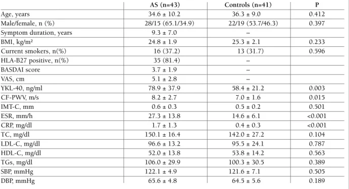
The YKL-40 level in the AS patients also demonstrated a strong correlation with CF-PWV values (r=0.773,
P<0.001; Figure 1).
A multiple backward linear regression analysis was used in order to adjust for any potential confounding influences of factors such as age, symptom duration, CRP, BASDAI, YKL-40, and IMT-C on AS patients. The linear regression analysis revealed that CF-PWV values were explained by serum YKL40 levels and IMTC va lues (adjusted R²= 0.707, P=0.013, and P=0.001, res -pectively; Table III).
AS patients with a higher disease activity score (BASDAI≥4) had higher YKL40, IMTC, and CFPWV va -lues than those with a low disease activity score (BAS-DAI<4) (P<0.001, P=0.008 and P<0.001, respectively; Table IV).
dIscussIon
Atherosclerosis is a strong predictor of future CVDs. AS patients have an increased risk of CVDs and mor-tality compared with the general population16. Mecha-nisms underlying AS-associated atherosclerosis are not
fully known. Studies have shown that chronic inflam-mation significantly contributes to accelerated athero-genesis in AS17,18. Arterial stiffness is regarded as a mar ker of CV risk and can be affected by chronic in -flam mation19. It is important to identify atherosclerosis in the early stages of AS by using biochemical mar -kers and noninvasive assessments, such as YKL-40, CRP, PWV, and IMT-C. Thus, the current study was de-signed to examine if arterial stiffness measured by CF--PWV and serum YKL-40 levels predicted subclinical CVDs.
Atherosclerosis is caused by lipoprotein infiltration into the intima and is considered to be an inflammato-ry disease. Previous studies have demonstrated that in-creased common carotid artery IMT is a good indica-tor of subclinical stages of atherosclerosis20,21. A study by Peters et al., which evaluated subclinical atherosclerosis in AS patients by measuring the IMTC, demons -trated higher IMT-C values in AS patients than in con-trols. Additionally, there was an association between IMT-C and CRP or BASDAI in AS patients22. Another
tAble I. clInIcAl chArActerIstIcs, cArdIovAsculAr And lAborAtory PArAmeters of PAtIents wIth AnKylosIng sPondylItIs (As) And control subjects (meAn ± sd)
AS (n=43) Controls (n=41) P
Age, years 34.6 ± 10.2 36.3 ± 9.0 0.412
Male/female, n (%) 28/15 (65.1/34.9) 22/19 (53.7/46.3) 0.397
Symptom duration, years 9.3 ± 7.0 –
BMI, kg/m² 24.8 ± 1.9 25.3 ± 2.1 0.233 Current smokers, n(%) 16 (37.2) 13 (31.7) 0.596 HLA-B27 positive, n(%) 35 (81.4) – BASDAI score 3.7 ± 1.9 – VAS, cm 5.1 ± 2.8 – YKL-40, ng/ml 78.9 ± 37.9 58.4 ± 21.2 0.003 CF-PWV, m/s 8.2 ± 2.7 7.0 ± 1.6 0.015 IMT-C, mm 0.6 ± 0.3 0.5 ± 0.2 0.501 ESR, mm/h 27.3 ± 13.8 14.6 ± 6.1 <0.001 CRP, mg/dl 1.7 ± 1.3 0.4 ± 0.3 <0.001 TC, mg/dl 150.1 ± 16.4 142.0 ± 27.2 0.104 LDL-C, mg/dl 96.6 ± 13.2 95.5 ± 24.1 0.787 HDL-C, mg/dl 52.0 ± 13.8 53.8 ± 14.2 0.563 TGs, mg/dl 106.0 ± 29.9 100.3 ± 30.5 0.389 SBP, mmHg 122.1 ± 4.9 121.6 ± 7.1 0.505 DBP, mmHg 65.6 ± 4.8 64.5 ± 5.6 0.189
BMI: Body mass index; VAS: Visual analogue scale; BASDAI: Bath Ankylosing Spondylitis Disease Activity Index; CF-PWV: Carotid–femoral pulse wave velocity; IMT-C: Intima-media thickness-carotid; LDL: Low-density lipoprotein cholesterol; HDL: High-density lipoprotein cholesterol; TGs: Triglycerides; TC: Total cholesterol; ESR: Erythrocyte sedimentation rate; CRP: C-reactive protein; SBP: Systolic blood pressure; DBP: Diastolic blood pressure.
tAble II. correlAtIon between dIseAse ActIvIty And cArotId-femorAl Pulse wAve velocIty (cf-Pwv), IntImA-medIA thIcKness-cArotId (Imt-c) In PAtIents wIth AnKylosIng sPondylItIs
CF-PWV IMT-C r r BASDAI 0.675* 0.461** VAS, cm 0.657* 0.485** YKL-40, ng/ml 0.773* 0.548* ESR, mm/h 0.554* 0.376** CRP, mg/dl 0.607* 0.546*
r=Spearman’s correlation coefficients; *P< 0.001; **P< 0.05; BASDAI: Bath Ankylosing Spondylitis Disease Activity Index; VAS: Visual analogue scale; ESR: Erythrocyte sedimantation rate; CRP: C-reactive protein.
tAble III. Influence of dIfferent fActors on cf-Pwv In PAtIents wIth AnKylosIng sPondylItIs
Dependent variable Independent variable B (SE) Beta P
CF-PWV, m/s
Panova <0.001 YKL-40 0.028 (0.011) 0.382 0.013
R²= 0.728, Adjusted R²= 0.707 IMT-C 3.585 (1.007) 0.349 0.001
The multivariate backward linear regression model was used for AS patients. P value < 0.05 indicates the presence of regression; Independent variables are as follows: Age, Symptom duration, C-reactive protein, Bath Ankylosing Spondylitis Disease Activity Index, YKL-40, IMT-C;Intima-media thickness-carotid
tAble Iv. comPArIson of AnKylosIng sPondylItIs (As) PAtIents AccordIng to dIseAse ActIvIty
AS group with BASDAI<4 AS group with BASDAI≥4
(n=23) (n=20) P
Age, years 36.9 ± 10.4 32.0 ± 9.4 0.114
Male/female, n (%) 13/10 (56.5/43.5) 15/5 (75/25) 0.205
Symptom duration, years 11.2 ± 7.6 7.1 ± 5.5 0.051
BMI, kg/m² 24.3 ± 1.8 25.2 ± 2.0 0.136 BASDAI score 2.2 ± 0.9 5.4 ± 1.1 <0.001 VAS, cm 2.9 ± 1.6 7.6 ± 1.4 <0.001 YKL-40, ng/ml 54.1 ± 21.8 107.4 ± 31.9 <0.001 CF-PWV, m/s 6.6 ± 1.6 10.1 ± 2.7 <0.001 IMT-C, mm 0.5 ± 0.3 0.7 ± 0.2 0.008 ESR, mm/h 17.8 ± 8.4 38.2 ± 10.3 <0.001 CRP, mg/dl 0.7 ± 0.5 2.7 ± 1.3 <0.001
BMI: Body mass index; VAS: Visual analogue scale; BASDAI: Bath Ankylosing Spondylitis Disease Activity Index; CF-PWV: Carotid–femoral pulse wave velocity; IMT-C: Intima-media thickness-carotid; LDL: Low-density lipoprotein cholesterol; HDL: High-density lipoprotein cholesterol; TGs: Triglycerides; TC: Total cholesterol; ESR: Erythrocyte sedimantation rate; CRP: C-reactive protein; SBP: Systolic blood pressure; DBP: Diastolic blood pressure.
0050,00CF-PWW (m/s)YKL-40 (ng/ml)15,0012,5010,007,505,00100,00150,00R Sq Linear=0,605r=0.733, p<0.001200,00 00 50,00 CF -P W W ( m /s ) YKL-40 (ng/ml) 15,00 12,50 10,00 7,50 5,00 100,00 150,00 R Sq Linear=0,605 r=0.733, p<0.001 200,00
fIgure 1.Correlation between carotid-femoral pulse wave velocity (CF-PWV) and serum YKL-40 level in patients with ankylosing spondylitis
study demonstrated higher carotid bulb IMT in AS pa-tients than in controls. A correlation between carotid bulb IMT and ESR suggested the possible role of in-flammation in accelerating the atherosclerotic process in AS patients. However, there was no correlation be-tween IMT-C and disease activity in the AS patients in this study23. A study by Gonzales-Juanatey et al. demonstrated higher IMT-C values in AS patients with no associated cardiovascular risk factors than in con-trols14. However, a study by Capkin et al. revealed that AS patients have higher PWV values but no statistical-ly significant differences in IMT-C levels compared to controls13. In another cross-sectional study, the authors demonstrated that IMT-C in young AS patients without clinically evident cardiovascular risk factors did not dif-fer from that in healthy controls24. This is consistent with our findings, since we found no differences in IMT-C between AS patients and controls. In this study,
may be caused by other factors, such as NSAID use or chronic inflammation38. Furthermore, it has been re-ported that endothelial dysfunction was correlated with the degree of inflammation in AS39. In a study by Cap-kın, which investigated subclinical atherosclerosis by assessing the PWV and IMT-C in AS patients, AS pa-tients were demonstrated to have higher PWV values. However, there were no differences at IMT-C levels be-tween patients with AS and the controls13. Their fin -dings are consistent with those in our own study. In the present study, CF-PWV was greater in AS patients than in healthy individuals. In addition, we found a corre-lation between CF-PWV and YKL-40, ESR, and CRP values, suggesting that inflammation may accelerate the atherosclerotic process in AS patients.
We also performed a multiple regression analysis using CF-PWV as the dependent variable, while the predictors were age, symptom duration, CRP, BASDAI, YKL-40, and IMT-C. The final sample size (43 AS pa-tients) did not allow us to analyze certain predictors, such as traditional cardiovascular risk factors. The results showed that CFPWV was explained by the mo -del composed of YKL-40 levels and IMT-C. Thus, our results indicate that YKL-40 contributed to explaining subclinical atherosclerosis as assessed by CF-PWV.
A limitation of this study is that there we lacked data regarding prior medication use, such as use of NSAIDs, DMARDs, and anti-TNF. We also cannot fully exclude the possible confounding effects of active disease, such as elevated ESR, CRP, and BASDAI score. Another limi -tation of our study was the relatively small patient num-ber. Therefore, further research is needed in larger pa-tient populations to confirm our findings.
Since the development of coronary atherosclerosis in patients with AS can occur independently of traditio -nal risk factors, additio-nal markers of coronary artery disease are required. This study showed that serum YKL-40 levels were increased in patients with AS. In addition, we found increased arterial stiffness, as indi-cated by increased CF-PWV, in AS patients compared to that in controls. Moreover, CF-PWV correlated with IMT-C, which is a reliable indicator of subclinical atherosclerosis. Hence, our results demonstrate that the development of atherosclerosis in AS may be shown by increased arterial stiffness. Additionally, YKL-40 might be used as a screening marker for coronary atheroscle-rosis in patients with AS because of its correlation with CF-PWV and IMT-C.As a result, we suggest that serum YKL-40 level and CF-PWV may predict early and sub-clinical atherosclerosis in patients with AS.
we also demonstrated that the IMT-C of AS patients was correlated to the indexes of disease activity, such as CRP, ESR, VAS, and BASDAI.
YKL-40 is a new potential biomarker of inflammation and vascular dysfunction in patients with coronary artery disease. It is expressed by local inflammatory cells (such as macrophages, neutrophils, and endothe-lial and vascular smooth muscle cells) during inflam-mation25. Unlike CRP, which is produced in the liver in response to systemic inflammation, YKL-40 is pro-duced locally at the site of inflammation, suggesting that YKL-40 could be superior to CRP in predicting disease progression and could possibly be used for this purpose26. It plays an important role in angiogenesis and remodeling of the extracellular matrix27,28. YKL-40 expression has been detected in macrophages and vas-cular endothelial cells during the early development of atherosclerosis29. Recently, several clinical studies have described an association between increased YKL-40 levels and CVDs30,31. Previous studies support the idea that soluble biomarkers can predict CVDs in AS10,32. In our previous study, we found that YKL-40 levels were higher in patients with early rheumatoid arthritis than in healthy controls33. We also observed a strong rela-tionship between YKL-40 levels and the severity of disea se activity. In another study, biomarkers of en-dothelial damage and disease activity were evaluated in AS patients. Serum levels of YKL-40 were increased and weakly associated with radiographic progression34. In our study, YKL40 was correlated with factors rela -ted to disease activity indicators such as CRP, ESR, VAS, and BASDAI. Furthermore, YKL-40 levels in AS pa-tients also demonstrated a strong correlation with CF--PWV and IMT-C. Based on our results, we believe that YKL-40 may be a new biomarker of disease activity that can also be used to predict the development of atherosclerosis in patients with AS.
Arterial stiffness, which is assessed by PWV, is im-portant for diagnosing the early stages of atherosclero-sis35. Many studies have demonstrated higher aortic PWV in patients with inflammatory arthritis16,36. Demi-ralp et al. assessed aortic elasticity and the stiffness in-dex in AS patients without cardiovascular involvement and reported decreased aortic elasticity and an in-creased aortic stiffness index37. In a cohort of AS pa-tients, CVDs risk factors, including blood lipids, hy-pertension, smoking, diet, and physical inactivity, were evaluated. No differences were demonstrated between AS patients and the general population. The authors suggested that the increased presence of CVDs in AS
corresPondence to
Aysegul Kucukali Turkyilmaz
Recep Tayyip Erdogan University Education and Research Hospital, Physical Medicine and Rehabilitation Department
E-mail: aysegulkemal@gmail.com
references
1. Mathieu S, Joly H, Baron G, et al. Trend towards increased ar-terial stiffness or intima-media thickness in ankylosing spondylitis patients without clinically evident cardiovascular disease. Rheumatology 2008;47:1203-1207.
2. Lehtinen K. Mortality and causes of death in 398 patients ad-mitted to hospital with ankylosing spondylitis. Ann Rheum Dis 1993;52:174-176.
3. Nurmohamed MT, van der Horst-Bruinsma I, Maksymowych WP. Cardiovascular and cerebrovascular disease in ankylosing spondylitis:current insights. Curr Rheumatol Rep 2012;14:415--421.
4. Kullo IJ, Ballantyne CM. Conditional risk factors for atheroscle-rosis. Mayo Clin Proc 2005;80:219-230.
5. Hahn BH, Grossman J, Chen W, McMahon M. The pathogene-sis of atheroscleropathogene-sis in autoimmune rheumatic disease:roles of inflammation and dyslipidemia. J Autoimmun 2007;28:69-75. 6. Maksimowicz-McKinnon K, Bhatt DL, Calabrese LH. Recent advan ces in vascular inflammation:C-reactive protein and oth-er inflammatory biomarkoth-ers. Curr Opin Rheumatol 2004; 16(1):18-24.
7. Rathcke CN, Vestergaard H. YKL40, a new inflammatory mar -ker with relation to insülin resistance and with a role in en-dothelial dysfunction and atherosclerosis. Inflamm Res 2006;55:221-227.
8. Kucur M, Isman FK, Karadag B, et al. Serum YKL-40 levels in patients with coronary artery disease. Coron Artery Dis 2007;18:391-396.
9. Rathcke CN, Vestergaard H. YKL-40 an emerging biomarker in cardiovascular disease and diabetes.Cardiovasc Diabetol 2009;8:61.
10. Pedersen SJ, Hetland ML, Sørensen IJ, et al. Circulating levels of interleukin-6, vascular endothelial growth factor, YKL-40, matrix metalloproteinase-3, and total aggrecan in spondy-loarthritis patients during 3 years of treatment with TNFα in-hibitors.Clin Rheumatol 2010;29(11):1301-1309.
11. Simon A, Megnien JL, Chironi G. The value of carotid intima-media thickness for predicting cardiovascular risk. Arterioscler Thromb Vasc Biol 2010;30(2):182-185.
12. Laurent S, Cockcroft J, Van Bortel L, et al. Expert consensus document on arterial stiffness: methodological issues and clin-ical applications. Eur Heart J 2006;27(21):2588-2605. 13. Capkin E, Kiris A, Karkucak M, et al. Investigation of effects of
different treatment modalities on structural and functional ves-sel wall properties in patients with ankylosing spondylitis.Joint Bone Spine 2011;78(4):378-382.
14. Gonzalez-Juanatey C, Vazquez-Rodriguez TR, Miranda-Filloy JA, et al. The high prevalence of subclinical atherosclerosis in pa-tients with ankylosing spondylitis without clinically evident car-diovascular disease. Medicine (Baltimore) 2009;88(6):358-365. 15. van der Linden S, Valkenburg HA, Cats A. Evaluation of diag-nostic criteria for ankylosing spondylitis. A proposal for modi-fication of the New York criteria.Arthritis Rheum 1984;27 (4):361-368.
16. Capkin E, Karkucak M, Kiris A, et al. Anti-TNF-α therapy may
not improve arterial stiffness in patients with AS: a 24-week fol-low-up. Rheumatology (Oxford) 2012;51(5):910-4.
17. Moyssakis I, Gialafos E, Vassiliou VA, et al. Myocardial perfor-mance and aortic elasticity are impaired in patients with anky-losing spondylitis. Scand J Rheumatol 2009;38(3):216-221. 18. Heeneman S, Daeman MJ. Cardiovascular risks in
spondy-loarthritis. Curr Opin Rheumatol 2007;19:358-362.
19. McEniery CM, Spratt M, Munnery M, et al. An analysis of prospective risk factors for aortic stiffness in men:20-year fol-low up from the Caerphilly prospective study. Hypertension 2010;56:36-43.
20. Lorenz MW, Markus HS, Bots ML, Rosvall M, Sitzer M. Predic-tion of clinical cardiovascular events with carotid intima-media thickness: a systematic review and meta-analysis.Circulation 2007;115(4):459-467.
21. Simons PC, Algra A, Bots ML, et al. Common carotid intima-me-dia thickness and arterial stiffness: indicators of cardiovascular risk in high-risk patients. The SMART Study (Second Manifes-tations of ARTerial disease).Circulation 1999;100(9):951-957. 22. Peters MJ, van Eijk IC, Smulders YM, et al. Signs of accelerated preclinical atherosclerosis in patients with ankylosing spondyli-tis.J Rheumatol 2010;37(1):161-166.
23. Perrotta FM, Scarno A, Carboni A, et al. Assessment of sub-clinical atherosclerosis in ankylosing spondylitis: correlations with disease activity indices. Reumatismo 2013;65(3):105-112. 24. Choe JY, Lee MY, Rheem I, et al. No differences of carotid inti-ma-media thickness between young patients with ankylosing spondylitis and healthy controls. Joint Bone Spine 2008;75 (5):548-553.
25. Kushner I. The phenomenon of the acute phase response. Ann N Y Acad Sci 1982;389:39-48.
26. Kjaergaard AD, Johansen JS, Bojesen SE, et al. Role of inflam-matory marker YKL-40 in the diagnosis, prognosis and cause of cardiovascular and liver diseases. Crit Rev Clin Lab Sci 2016; DOİ:10.1080/10408363.2016.1190683
27. Lee CG, Da Silva CA, Dela Cruz CS, et al. Role of chitin and chi ti nase/chitinase-like proteins in inflammation, tissue re-modeling, and injury. Annu Rev Physiol 2011;73:479-501. 28. Shao R. YKL-40 acts as an angiogenic factor to promote tumor
angiogenesis. Front Physiol 2013;4:122.
29. Michelsen AE, Rathcke CN, Skjelland M, et al. Increased YKL40 expression in patients with carotid atheros clero -sis.Atherosclerosis 2010;211(2):589-595.
30. Kastrup J, Johansen JS, Winkel P, et al. High serum YKL-40 con-centration is associated with cardiovascular and all-cause mor-tality in patients with stable coronary artery disease. Eur Heart J 2009;30:1066-1072.
31. Wang Y, Ripa RS, Johansen JS, et al. YKL-40 a new biomarker in patients with acute coronary syndrome or stable coronary artery disease. Scand Cardiovasc J 2008;42:295-302. 32. Azevedo VF, Faria-Neto JR, Stinghen A, et al. IL-8 but not ot her
biomarkers of endothelial damage is associated with disease ac-tivity in patients with ankylosing spondylitis without treatment with anti-TNF agents.Rheumatol Int 2013;33(7):1779-1883. 33. Turkyilmaz AK, Devrimsel G, Kirbas A, et al. Relationship
be-tween pulse wave velocity and serum YKL-40 level in patients with early rheumatoid arthritis.Rheumatol Int 2013;33(11): 2751-2756.
34. Maksymowych WP, Landewé R, Conner-Spady B, et al. Serum matrix metalloproteinase 3 is an independent predictor of struc-tural damage progression in patients with ankylosing
spondyli-tis. Arthritis Rheum 2007;56:1846-1853.
35. Arnett DK, Evans GW, Riley WA. Arterial stiffness: a new car-diovascular risk factor?Am J Epidemiol 1994;140(8):669-682. 36. Maki-Petaja KM, Hall FC, Booth AD, et al. Rheumatoid arthri-tis is associated with increased aortic pulse wave velocity, which is reduced by anti-tumor necrosis factor-alpha therapy. Circu-lation 2006;12;1185-1192.
37. Demiralp E, Kardesoglu E, Kiralp MZ, et al. Aortic elasticity in pa tients with ankylosing spondylitis.Acta Cardiol 2004;59(6):630-634.
38. Sundström B, Johansson G, Johansson I, et al.Modifiable car-diovascular risk factors in patients with ankylosing spondylitis. Clin Rheumatol 2014;33(1):111-117.
39. Peters MJ, Visman I, Nielen MM, et al. Ankylosing spondylitis: a risk factor for myocardial infarction?Ann Rheum Dis 2010;69(3):579-581.