Universidade de Lisboa
Faculdade de Ciências
Departamento de Informática
Genome-wide profiling of RNA polymerase II and associated
co-transcriptional processes using advanced NET-seq data
Tomás Pires de Carvalho Gomes
Dissertação
Mestrado em Bioinformática e Biologia Computacional
Especialização em Biologia Computacional
Orientadores:
Doutora Ana Rita Fialho Grosso
Prof. Doutora Lisete Maria Ribeiro De Sousa
I
Agradecimentos
Este foi um ano preenchido, de trabalho e pessoas.
Quero começar por agradecer à Doutora Ana Rita Grosso pela disponibilidade constante, pelo seu apoio crítico e pelo que me ensinou, não só este ano como também durante a sua cadeira no mestrado. Mas também pelos incentivos e atitude positiva que ofereceu, quer o projeto corresse melhor ou pior.
À professora Maria do Carmo Fonseca, pelas suas ideias, discussão crítica, pelo delinear do projeto, e também pelo entusiasmo constante demonstrado pelos resultados obtidos.
À Ana Paula Leite, que me trouxe inicialmente para este projeto, e que embora me tenha orientado pouco tempo me deu conselhos valiosos.
Agradeço a todos os meus colegas de gabinete, Célia, Bruno, Robert, Paco, Marina, Jorge, pela hospitalidade na introdução no grupo e no mundo científico, através de discussões e reflexões científicas, sociais e por vezes ambas. E estendo o agradecimento a toda a equipa, todos com trabalhos interessantes em diversos temas, o que me abriu vários horizontes de investigação.
Obrigado também à professora Lisete de Sousa, principalmente pelas aulas e tudo o que ensinou, mas também pela coorientação deste trabalho.
Uma nota especial para a comunidade mundial de bioinformáticos/biólogos computacionais, que com a sua grande partilha de problemas e ideias online me ajudou com muitas dúvidas. Agradeço também a um valioso grupo de amigos. Precisaria de outra tese para indicar um a um quem são e como contribuíram, não só neste ano, mas nos quatro anteriores também, e alguns até antes. Mas os últimos cinco anos foram verdadeiramente de grande mudança de vida, e muito se deveu a vocês.
À minha família, pelo acompanhamento que sempre me deram. Ao meu pai pelo constante interesse e incentivo a continuar a fazer o que gosto; à minha mãe pela constante preocupação e dedicação sobre o meu trabalho; ao meu irmão pela distração no dia-a-dia; aos meus avós pela sua sabedoria e paciência desde sempre; à minha tia Fernanda pelo apoio e encorajamento a procurar fazer mais e melhor. Por tudo, no fundo, desde que me lembro. Por último, e em jeito de dedicatória, um agradecimento à Hajrabibi, que tem a distinta característica de saber sempre o que dizer quando é preciso. Que eu te possa ajudar tanto ou mais.
II
Nota Prévia
A presente tese encontra-se escrita em inglês e em formato de publicação. A língua inglesa foi escolhida para ser usada por ser hoje em dia a língua franca da comunidade científica, e ao pretender seguir uma carreira de investigação impõe-se a necessidade de um domínio crescente dessa língua.
Adicionalmente, o inglês é também utilizado devido ao formato de publicação científica desta tese. O projeto desenvolvido ao longo do último ano resultou num artigo científico submetido à revista Cell. Como tal, a presente tese inclui não só resultados das análises computacionais realizadas, mas também validações biológicas experimentais complementares ao trabalho desenvolvido, que permitem uma melhor compreensão da questão biológica abordada. No entanto, este relatório pretende salientar o trabalho desenvolvido pelo autor no referido projeto, isto é, a análise de dados de sequenciação de alto rendimento usando software adequado a cada teste, como será descrito mais à frente. Os capítulos desta tese, incluindo não só o Capítulo 2, que contém o artigo, mas também os restantes, estão assim escritos em formato idêntico ao utilizado na submissão de manuscritos à referida publicação, mas com a inclusão de figuras ao longo do texto, a fim de facilitar a leitura e compreensão. Para facilitar a compreensão, foi também dado um estilo diferente aos títulos e subtítulos.
III
Resumo
A transcrição é o processo, presente em todos os seres vivos, em que a partir de uma cadeia molde de DNA se sintetiza uma cadeia complementar de RNA. A grande maioria dos genes em eucariotas é transcrita pela RNA polimerase II. A cadeia de RNA sintetizada não é, no entanto, o produto final, já que pode ser alvo de vários tipos de processamento, como splicing, poliadenilação ou edição de bases. Estes fenómenos foram já descritos como ocorrendo co ou pós-transcricionalmente. No entanto, não são ainda conhecidos todos os componentes, nem como são regulados estes processos ou qual a sua interação com a RNA polimerase II, em particular com o seu domínio carboxi-terminal (CTD).
Para abordar estes problemas de uma forma não enviesada, optou-se por adaptar uma técnica anteriormente descrita, que abrange todo o genoma, de alto rendimento e precisão, a native elongating transcript sequencing (NET-seq); sendo ela modificada de modo a poder detetar qual o estado de fosforilação do domínio carboxi-terminal da polimerase isolada em cada ensaio. Ao novo protocolo chamou-se advanced NET-seq (ANET-seq). Para além dos dados gerados por este protocolo, foram também obtidos dados de RNA ligado à fração de cromatina (ChrRNA). Todos os dados foram obtidos de células HeLa, sendo esta a primeira instância em que um estudo de nível genómico com esta precisão de mapeamento foi aplicado em mamíferos.
Análise inicial destes dados revelou uma distribuição das isoformas do CTD nos genes idêntica ao previamente descrito por outras técnicas. Adicionalmente, verificou-se também a captura de precursores do splicing, nomeadamente do 3’ do exão upstream, distintamente nos casos em que este é incluído no transcrito final. Estes exões aparecem principalmente associados a polimerase fosforilada na serina 5 do seu CTD. Outra observação curiosa foi a deteção de precursores do processamento de micro RNAs pelo complexo Drosha/DGCR8. Diferenças na deteção destes precursores permitiu postular diferentes dinâmicas para o processamento destes RNAs não codificantes.
Também se obtiveram dados de ANET-seq (com anticorpo para fosforilação da serina 2) e ChrRNA de células HeLa transfetadas com siRNA contra fatores de terminação – Xrn2 - e processamento do terminal 3’ do pre-mRNA – CPSF73 e CstF64+CstF64τ. Análise destes dados permitiu concluir que os fatores de processamento, mas não o Xrn2, influenciam significativamente a dinâmica da polimerase na região 3’ do gene, no final da transcrição, promovendo a sua pausa e subsequente desassociação do DNA. Constatou-se também que
IV
estes fatores afetam a acumulação de polimerase junto ao promotor dos genes, afetando igualmente a produção de transcritos upstream do promotor (PROMPTs), podendo concluir-se que estes fatores participam na regulação da transcrição não-produtiva.
Os resultados obtidos foram satisfatórios e também surpreendentes. Com este trabalho, é apresentada uma nova forma de estudar, ao nível do genoma, como ocorre a regulação da transcrição pelo CTD. Mostram-se também novas provas sobre processamento co-transcricional do RNA e a sua ligação à fosforilação do CTD. Foram igualmente elucidados os papéis de alguns fatores envolvidos na fase final da transcrição. Finalmente, ficou outra vez demonstrada a importância de estudos abrangentes na área da transcrição, em complemento dos trabalhos moleculares e bioquímicos já desenvolvidos há décadas. Espera-se, de futuro, um aprofundamento das técnicas de alto rendimento, e uma consequente adequação das ferramentas bioinformáticas a estes estudos.
Palavras-chave: transcrição; ANET-seq; sequenciação de RNA; CTD; splicing; micro RNA; clivagem e poliadenilação; terminação.
V
Abstract
Transcription is a process present in all living beings where, from a DNA template, a complementary RNA strand is synthesized. Most eukaryotic genes are transcribed by RNA polymerase II. The resulting RNA strand is not, however, the final product, since it’ll still be subject to various processing steps, such as splicing, polyadenylation or base editing. These modifications have been described as occurring co or post-transcriptionally. Yet, it is still not known how these processes are regulated, nor what all of their interveners are or how do they interact with RNA polymerase II, in particular with its C-terminal domain (CTD).
To address these problems in an unbiased way, a previously described genome-wide and high-precision technique, native elongating transcript sequencing (NET-seq), was adapted so it could detect what was the phosphorylation isoform from the isolated polymerase’s CTD. The new protocol was called advanced NET-seq (ANET-seq). In addition to the data generated by this protocol, RNA associated with the chromatin fraction was also sequenced. All data was obtained from HeLa cells, applying this genome-wide high-resolution technique to a mammalian system.
Initial analysis of ANET-seq data revealed that distribution of CTD isoforms in genes was similar to previously described profiles obtained by other protocols. Additionally, it was also verified the capture of splicing intermediates, in particular the 3’ end of the upstream exon, distinctively in cases where it was included in the final transcript. These exons are mainly associated with polymerase phosphorylated in the CTD’s Ser5. Another curious observation was the detection of micro RNA precursors, resulting from Drosha/DGCR8 processing. Differences in the detection of these precursors allowed the proposal of different processing dynamics for this type of non-coding RNAs.
ANET-seq data (with a Ser2-directed antibody) and ChrRNA from HeLa cells transfected with siRNA for termination factor Xrn2 and 3’ processing factors CPSF73 and CstF64+CstF64τ were also obtained. The analysis of this data showed that 3’ processing factors, but not Xrn2, significantly influence Pol II dynamics in the gene’s 3’ region, at the end of transcription, promoting its pause and dissociation from the DNA template. It was also observed that these factors influence polymerase accumulation near gene’s promoters, and equally affect promoter upstream transcripts (PROMPTs), leading to the conclusion that these factors regulate termination of unproductive transcription.
VI
Obtained results were satisfactory and also sometimes surprising. This work presents a novel genome-wide way to study how transcription is regulated by the CTD. New evidence of co-transcriptional RNA processing arouse, as well as their connection with CTD isoforms. There were also new revelations about transcription termination factor’s functions. Finally, it was once again demonstrated the importance of genome-wide techniques in transcription study, which complete molecular and biochemical work in the same area that has been developed for decades. In the future, a greater development of high-throughput techniques, and a constant adaptation of bioinformatical tools to these studies is expected.
Keywords: transcription; ANET-seq; RNA sequencing; CTD; splicing; micro RNA; cleavage and polyadenylation; termination.
Table of Contents
Agradecimentos ... I Nota Prévia ... II Resumo ... III Abstract ... VChapter 1
1. Transcription ... 1 1.1 Overview ... 11.2 The RNA Polymerase II and the C-Terminal Domain ... 2
1.3 Stages and players of transcription ... 5
2. Pre-mRNA Processing ... 6
2.1 Splicing ... 6
2.2 3’ end processing and transcription termination ... 9
3. Micro RNAs ... 11
3.1 Overview ... 11
3.2 Micro RNA transcription and processing ... 12
4. Genome-wide study of transcription ... 14
4.1 High-throughput sequencing approaches ... 14
4.2 Data Analysis ... 17 5. Objectives ... 20
Chapter 2
1. Summary ... 24 2. Highlights ... 25 3. Introduction ... 26 4. Results ... 28 4.1 ANET-seq strategy ... 284.2 Pol II CTD phosphorylation-specific nascent RNA profiles at TSS and TES ... 30
4.3 Exon tethering to Pol II S5P for co-transcriptional splicing ... 32
4.4 Co-transcriptional pre-miRNA biogenesis ... 35
4.5 Pol II pausing regulated by CPA factors at TES ... 37
4.6 3' end termination machinery regulates metabolism of promoter-associated RNA ... 41
5. Discussion ... 44
7. References ... 49
8. Supplementary Material ... 53
8.1 Extended experimental procedures ... 53
8.2 Supplementary Figures ... 59
8.3 Supplementary References ... 66
Chapter 3
Conclusions and Perspectives ... 681
1. Transcription
1.1 OverviewIn 1956, Francis Crick first proposed what he called the “Central Dogma of Molecular Biology”. The Dogma not only stated, in his own words, that “Once information has got into a protein it cannot get out again”, but
also outlined the possible ways this information would be transferred between nucleic acids and proteins. Later, in 1970, Crick developed these ideas, and classified the nine possible
ways information could be
transferred between the intermediates (DNA, RNA and protein) into three categories: General Transfers, Special Transfers and Unknown Transfers (Francis Crick, 1970) (Figure 1). General Transfers refer to reactions present in all cells,
whereas Special Transfer refers to reactions that were postulated to exist, and later identified only in a subset of life forms or in vitro (Uzawa et al., 2002). Unknown Transfers are reactions postulated not to exist since they would require very complex machinery. Although great advancements and discoveries have been made in the field of molecular biology, the core message of the dogma still holds, yet it does not address certain details of the described phenomena, such as gene expression regulation or post-translational modifications.
According to the Dogma, information is stored in the DNA nucleotide sequence, and to be effectively used to produce proteins uses an intermediary, RNA. Transcription is the synthesis of RNA using DNA as a template. Although the interveners vary greatly between prokaryotes and eukaryotes, the core process is very similar. The main player in transcription is RNA polymerase, which reads the DNA strand and synthesizes a complementary RNA molecule (Chamberlin and Berg, 1962). Like any complex biochemical reaction, eukaryotic transcription can be divided in several steps. These steps are defined by the different factors that associate with the polymerase and the transcribed gene’s sequence in a given moment.
Figure 1: A representation of the Central Dogma of Molecular Biology,
including DNA (top), RNA (bottom left) and protein (bottom right), and the possible information transfers between them (arrows). Green, General Transfers; Yellow, Special Transfers; Red, Unknown Transfers.
2
The progression of transcription in each phase, however, depends not only on these factors, but also on chromatin conformation and components, and also the gene sequence (Nojima et al., 2013; Grosso et al., 2012; Peterlin et al., 2006; Jonkers et al., 2014). In addition, eukaryotes possess different RNA polymerases, each responsible for the transcription of a subset of genes. This results in a highly regulated process, allowing the cell to precisely adjust its components’ concentration in response to diverse stimuli.
1.2 The RNA Polymerase II and the C-Terminal Domain
As previously mentioned, RNA polymerase is the main agent involved in transcription, synthesizing RNA in a DNA-dependent manner. It is an essential enzyme for all organisms - even virus, which may use the host’s polymerase -, but despite that there are many differences, mainly structural but
some also functional, between
prokaryotes and eukaryotes.
In animals, three RNA polymerases exist (Roeder and Rutter, 1969). RNA polymerase I (Pol I) is responsible for transcription of pre-45S rRNA, which generates all the other mature rRNA except 5S (Jacob, 1995), whereas RNA polymerase III (Pol III) is involved in the production of tRNAs, rRNA 5S, a small subset of micro RNAs and other small RNAs found in the nucleus and cytosol (Weinman and Roeder, 1974; Willis, 1993). As for all the other transcripts,
Figure 2:Top -Back view of the RNA polymerase II complex structure, and a scheme of the CTD sequence. RPB1 in grey, RPB2 in bronze, RPB4 in red, RPB6 in green, RNP domain of RPB7 in blue, C-terminal of RPB7 in light blue and the rest in black. Structure from Bushnell and Kornberg, 2003. Bottom – CTD post-translational modifications. Filled circles indicate existence, open circles indicate inexistence. Yellow, phosphorylation; Blue, glycosylation; Green, proline cis-trans isomerization.
3
including all mRNAs, their production is attributed to RNA polymerase II (Pol II).
Human Pol II is a 550kDa protein complex, composed of 12 different subunits (Acker et al., 1997). The whole complex is highly conserved among eukaryotes, and most of its subunits are interchangeable among species without any prejudice for transcription (Shpakovski et al., 1995). Therefore, many structural and functional studies are conducted using yeast Pol II, considered the archetype for eukaryotic RNA polymerases. Some subunits have a function of their own, whereas others interact to give rise to a function, as is the case for the subunits that constitute the active site (Acker et al., 1997; Woychik and Hampsey, 2002). In addition to the enzyme itself, there are also other components that constitute the RNA polymerase II holoenzyme (Myer and Young, 1998). The holoenzyme is the complex recruited to eukaryotic promoters, including the core enzyme and the proteins that recognize promoters or enhancers, and also factors whose function is to remodel the chromatin, allowing transcription to proceed.
From the 12 subunits that make Pol II, RPB1 is the largest, and, in interaction with others, constitutes part of the enzyme’s active site (Cramer et al., 2001). But RPB1 has other important regions, like its C-Terminal Domain (CTD) (Figure 2). This is a structurally disordered region, composed of a repetition of the heptapeptide Tyrosine-Serine-Proline-Threonine-Serine-Proline-Serine (Y-S-P-T-S-P-S). Although the heptapeptide itself is highly conserved among eukaryotes, the number of tandem repetitions varies greatly, from 26 in Saccaromyces cerevisiae, to 42 in Drosophila melanogaster, 34 in Arabidopsis thaliana and 52 in vertebrates. The CTD amino acids serve as targets for reversible post-translational modification of Pol II. These changes are intrinsically linked to the dynamics of transcription and its associated phenomena, yet some modifications assume more preponderant roles than others in the progression of transcription. The most common modifications are the phosphorylation of Ser2 or Ser5 (Phatnani and Greenleaf, 2006), but other residues can be modified in various ways (Figure 2, bottom).
The main, general role of the CTD, along with its modifications, is to act as a scaffold, recruiting different interveners of transcription and RNA modifiers. The post-translational modifications are the cause behind the multitude of CTD interactions, allowing for a fine tune of RNA synthesis and modification. All amino acids of the heptad have modifications associated to them, and although some of these are mutually exclusive (phosphorylation and glycosylation, for example), the number of possibilities allows for a wide range of combinations. In addition, other non-consensus residues may also be modified, as is the case for arginine 1810 methylation (Sims et al., 2011), which regulates CARM1 activity, involved
4
in some snRNA and snoRNAs expression. Non-consensus lysines were also demonstrated to be the target of an ubiquitin-protein ligase in mice (Li et al., 2007).
From the most studied marks – serine phosphorylations – the first to appear in a gene’s transcription is Ser5, highly associated with the promoter, although it can still be found in the rest of the gene. This mark has been particularly linked to 5’ capping, H3K4 trimethylation and early termination events (Terzi et al., 2011; Komarnitsky et al., 2000). Ser7 phosphorylation is also an early mark in transcription, but it generally extends further than Ser5, despite their ChIP pattern being very similar (Kim et al., 2009). Ser7 is associated with the Integrator machinery, responsible for snRNA processing (Egloff et al., 2007). However, Ser7 is a less conserved position in the CTD heptad, sometimes replaced by arginine or lysine. Usually after promoter clearance, Ser2 phosphorylation begins to be observable. This does not mean that other marks disappear, since it is well described the double marking Ser2P-Ser5P along the gene body, and is responsible for recruiting SET2, inducing the methylation of H3K36. Phosphorylation of Ser2 is carried out by CDK9, a Ser5P-dependent kinase part of the P-TEFb complex, but only when there’s a relation with splicing and termination events (Napolitano et al., 2013). CDK9-driven phosphorylation was once also thought to be related to transition to productive elongation, but it in fact drives such transition by catalyzing the phosphorylation of SPT5, a subunit of the DSIF complex (Garber et al., 2000). It is now believed that CDK12 is the kinase responsible for elongation-associated phosphorylation of the CTD (Bartkowiak et al., 2010).
Other CTD modifications seem to have more specific roles, and consequently they’re function is not well known or studied. Thr4 phosphorylation, for example, is known to be involved in histones 3’end processing (Hsin et al., 2011). Glycosylation is also not very well described, but it is postulated to regulate phosphorylation, as the two are mutually exclusive.
The fact that so many transcription-related processes seem to have elements interacting with the CTD repeats of the largest Pol II subunit makes them a prime target for studies in transcription regulation and dynamics. But, in spite of the knowledge gained from genome-wide ChIP studies about where in the gene each CTD isoform appears, it is still fairly misunderstood when in a gene’s transcription the phosphorylation/dephosphorylation of some of these amino acids happens, in particular the widely studied Serine 2 and Serine 5 phosphorylations. It can be concluded that a technology that can map these CTD patterns with elevated precision and in a genome wide fashion is essential to reveal their relationship with gene sequence, co-transcriptional processing or regulation events.
5
1.3 Stages and players of transcription
It is possible to define at least eight steps for the whole transcription process in eukaryotes (Fuda et al., 2009): chromatin opening, pre-initiation complex formation, initiation, promoter clearance, escape from pausing, productive elongation, termination and recycling. These can also be summarized in initiation (comprising the aforementioned initiation, promoter clearance and escape from pausing), elongation and termination, in order to highlight the beginning, development and end of the RNA molecule synthesis. As previously stated, these stages are characterized by specific elements, resulting in a fine regulation of transcription.
Initiation, the first stage of active transcription, depends on the opening of chromatin and pre-initiation complex assembly. Chromatin opening consists on unwinding DNA from nucleosomes, mainly by histone acetylation, a modification very early described to promote RNA synthesis (Allfrey et al., 1964; Hebbes et al., 1988). Conversely, gene silencing is usually promoted by histone methylation (Chen et al., 1999), as is the case for H3K9me3 histone mark (for silenced promoters), but not for H3K4me3 (active promoter, present at the transcription start site), H3K36me3 and H3K79me2 (active gene body), which collectively indicate the presence of an actively transcribed gene (Kouzarides, 2007). After chromatin remodeling, pre-initiation complex assembly – which corresponds to RNA polymerase and general transcription factors – occurs, according to the core promoter elements present, and is regulated by distal and proximal enhancers (Stargell and Struhl, 1996).
Although pre-initiation complex assembly makes Pol II essentially ready to start transcribing, it won’t always occur. Many times initiation is a rate-limiting step in transcription, resulting in an accumulation of RNA polymerase at the transcription start site (TSS). Initiation can be regulated in the open complex formation step, promoter clearance by dethatching from pre-initiation complex factors, or escape from promoter-proximal pausing (Saunders et al., 2006). In particular, promoter-proximal pausing is responsible for most of the accumulation of polymerase in the TSS region. This stage is known to be regulated by P-TEFb, a complex that includes a Ser2 kinase for the C-terminal domain (CTD) of RNA polymerase II (Pol II), and that enables its transition to productive elongation (Ni et al., 2008). P-TEFb not only phosphorylates Pol II, but also some factors that contribute negatively to elongation, such as DRB Sensitivity Inducing Factor (DSIF). Shortly, DSIF interaction with the Negative Elongatin Factor (NELF) and Pol II is disrupted by the kinase activity of P-TEFb, thus allowing for the polymerase to advance, with a hyperphosphorylated CTD (Yamaguchi et al., 1999). This promoter-pausing regulation mechanism allows for a fast
6
response to environmental changes in terms of gene expression, as was attested by the description of this mechanism in Drosophila melanogaster hsp70 gene (Boehm et al., 2003).
During elongation, fewer factors seem to be involved in transcription regulation. However, polymerase progression is not constant. Productive elongation requires chromatin remodeling by removing nucleosomes out of the way (Orphanides et al., 1998; Belotserkovskaya et al., 2003). More recently, it has been demonstrated that pausing is highly correlated with nucleosomes and sequence (Chruchman and Weissman, 2011, Grosso et al., 2012), and also that elongation rates are correlated to GC content, DNA methylation and exon density, suggesting a connection to splicing (Jonkers et al., 2014).
Transcription termination is the hardest phase of transcription to study, due to its many interveners, its variability between genes and the difficulty to establish an in vitro system that replicates it. Nevertheless, it has been described that transcription proceeds after the polyadenylation (pA) site, peaking on average about 1.5kb after this sequence (Core et al., 2008). However, evidence also shows that this is not a general feature, and depends on the gene’s transcription rate and magnitude (Grosso et al., 2012). A more detailed and mechanistic description will be presented next.
2. Pre-mRNA Processing
2.1 SplicingWhen the Human Genome Project started in 1990, no one would still believe that the human genome contained the 6.7 million genes proposed in 1964 by Friedrich Vogel. However, the estimate at the time would still be about 5 times larger than the most recent number of about 19000 (Pertea and Salzberg, 2010; Ezkurdia et al., 2014). More so, being this value very close to the predicted number of genes in other invertebrates (and, in general, less complex life forms), there was a realization that phenotype diversity wasn’t that much dependent of protein-coding genes number. However, protein diversity – which accounts for part of phenotype diversity, together with expression regulation - can be achieved by other means, such as post-translational modification and alternative splicing. It has been shown that alternative splicing patterns divergence has a relevant role in determining differences between vertebrate species (Barbosa-Morais et al., 2012). Exon skipping seems to be the most prevalent form of alternative splicing in this clade – especially in humans –, and more
7
relevant than in invertebrates (Kim et al., 2007), but there can are also be other types, like alternative donor or acceptor sites and retained introns (Sammeth et al., 2008).
But not all introns are subjected to alternative splicing, meaning that generating diversity is not the sole reason why introns exist. While their origin is highly debatable, they are maintained in large genomes because the organism can support their energetic cost and because they are not very disadvantageous, even when suffering insertions or deletions (Lynch and Conery, 2003). Introns can then be made useful to genomes, as is the case with alternative splicing previously explained. Introns are also associated with many non-coding RNAs (ncRNA), which can be excised after splicing occurs (Rearick et al., 2011), and with regulatory roles (Jonsson et al., 1992; Hughson and Schedl, 1999).
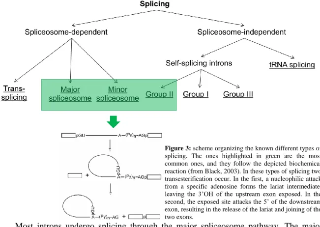
Figure 3 shows how the known types of splicing can be organized. Most introns depend on the spliceosome for their excision, but some are capable of it by themselves, through more or less similar mechanisms. Trans-splicing is a distinct case in spliceosome-dependent splicing, occurring only in a restricted number of species, and it involves splicing together two exons from different genes (Bonen, 1993).
Most introns undergo splicing through the major spliceosome pathway. The major spliceosome is composed of several ribonucleoproteins (RNPs), consisting in associations of one or two small nuclear RNA (snRNA) and proteins. The snRNA that are part of this structure are U1, U2, U4, U5 and U6. U4 and U6 are assembled together in the same RNP,
Figure 3: scheme organizing the known different types of
splicing. The ones highlighted in green are the most common ones, and they follow the depicted biochemical reaction (from Black, 2003). In these types of splicing two transesterification occur. In the first, a nucleophilic attack from a specific adenosine forms the lariat intermediate, leaving the 3’OH of the upstream exon exposed. In the second, the exposed site attacks the 5’ of the downstream exon, resulting in the release of the lariat and joining of the two exons.
8
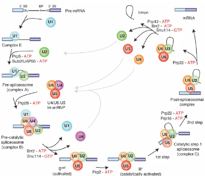
whilst the others are each on a different RNP. The association between RNPs and the intron varies during splicing, as well as the conformation of the snRNA and proteins (Will and Lührmann, 2011) (Figure 4). This process depends on the conservation of the components of the spliceosome, but also sequence components of the intron. A mutation in the branch point or any of the other conserved sequences will result in defective splicing (Reed and Maniatis, 1988; Talerico and Berget, 1990). This is also one of the keys to alternative splicing, as certain factors may enhance the detection of weaker splice sites, i.e., sequences that are only partially similar to the canonical splice sequences (Guiner et al, 2001).
The minor spliceosome is responsible for the splicing of only about 1 in every 300
introns (Steitz et al., 2008). This spliceosome contains the unique snRNA U11, U12, U4atac and U6atac, and also shares the U5 snRNA with the major spliceosome (Tarn and Steltz, 1996). This pathway is also characterized by splicing AT-AC introns, which have different conserved sequences. Despite the differences, the mechanism employed is very similar to the major spliceosome. There is a functional equivalence between U1 and U11, U2 and U12, U4
Figure 4: Assembly dynamics of the major spliceosome during intron splicing, highlighting interactions between snRNPs
9
and U4atac, and U6 and U6atac (Will and Lührmann, 2005). However, the minor spliceosome includes some proteins not involved in RNP complexes.
It has been shown that splicing can occur not only post-transcriptionally, but also co-transcriptionally (Beyer and Osheim, 1988). Although there is much evidence that this phenomenon is widespread, and that the spliceosome actually co-localizes with nascent transcripts (Lacadie et al., 2006), it has proven difficult to establish how it is regulated. Like other transcript modifications (such as capping and 3’ end processing) there is a known correlation between co-transcriptional splicing and CTD modification (Fong and Bentley, 2001), but the exact interaction with the spliceosome is not known. Some evidence, although not definite, points to the recruitment of spliceosome RNPs to the nascent RNA by interaction of these with newly synthesized splicing signals and by elongation factors, this last point explaining the correlation with the CTD dynamics (Neugebauer, 2002). Evidence also points to there being no distinction between splicing of constitutive or alternative exons happening co or post transcriptionally, although there seems to be some lag in transcription and splicing (Johnson et al., 2000; Pandya-Jones and Black, 2009). In another recent study, spliced intermediates were sequenced together with nascent transcripts associated with polymerase in yeast, allowing for a new way of studying these events (Churchman and Weissman, 2011). Novel approaches are now needed to understand transcription and splicing’s mutual influence.
2.2 3’ end processing and transcription termination
Compared with transcription initiation, less is known about termination. This is in part due to difficulties in studying termination, since it requires handling nascent transcripts, and also because of some neglect for being a process happening downstream of the encoding region, and hence it could be concluded to not have any role in gene expression regulation. Termination is characterized by detachment of Pol II from the DNA template, after transcription of the polyadenylation (pA) site. Since it was discovered that an intact pA site was essential for transcription termination (Connelly and Manly, 1988), evidence for a connection between termination and pre-mRNA 3’ end processing - which includes cleavage and polyadenylation (CPA) – has been increasing (Proudfoot et al., 2002).
3’ end processing mechanisms depend on large protein complexes (Shi et al., 2009), with elements involved in binding to specific RNA sequences, cleaving the RNA and polyadenylation of the new transcript’s generated end. Recognition of the AAUAAA motif, which has long been proven to be required for cleavage and polyadenylation (Zarkower et al.,
10
1986), is performed by the Cleavage and Polyadenylation Specificity Factor (CPSF), a complex with five subunits, including the endonuclease CPSF73. Downstream of this motif, the Cleavage stimulation Factor, specifically its subunit CstF64, will bind to a U/GU rich motif. These two factors interact with each other through various other elements of the complex, ultimately promoting the endonucleolytic activity of CPSF73 between the two motifs, 10 to 30 bases downstream of the pA site (Liu et al., 2007; Mandel et al., 2006; Lutz, 2008). This process and its components are highly conserved in eukaryotes but, despite being functionally very relevant, it is interesting to point out that CstF64 has a redundant role, since another protein, CstF64τ, is capable of performing the same function. Both proteins are very conserved, and seem to have different affinities with their interaction partners, but the biological reason for this functional duplication is not still fully understood (Yao et al., 2013). In the late 1980’s, two models emerged to explain transcription termination. The allosteric model (Logan et al., 1987) postulates that transcription of the pA site leads to conformational changes in Pol II or associated elongation factors, which causes dissociation of said factors and/or the association of termination factors, leading to 3’ end processing and downstream pausing of the polymerase after the release of the downstream transcript. The torpedo model (Connely and Manly, 1988) advocates for a termination-dependent degradation of the transcript downstream of the cleavage site by an exonuclease (the “torpedo”), later revealed to be Xrn2 (West et al., 2004). This enzyme’s activity requires a 5’ entry point, which is generated by co-transcriptional cleavage (CoTC), a process in which RNA cleaves itself once it is transcribed (Teixeira et al., 2004). However, CoTC activity was only so far identified in a small subset of genes (Nojima et al., 2013), making it hard to generalize this mechanism for now. But importantly, there is evidence that the two mechanisms may act together, since it has been described that cleavage after the pA can happen with Pol II still bound to the template, and that degradation by Xrn2 precedes the polymerase release from the template (West et al., 2008). This implies that pA site recognition is needed for the success of termination, hence pointing to a mixed model. Finally, it is also worth referring that pausing after the pA site might also play a role in termination by slowing down the polymerase.
The above descriptions of transcription termination and 3’ end processing refer to protein-coding genes in general, but replication-dependent histones are a notable exception. Histones are a highly conserved class of proteins responsible for chromatin packing in nucleosomes. They are subject to modifications, leading to very diverse roles in transcription regulation. Their genes are especially upregulated at the start of the S phase (Stein et al., 2006) because of DNA synthesis. Genes coding for replication-dependent histones are typically less than
11
2000 base pairs and intronless. The promoter region of H4 histone gene – the most studied – has regulatory sequences unique to other protein-coding genes (Ramsey-Erwing et al., 1994), but studies in Drosophila suggest that not all genes from this family are regulated by the same factors (Isogai et al., 2007). Similarly to other genes, histone mRNA has a 5’ 7-methylguanosine cap. However, these transcripts are not polyadenylated, relying instead on an exclusive 3’ end processing mechanism. It involves an RNA hairpin formed in the 3’ untranslated region (UTR), where a hairpin-binding protein (HBP) binds, so that CPSF73 can cleave the pre-mRNA (Dominski et al., 2005). Recognition of the cleavage site and positioning of the nuclease is thought to be made by U7 snRNP, which also helps in degradation of the 3’ cleaved portion (Cotten et al., 1988). These specificities, and the relevance of histones in the cellular context, may translate into particular Pol II dynamics and profiles in the synthesis of histone mRNA, not observed in other protein-coding genes.
The complexity of 3’ processing and termination mechanisms, allied to the co-transcriptionality and diversity of functions of its components - as shown for CPSF73, but also Xrn2, that has a role in premature termination (Brannan et al., 2012) – hints at a link with transcription regulatory mechanisms. It can be postulated that Ser2 phosphorylation of the CTD, a mark often found at the end of genes, may be related to these processes. An in-depth study tracking the polymerase in CPA factors-depleted cells can certainly shine a light on mRNA 3’ end determination and effects of the downstream processing events in transcription.
3. Micro RNAs
3.1 OverviewMicro RNAs (miRNA) are RNA strands of about 22 nucleotides that are involved in gene expression regulation by transcriptional silencing. They were first discovered in C. elegans in 1993, when it was described that the gene lin-4 produced a short non-coding RNA with an almost complementary sequence to the 3 ’end of the mRNA of lin-14 (Lee et al., 1993). Since then, miRNA have been discovered in all superior eukaryotic organisms (Maxwell et al., 2012). miRNA derive from pre-miRNA, which is an RNA hairpin.
Regulation by miRNA is preformed through the RNA interference (RNAi) pathway. Their function is accomplished together with other proteins in the RNA-induced silencing complex (RISC). In the RNAi pathway, pre-miRNA is exported to the cytoplasm via the Exportin
5-12
RanGTP complex (Lund et al., 2004). Upon arriving, the hairpin is cleaved by Dicer, a type III RNase, generating a double-strand RNA (dsRNA) of about 22nt (Bernstein et al., 2001). Dicer is one of the elements of RISC, the others being HIV-1 transactivation responsive element (TAR) RNA-binding protein (TRBP), PACT and proteins of the Argonaut family (Rana, 2007). The complex then selects only one of the strands of the dsRNA to be used. The selection is not yet fully comprehended, but some evidence points to a selection based on the strand thermodynamic stability, discarding the most stable strand (Siomi and Siomi, 2009). The complete ribonucleoprotein complex is called miRISC. Finally, the miRNA incorporated in RISC will find its target and bind to it. Binding can be partial (only some nucleotides pair with the target) or complete. The second is more common in plants, although it can also happen in animals. These two mechanisms are functionally different, since incomplete pairing only leads to translational silencing (which can be transient), whereas complete pairing leads to target mRNA degradation (by the C-terminal PIWI domain of Argonaut proteins), although it might not always be the case (Bartel et al., 2004). Many studies point to the pairing of some positions having more effect in the mRNA’s fate, rather than the whole miRNA. Additionally, in cases of degradation, it has been shown that miRNA can proceed to a different target to fulfill the same function (Hutvágner and Zamore, 2002). As for silenced mRNAs, in some instances they are clustered in sub-cellular regions called Processing bodies (P-bodies), where other interveners may eventually, but not certainly, mediate RNA turnover, generally by decapping mRNA (Brengues et al., 2005).
3.2 Micro RNA transcription and processing
As more miRNAs were discovered, it became possible to classify them into distinct categories according to their gene structure. Some miRNA are intragenic (Figure 5A), whereas others are intergenic (Figures 5B and C). Intragenic micro RNAs are found in introns, and usually have the same orientation than the host gene. They can be found in introns of protein-coding and long non-coding RNA genes (He et al., 2008). Intergenic miRNA can be near or far from other genes. They can be classified into clustered miRNA (Figure 5B) or single miRNA (Figure 5C). Micro RNAs belonging to the same cluster can have similar functions or related targets, with some clusters being associated with tumors (Mendell, 2008). It is thought that all intergenic miRNA have a larger associated transcript called primary-miRNA (pri-miRNA), and some were already described, as seen in Figure 5B (Lee et al., 2002). Like their intragenic counterparts, most intergenic miRNA are transcribed
13
by Pol II (Lee et al., 2004), although some miRNA clusters, because of their close association with Alu elements, are transcribed by RNA polymerase III (Borchert et al., 2006).
In order to obtain the pre-miRNA, introns (after being debranched) and pri-miRNA have to be cleaved so that the hairpin sequence can be obtained. The nuclear RNase III Drosha is responsible for the primary transcript cleavage (Lee et al., 2003), acting together with DGCR8 (Yeom et al., 2006). However, the mechanism Drosha uses for recognizing the cleavage site is still very debated (Zeng et al., 2004; Ma et al., 2013), and so prediction of these sites has a large value in understanding the mechanism and recognizing novel miRNA (Hu et al., 2013). In addition to this, some micro RNAs differ from their reference in very few nucleotides. These are called isomiRs (Morin et al., 2008), and although their biogenesis is still poorly understood, there is evidence pointing to some of them owing their variability to multiple cleavage by Drosha (Ma et al., 2013).
Just like every type of gene, micro RNAs must also have proper nomenclature for organization purposes. In earlier times, naming was more similar to genes, and the standardization to include “mir” on their name (still following the formatting for a species gene’s name) was only included later (Ambros et al., 2003). Also, miRNAs from the same hairpin used to be distinguished by their expression level, but nowadays that is done based on strand. Naming also depends on homology with miRNA found in other organisms, on the genomic locus they’re included, on base differences between two sequences and on the order of discovery (Griffiths-Jones, 2004). Most of the current information about micro RNAs is on miRBase (current version is v21, most recent description in Kozomara and Griffiths-Jones, 2014). This database includes deep sequencing datasets, genomic coordinates, sequence and
Figure 5: Three distinct types of miRNA genes. A – intronic miRNA; B – Intergenic clustered miRNA; C – intergenic
14
biological information, among other relevant details. In this database, miRNA are named like has-mir-17-5p, where the first three letters describe the species, and 5p tells the strand. When new miRNAs are discovered they can be added to the database and the attributed name can be then included in the publication.
4. Genome-wide study of transcription
4.1 High-throughput sequencing approachesUnderstanding transcription is historically associated with molecular, genetic or biochemical studies of single genes or components. The first change in this paradigm came with the introduction of microarrays (Schena et al., 1995), allowing for the quantification of the RNA from several genes at the same time. With development of the technology, it became possible to identify alternative splice sites (Johnson et al., 2003) and to correlate co-expression of genes with promoter motifs (Veerla and Höglund, 2006). In spite of the huge breakthroughs they allowed in gene expression studies, microarrays have some inherent bias when it comes to coverage and amplification (Boelens et al., 2007). The development of next generation sequencing (NGS) technologies allowed for the development of less biased, albeit more expensive, techniques, with greater coverage. RNA-seq was capable of providing the same type of information as microarrays, but in an even larger scale, driving the discovery of novel transcripts and isoforms. Analyzing cell-fraction RNA-seq data also revealed a generalized presence of co-transcriptional splicing in protein-coding genes, but not so much in lncRNAs (Tilgner et al., 2012). But because it is a dynamical process, transcription ought to be studied using methodologies that focus not only on the end product, the mRNA, but also on the intermediates in its synthesis – nascent RNA and Pol II.
Chromatin immunoprecipitation (ChIP) protocols have been developed to assess interactions between proteins and DNA (Kim and Ren, 2006). The binding sites in DNA were initially analyzed using microarrays, but quickly became evident that the coverage offered by next generation high-throughput sequencing would provide more accurate and robust results, which led to the development of ChIP-seq protocols (Johnson et al., 2007; Robertson et al., 2007). Later, ChIP-seq was used to assess Pol II distribution across genes (Baugh et al., 2007), which attested the large accumulation of stalled Pol II in a promoter proximal region previously described in microarray studies (Kim et al., 2005; Guenther et al., 2007). ChIP-seq
15
assays were also performed using antibodies that specifically target different phosphorylation patterns, thus showing a broad genome-wide picture of where in genes each phosphorylation mark could be found (Rahl et al., 2010, Grosso et al., 2012).
While ChIP-seq profiles rendered a good image of gene occupancy by Pol II, resolution was still low, there was no distinction of whether the enzyme was actively transcribing or paused, and the focus in the relevant part of transcription – RNA synthesis – was not being captured. This resulted in the development of four techniques that aimed to solve these problems, all of them targeting Pol II-associated RNA (Figure 6).
Short capped RNA-seq (Nechaev et al., 2010) was introduced to study promoter-proximal pausing. It captures RNAs with a 7-methylguanosine cap, selecting those with between 25 and 120 bp. Those short RNAs are then sequenced, and it is possible to determine the first base transcribed - by using a 5’ sequencing primer – or the last base – using a 3’ sequencing primer. The last base sequenced is the last base incorporated by the polymerase, so the technique allows determination of paused Pol II position at a single-nucleotide resolution by aligning the whole reads and then extracting that base. However, this protocol can only provide knowledge on the position of paused polymerases in early elongation, because of the size and cap selection steps, and it cannot also accuratly distinguish between Pol II associated nascent transcripts and released short transcripts.
The sequencing of native elongating transcripts (NET-seq) associated with Pol II (Churchmand and Weissman, 2011) was able to expand the single-nucleotide resolution of polymerase tracking to the whole gene. This method is based on the immunoprecipitation of a
Figure 6: RNA-based genome-wide transcription tracking protocols. Short-capped RNA sequencing selects the target
RNAs by the presence of a 7-methylguanosine cap; NET-seq selects the RNA attached to the polymerase by immunoprecipitaton targeting a synthetic flag; and GRO-seq and PRO-seq are based on in vitro run on reactions using labeled nucleotides. Description of each technique in the main text.
16
flag-tagged Pol II. Due to the stability of the trenary complex, it is possible to extract the RNA associated with the polymerase. Aligning the reads and extracting the position of the last base will tell which was the last nucleotide incorporated. The method was also designed as strand-specific, giving information about sense and antisense nascent transcripts. This is an important distinction as it reduces noise in the data and allows the discovery of new transcriptional units. Finally, NET-seq also captures splicing intermedates from co-transcriptional splicing. While this has to be considered while analyzing the data, it also allows the study of splicing, and perhaps other co-transcriptional events that generate intermediates, if they’re captured by the protocol.
Both of the methods described above unbiasedly capture Pol II-associated transcripts. But they make no distinction of which polymerases are paused or actively engaged in transcription. Acquiring the position of engaged Pol II can be achieved through nuclear run-on reactirun-ons coupled to sequencing, as it is drun-one in global run-run-on sequencing (GRO-seq) and precise run-on sequencing (PRO-seq) (Core et al., 2008; Kwak et al., 2013). The first method uses Br-UTP to mark the run-on synthesized transcripts so they can be isolated and then sequenced. Yet this method lacks precision when compared to the others described, so it was upgraded to PRO-seq. In this protocol, four libraries are prepared from four run-on reactions, each of them using only one nucleotide as substrate. The nucleotides used are biotinylated so that the transcripts can be selected. The libraries are then sequenced and merged, and the last base of each read is considered the position where the engaged Pol II is. This method does not guarantee single nucleotide resolution as it is possible to find the same nucleotide repeated, which would drive the incorporation of two nucleotides in the nuclear run-on reaction. Also, the added manipulation required for isolation of nuclei and run-on may also disrupt some features of transcription.
Even though these genome-wide protocols show data for virtually all the genes in the genome, there are still biases that need to be considered when presenting the results for these experiments. First of all, the protocols are designed to be preformed in a collection of cells. While this captures the biological variability between individuals, it does not express exactly how a single individual behaves. Specifically, when looking at a single-nucleotide resolution gene profile, we may identify two consecutive bases with signal, yet it is physically impossible to have two polymerases in consecutive bases. Development of single-cell protocols will bring this individuality to the analysis, but conversely sacrificing the patterns only observable when looking to many subjects. Second, it is customary to look at metagene profiles (i.e. a global average profile for all genes) when interpreting this kind of data. While
17
it may be informative of general features, it may be necessary to subset the genes by some feature, and ultimately look at many individual genes, so as to identify differences between them that may be lost while averaging the full set. Finally, a constant adaptation of computational biology’s protocols and tools is needed, as more advanced and singular arise. While some steps in the pipelines are well established, especially alignments, other steps are specifically adapted to the protocol in question and may therefore not be fully optimized.
4.2 Data Analysis
The exponential increase of available biological datasets from high-throughput techniques – and, in particular, NGS protocols – has stimulated the development of bioinformatical tools that can accurately, but also efficiently, process the large volumes of data produced. These datasets are not only abundant, but also diverse, as all biological research fields have realized the importance of more comprehensive data, rather than focusing on individual components of the system studied. This led to the development of many protocols, like the ones described in the previous section, to which the data analysis must adapt.
Although the output of sequencing platforms can differ between them, the most common type of files – and the one used in this project - is FASTQ (Cock et al., 2010). In this kind of files, each read is named uniquely and accompanied by the quality of each base. It is important to know what is the encoding for the quality scores, since some tools do not automatically recognize it. Read quality should also be assessed before starting any analysis, to account for possible biases. A commonly used tool that collects relevant quality statistics is fastqc (http://www.bioinformatics.babraham.ac.uk/projects/fastqc/, last accessed on August 23rd 2014). This allows the identification of overrepresented sequences, such as adapters or RNA sequences that either hinder the analysis or introduce bias, leading to wrong conclusions.
There is a plethora of adaptor trimming tools to choose from. The choice varies with read type and size, as well as features offered by each software. Cutadapt (Martin, 2011) is one of these tools, and it allows the user to trim reads from both ends and considering as many adapter sequences as needed. Besides, it does not require the complete identification of the adaptor to perform trimming, allowing the user to set a minimum of nucleotides corresponding to the adapter to be trimmed with a certain error rate. When sequencing short nucleotide strands using a pair-end protocol, it is possible that the read size is greater than the actual length of what is being sequenced. In this case, a variable number of bases
18
corresponding to the opposite strand’s adaptor might be sequenced, resulting in a read that cannot be aligned with the reference genome. The referred features offered by Cutadapt present a way to solving this issue.
Aligning the obtained reads to a reference genome is arguably the most important step in a sequencing analysis. However, it is usually also the most computationally demanding stage, even more considering the need of high coverage data. This happens because of the elevated number of comparisons that would have to be made between the bases of millions of reads and the billions of bases in a genome, and even more considering the quality of each base. Luckily, it is possible to implement some heuristics in order to speed up the algorithms. Many aligners have been developed over the years, with the purpose of finding the best balance between an accurate alignment and a fast execution. TopHat2 (Kim et al., 2013) is the latest version of a widely-used tool for aligning RNA-derived reads to reference genomes. TopHat2 resorts to Bowtie2, a tool from the same lab (Langmead and Salzberg, 2012), to align the reads, and then performs splice junction finding. Many parameters are customizable so that the user can fit them to the data. It is usually important to get uniquely aligned reads, as multiple aligned RNA reads cannot be interpreted as a product of a single DNA sequence transcription.
TopHat2 outputs a BAM file with information about where the reads aligned to the reference. It is a useful way of storing the alignment, but cannot be directly worked with. To work with BAM files (and their non-binary counterpart, SAM files), SAMtools is available (Li et al., 2009). This collection of tools enables visualization, filtering, sorting and indexing of these files, among other features. They can also be used together with other tools to find spliced reads, or to isolate the last nucleotide as single-nucleotide resolution techniques require. However, BAM and SAM formats do not supply appropriated data that can be worked on, such as read counts in genome intervals, and also don’t provide effective means to make operations in the data, like intersections or subtractions. For these cases, BEDTools (Quinlan and Hall, 2010) come in handy. This toolkit, that relies mostly on the BED format (Kent et al., 2002), provides the user with a framework for intersecting datasets, separating data by location, calculating local or genome-wide coverage, and can be articulated with other tools if needed. The output BED format files are fairly more readable than the SAM format ones, and information can be easily extracted to be processed. BEDTools is also compliant with other file formats, such as GTF of BAM files, which makes the analysis much more agile since there is no need of converting these files to another format. For instance, a BAM file output by an aligner may be directly used to assess coverage of desired features.
19
Visualization is of great importance when dealing with genomic data. The UCSC Genome Browser (Kent et al., 2002) is a useful online resource for visualizing data in a genomic context. It provides several tracks of annotations or data from other sources, so as to facilitate interpretation. It is possible to see, gene by gene, the presence of RNA-seq reads or polymerase distribution. But to get a sense of what is happening in the average gene, data can be compiled in metagene profiles. These profiles divide genes in windows, and plot the mean read counts - or a normalized version like reads per kilobase per million reads (RPKM) – of every window of all genes. This results in an average profile that makes it possible to identify features present in many genes, for instance the accumulation of polymerase in the promoter region (Core et al., 2008; Nechaev et al., 2010). However, construction and interpretation of these must be done carefully. First, not all genes can be included in a profile. Many genes overlap each other, and that can give rise to features that may not exist. Other sets of genes may also have their unique features - for example, replication-dependent histones, which are shorter than average genes and have unique Pol II occupation profiles -, and so should be removed from the analysis. Second, genes can also have unique features that are not displayed or that interfere with the profile, and therefore some manual selection of the genes included has to be preformed, and individual examples should always be shown. Other metrics can be presented to highlight differences between sets of genes or conditions. A logarithm of the quotient of the number of reads in two windows is a useful means for making such comparisons, and a distribution of the values for the genes can also be shown as a boxplot. Further statistical testing can be applied to confirm those differences.
From an RNA-seq experiment we can also identify which transcript isoforms are more or less expressed. The Cufflinks software (Trapnell et al., 2010) is widely used as a tool to attribute read counts to genes and isoforms, allowing inferences to be made about their expression. But it is also possible to identify which exons are alternatively being expressed. MISO (Mixture-of-Isoforms) (Katz et al., 2010) uses RNA-seq data to quantitatively predict which alternative splicing phenomena occur in a sample or between samples. This program uses its own database of alternative splicing events, and follows a Bayesian framework to attribute a read either to one isoform or another. The database are divided by their type of event (skipped exons, alternative 3’/5’ splice sites, mutually exclusive exons, tandem 3’
UTRs, retained introns, and alternative first or last exons), and processing results in a value that indicates whether one event or the other is selected. For instance, in a larger scale, this allows seeing differences between included or skipped exons in polymerase occupation, but these and other features can also be shown individually.
20
NGS technologies opened the door to large genomic studies, and to high coverage sequencing data. Yet, constant adaptation of methodologies is required for capturing not only the broad patterns but also the fine details provided by these approaches.
5. Objectives
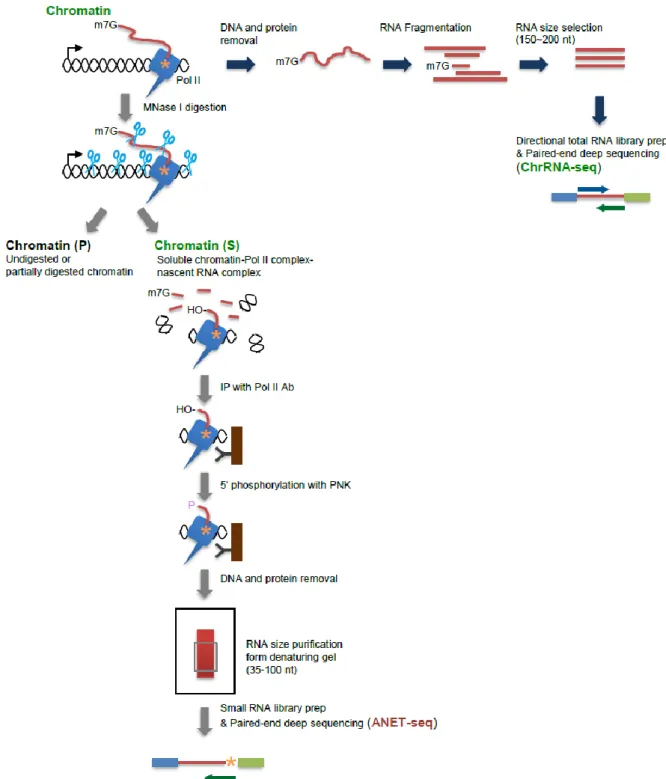
Considering the complexities of RNA transcription and processing, this work aims to elucidate more about the interactions between these two processes in a genome-wide scope. The focus of this study is the Pol II CTD, since it is one of the key regulators of interactions involving Pol II during transcription. To achieve this, the NET-seq protocol (Churchman and Weissman, 2012) was modified to enable usage of CTD isoform-specific antibodies. An additional step was also included where the chromatin fraction from which Pol II was precipitated was treated with micrococcal nuclease (MNase), in order to degrade exposed RNA sequences, thus increasing the specificity for RNA protected by Pol II. This new protocol was termed “advanced NET-seq” (ANET-seq), and allows for the first time for a single-nucleotide resolution mapping of CTD isoforms in the genome. To further support ANET-seq results and interpretation, chromatin-fraction RNA (ChrRNA) was sequenced. This would show some unstable RNAs, such as promoter upstream transcripts (PROMPTs) introns, and transcription downstream of the 3’ end. All samples were sequenced in Illumina HiSeq 2000 or 2500 sequencers.
Two sets of data were produced for this project. The first includes one ChrRNA sample and ANET-seq samples generated using antibodies for unphosphorylated Pol II CTD, Ser2-phosphorylated CTD, Ser5-Ser2-phosphorylated CTD and all CTD isoforms. These aimed at showing differences between CTD isoforms in transcription. The second includes ChrRNA and ANET data from three 3’ end processing and termination factors knock-downs and a control. These are meant to show the effects that each factor has on Pol II transcription dynamics and changes in newly synthesized transcripts. The ANET-seq from this set used an antibody targeting Ser2-phosphorylated CTD, the predominant isoform at the end of genes.
The main objectives of this project were:
1. Define an analysis pipeline for ANET-seq data.
21
3. Examine the CTD phosphorylation dynamics associated with splicing and miRNA biogenesis.
4. Elucidate the different roles of the termination factors Xrn2, CPSF73 and CstF64+CstF64τ during transcription.
Being a frontier discipline, computational biology requires the input and collaboration specialists from different fields. It is from the combination of these different skill sets that complex and relevant new discoveries can be made. In this work, I preformed all of the sequencing data analysis, such as trimming and aligning reads, making average gene and exon profiles, plotting single gene profiles or calculating Escaping Indices. The experimental work was performed by other scientists specialized in those protocols and they are duly credited in the final manuscript.
The funding for the work here presented was granted by Wellcome Trust Programme, ERC Advanced Grants and Fundação Ciência e Tecnologia.
22
Chapter 2
23
Human genome-wide profiles of nascent transcription and
co-transcriptional processing: advanced NET-seq technology
Takayuki Nojima1§, Tomás Gomes2§, Ana Rita Fialho Grosso2, Hiroshi Kimura3, Michael J. Dye1, Somdutta Dhir1, Maria Carmo-Fonseca2* and Nicholas J. Proudfoot1*
1
Sir William Dunn School of Pathology, University of Oxford, South Parks Road, Oxford OX1 3RE, United Kingdom.
2
Instituto de Medicina Molecular, Faculdade de Medicina, Universidade de Lisboa, 1649-028 Lisboa, Portugal
3
Department of Biological Sciences, Graduate School of Bioscience and Biotechnology, Tokyo Institute of Technology, Yokohama, Japan
§
These authors have contributed equally to the work. * Joint communicating authors
24
1. Summary
RNA polymerase II (Pol II) transcribes nascent RNA throughout the mammalian genome. However, many aspects of nascent RNA metabolism are poorly understood due to RNA instability and technical limitations. We have employed high throughput sequencing at single-nucleotide resolution to characterize nascent transcription in HeLa cells; advanced native elongating transcript-sequencing (ANET-seq). This provides precise maps of nascent RNA within the Pol II elongation complex that correlate with the C-terminal domain (CTD) phosphorylation state of the Pol II large subunit. We detect substantial Pol II bidirectional pausing at transcription start sites (TSS). We also demonstrate exon tethering to the CTD Ser5 phosphorylated Pol II complex and co-transcriptional pre-miRNA biogenesis. Depletion of cleavage and polyadenylation (CPA) factors causes termination defects, reducing Pol II pausing at transcription end site (TES). Additionally the 3' end termination machinery plays a promoter role by restricting non-productive RNA synthesis at the TSS in both sense and antisense directions.
25
2. Highlights
ANET-seq monitors nascent RNA within the mammalian Pol II complex.
Pol II pausing at TSS and TES with different Pol II CTD phosphorylation states. Exon tethering during co-transcriptional splicing links CTD S5P to 5’SS cleavage. Diverse kinetics of co-transcriptional pre-miRNA biogenesis.
CPA factors are associated with Pol II pausing at TES.