Faculdade de Medicina
The role of IL-7 in the Homeostasis of
Human Naive and Memory
CD4
+
T cell subsets
Rita Isabel Silva de Azeved
o
Doutoramento em Ciências Biomédicas
Especialidade em Imunologia
Faculdade de Medicina
The role of IL-7 in the Homeostasis of
Human Naive and Memory
CD4
+
T cell subsets
Rita Isabel Silva de Azevedo
Tese orientada pela Doutora Maria V. D. Soares
Doutoramento em Ciências Biomédicas
Especialidade em Imunologia
Todas as afirmações efectuadas no
presente documento são da exclusiva
responsabilidade do seu autor, não
cabendo qualquer responsabilidade à
Faculdade de Medicina de Lisboa pelos
conteúdos nele apresentados.
A
impressão
desta
dissertação
foi
aprovada pela Comissão Coordenadora
do Conselho Científico da Faculdade de
Medicina de Lisboa em reunião de 18 de
Janeiro de 2011.
Dissertação apresentada à Faculdade de
Medicina da Universidade de Lisboa,
para obtenção do grau de Doutor em
Ciências Biomédicas.
A presente dissertação foi realizada na
Unidade de Imunologia Clínica do
Instituto de Medicina Molecular,
Faculdade de Medicina da
Universidade de Lisboa e
Division of Infection & Immunity,
University College London.
O trabalho aqui apresentado foi
realizado com âmbito nos projectos
PPCDT/BIA-BCM/61079/2004 e
PTDC/SAU-MII/67662/2006,
co-financiados pelo fundo
comunitário FEDER através dos
programas POCI 2010 e PTDC.
Bolsa de Doutoramento da Fundação
para a Ciência e a Tecnologia
ACKNOWLEDGEMENTS ... I
ABBREVIATIONS ... III
RESUMO ... VII
SUMMARY ... IX
INTRODUCTION ... 1
1. Interleukin-7: a key cytokine in T cell homeostasis ... 1
1.1. The role of γC cytokines in T cell homeostasis ... 1
1.2. IL-7 Receptor signalling in T cells ... 2
1.3. Therapeutic applications of IL-7 ... 4
1.4. Hematopoietic Stem Cell Transplantation: a major disturbance to T cell homeostasis ... 9
2. Immune response: Naive to Memory ... 11
2.1. T cell subsets: Markers & Nomenclature ... 11
2.2 Naive CD4+ T cell subsets defined by CD31 expression ... 12
2.3. CD45RA re-expressing memory T cells ... 15
3. Immune and Cellular senescence ... 18
3.1. Telomeres, Telomerase and Senescence ... 19
3.2. Immune-senescence in the elderly ... 22
3.3. CMV infection accelerates immune-senescence ... 23
3.4. Cellular Senescence ... 24
References ... 29
CHAPTER 1 ... 57
Role of IL-7 in the homeostasis of human CD31
+naive CD4
+T cells
Introduction ... 572. Purification of lymphocyte subsets ... 59
3. In vitro cell culture ... 59
4. Flow cytometric analysis ... 59
5. Signal-Joint TREC quantification by Real-Time PCR ... 61
6. TCR-chain CDR3 spectratyping ... 62
7. Statistical analysis ... 62
Results ... 63
Chapter 1.1 ... 63
IL-7 sustains CD31 expression in human naive CD4+ T cells and preferentially expands the CD31+ subsetin a PI3K-dependent manner Chapter 1.2 ... 76
Long Term Immune Reconstitution Following Haplotype-Mismatched Hematopoietic Stem Cell Transplantation Discussion ... 92
References ... 96
CHAPTER 2 ... 103
Characterization of human CD45RA
+CD27
-CD4
+T cells
Introduction ... 103Methods ... 104
1. Blood samples ... 104
2. Purification of Lymphocyte Subsets... 104
3. In vitro Cell Culture ... 104
4. Proliferation assessment by [3H]Thymidine Incorporation ... 105
5. Flow Cytometric Analysis ... 105
6. Measurement of Telomerase Activity by TRAP assay ... 108
7. Real-Time quantitative PCR (RT-qPCR) ... 108
8. Western blot analysis ... 109
IL-7-driven homeostatic mechanism induces CD45RA re-expression on CD45RA -CD27+ CD4+ T cells
Chapter 2.2... 126
CD45RA+CD27- CD4+ T cells exhibit p38 MAPK-regulated telomere-independent senescence Discussion ... 141 References ... 148
CONCLUSIONS ... 155
References ... 162LIST OF PUBLICATIONS ... 165
Peer-reviewed articles ... 165Manuscripts under submission ... 166
Manuscripts in preparation ... 166
Communications ... 167
Major Conferences Attended ... 168
APPENDIX ... 171
CHAPTER 1
Role of IL-7 in the homeostasis of human CD31
+naive CD4
+T cells
Chapter 1.1
IL-7 sustains CD31 expression in human naive CD4+ T cells and preferentially
expands the CD31+ subset in a PI3K-dependent manner
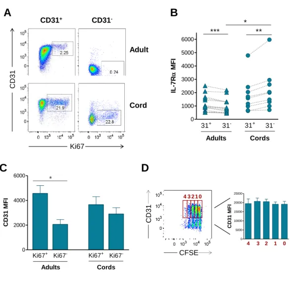
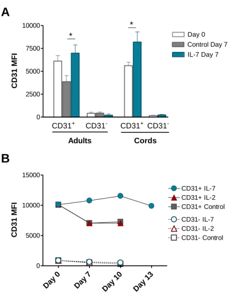
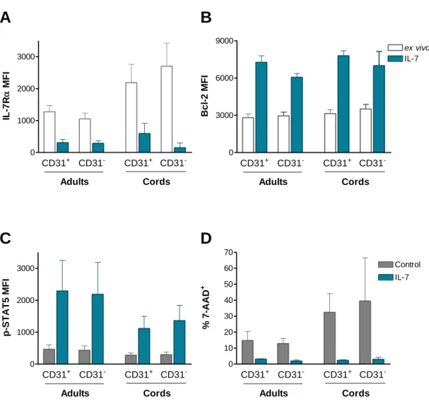
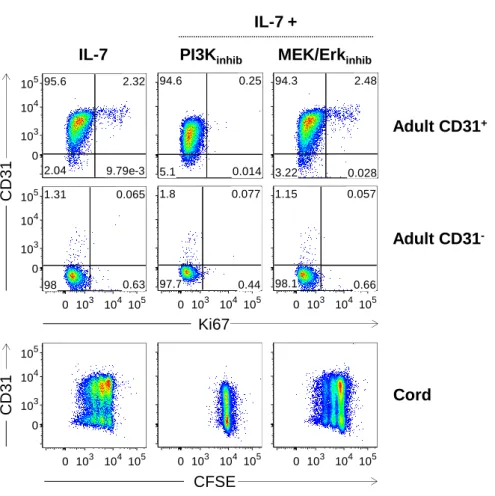
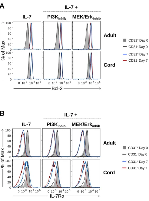
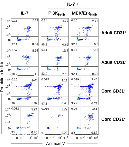
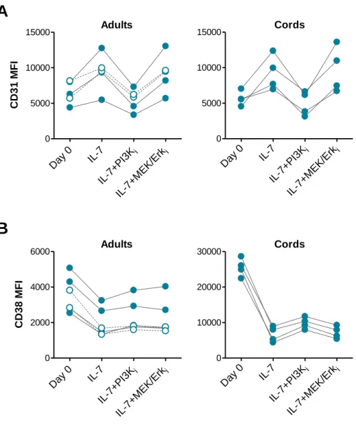
Figure 1: CD31 expression profiles and gating strategy used to purify CD31+ and CD31 -naive CD4+ T cell subsets from adult and cord blood. ... 64 Figure 2: IL-7-induced cycling of adult naive CD4+ T cells is restricted to the CD31+ subset. ... 65 Figure 3: IL-7 promotes the maintenance but not re-expression of CD31 on both adult and cord blood naive CD4+ T cells. ... 68 Figure 4: IL-7 stimulation leads to IL-7Rα down-modulation, Bcl-2 up-regulation, STAT5 phosphorylation and rescue from apoptosis in both CD31+ and CD31- naive CD4+ subsets. ... 69 Figure 5: IL-7-induced proliferation of adult CD31+ and cord blood naive CD4+ T cells is dependent on the PI3K pathway. ... 71 Figure 6: Bcl-2 and IL-7Rα expression on adult CD31+ naive CD4+ T cells is independent of the PI3K pathway. ... 72 Figure 7: IL-7-mediated survival of naive CD4+ T cell subsets is only minimally affected by PI3K inhibition. ... 74 Figure 8: IL-7-mediated CD31 maintenance on both adult and CB naive CD4+ T cells is dependent on the PI3K pathway. ... 75
Chapter 1.2
Long Term Immune Reconstitution Following Haplotype-Mismatched
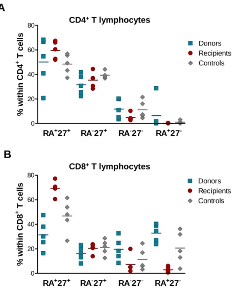
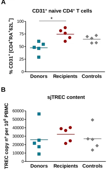
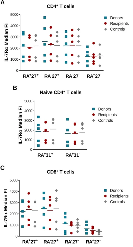
Figure 11: Frequency of naive and memory subsets within the CD4+ and CD8+ T cell pools. ... 80 Figure 12: Assessment of relative RTE levels through the expression of CD31 and sjTREC content. ... 82 Figure 13: Telomere length measurement within CD4+ and CD8+ T cells. ... 83 Figure 14: IL-7Rα expression within CD4+ and CD8+ T cell subsets. ... 85 Figure 15: Assessment of TCR repertoire by spectratyping analysis of the CDR3 Vβ regions of CD4+ T cells. ... 87 Figure 16: Spectratyping analysis of the CDR3 Vβ regions of CD4+ T cells from a representative recipient together with the respective donor and age-matched control. .... 88 Figure 17: Assessment of TCR repertoire by spectratyping analysis of the CDR3 Vβ regions of CD8+ T cells. ... 89 Figure 18: Spectratyping analysis of the CDR3 Vβ regions of CD8+ T cells from a representative recipient together with the respective donor and age-matched control. .... 90 Figure 19: Complexity score within CD4+ and CD8+ T cells. ... 91
CHAPTER 2
Characterization of CD45RA
+CD27
-CD4
+T cells
Chapter 2.1
IL-7-driven homeostatic mechanism induces CD45RA re-expression on CD45RA
-CD27+ CD4+ T cells
Figure 1: CD4+ CD45RA+ CD27- cells express high levels of differentiation markers and of cytolytic molecules. ... 111 Figure 2: CD4+ CD45RA+CD427- cells do not accumulate in culture following activation. ... 113
Figure 4: CD4+ CD45RA+CD427- cells have impaired cell survival following activation. ... 116 Figure 5: Detection of pAkt(Ser473) phosphorylation by flow cytometry. ... 118 Figure 6: CD4+ CD45RA+CD27- cells have impaired Akt(Ser473) phosphorylation. ... 119 Figure 7: CD4+ CD45RA-CD27+ cells stably re-express CD45RA following IL-7-driven proliferation. ... 121 Figure 8: CD4+ CD45RA-CD27+cells do not re-express CD45RA when stimulated with other γC cytokines nor do CD45RA
-CD27-. ... 122 Figure 9: Transcription factors involved in T cell differentiation are highly expressed in CD4+ CD45RA+CD27- cells but only T-bet is induced by IL-7. ... 124
Chapter 2.2
CD45RA+CD27- CD4+ T cells exhibit p38 MAPK-regulated telomere-independent
senescence
Figure 10: CD4+ CD45RA+CD27- cells do not have the shortest telomeres but have impaired telomerase activity. ... 127 Figure 11: Assessment of -H2AX expression by flow cytometry... 129 Figure 12: CD4+ CD45RA+CD27- cells express high levels of -H2AX following activation. ... 130 Figure 13: CD4+ CD45RA+CD27- cells express higher levels of total and phosphorylated p38. ... 132 Figure 14: CD4+ CD45RA+CD27- cells express lower levels of -H2AX when the p38 pathway is inhibited. ... 133 Figure 15: p38 inhibition improves cell recovery and survival but not proliferation of CD45RA+CD27- cells. ... 135 Figure 16: p38 inhibition increases Bcl-2 expression but not pAkt(Ser473) phosphorylation. ... 136
Figure 18: Inhibition of p38 abrogates the TNF-α-induced down-modulation of telomerase activity. ... 139
A
CKNOWLEDGEMENTS
I would like to start by acknowledging my supervisor Doutora Maria Soares for all her support and helpful discussions not only during the writing of this thesis but also throughout the six years that we have worked together. Despite all the peaks and valleys, I want to thank Maria for always believing in me.
I wish to thank Professor Arne Akbar for allowing me to develop part of my PhD project in his lab in the Division of Infection & Immunity, University College London, and for being incredibly supportive and enthusiastic about my work. I will be forever grateful to Arne for making me feel truly welcome in his group and for bestowing on me such a wonderful opportunity to work (and play) in London.
I also have to acknowledge Professora Doutora Ana Espada de Sousa and Professor Doutor Rui Victorino for giving me the opportunity to perform my PhD project in Unidade de Imunologia Clínica, Instituto de Medicina Molecular.
I want to thank Professor Doutor João Lacerda from Serviço de Hematologia, Hospital de Santa Maria, for allowing me to participate in a very interesting project that gave me a new perspective on research and for always showing support and trust in my work.
I must say a big thank you to my colleagues and true friends from Unidade de Imunologia Clínica who took me under their wing, teaching me all the lab techniques that were to be the foundations of my future work and, more importantly, personal lessons that will stay with me throughout my life: Adriana Albuquerque showed me how to juggle hard work and skilfulness, with endless generosity and kindness; Rita Cavaleiro taught me to stand up for myself and for what I think is right, and I’ll always be thankful for that; Russell Foxall selflessly shared his time and endless immunological wisdom… and he also starred in the most hilarious moments I have ever witnessed in a lab; Doutora Alcinda Melo always managed to put a smile on my face with her positive outlook on life and all its mishaps. I especially want to thank Rita Tendeiro, not only for being my trusted extra pair of hands during TREC quantification and the final experiments I performed in Lisbon, but most importantly for being a very close and dear friend, i.e. a
minh’ámiga . I also must thank Ana Luísa Caetano from Unidade de Citometria de
Molecular. Ana Luísa went far beyond the call of duty, working til the wee hours of the morning if necessary, to help me out and I’ll always cherish her friendship.
Thank you all for making it worth my while to get into work every day, even when the going got really tough indeed.
I wish to acknowledge my surrogate supervisors at the UCL and cari amici, Valentina Libri and Diletta di Mitri, for their support, companionship and all the lovely dinners they hosted. They both taught me a lot in the lab and were instrumental in making London feel like home. I must acknowledge Toni for making life in the lab run smoothly through her great organizational skills and generous helpfulness. But most importantly, I want to thank Toni for basically being Toni, a kind-hearted friend who features in my fondest and funnest memories of London. I also wish to thank Elaine, Nicky, Daisy and Simran for their friendly faces and kind words that really helped warm the cockles of my heart in cold London.
I wish to acknowledge Dário Ligeiro from Immunogenetics Laboratory, Centro de Histocompatibilidade do Sul, for performing the spectratyping analysis, and Rui Soares from Unidade de Imunologia Clínica for technical support during TREC quantification.
I want to acknowledge Doutora Helena Ferreira from Hospital Universitário de Santa Maria, Lisboa, for providing the umbilical cord blood samples. I also acknowledge Rémy Cheynier from Institute Pasteur, Paris for providing the pCD3-TREC plasmid, and Professor David Kipling from University of Wales College of Medicine for supplying BIRB796 aliquots.
I acknowledge Fundação para a Ciência e a Tecnologia for the financial support through the PhD Scholarship (SFRH/ BD/ 29120/ 2006), co-financed by POCI 2010 and Fundo Social Europeu.
Last but definitely not least, I would like to thank my friends and family for all their love and endless patience to put up with the crazy hours, weariness and quite frankly grumpiness that came with the territory of surviving this PhD. Most especially of all, I want to thank my parents for their unwavering love and support throughout my life, always motivating me to do my best.
Obrigada por tudo, Mãe e Pai, esta tese é para vocês porque sei que o meu sucesso passado, presente e futuro se deve ao vosso amor e apoio incondicionais.
A
BBREVIATIONS
7-AAD 7-Aminoactinomycin D
γC Common γ Chain
AIDS Acquired Immune Deficiency Syndrome
APC Antigen-Presenting Cell
ATM Ataxia Telangiectasia Mutated
ATR Ataxia Telangiectasia and Rad3-related Protein
BSA Bovine Serum Albumin
c-ART Combination Anti-retroviral Therapy
CCR CC Chemokine Receptor
CD Cluster of Differentiation
CDR3 Complementarity-Determining Region 3
CFSE Carboxyfluorescein Diacetate Succinimidyl Ester
CMV Cytomegalovirus
Con A Concanavalin A
DDR DNA Damage Response
DLI Donor Lymphocyte Infusion
DMSO Dimethyl Sulfoxide
DSB Double Strand Break
EBV Epstein-Barr Virus
Eomes Eomesodermin
ERK Extracellular Signal Regulated Kinase
FACS Fluorescence-Activated Cell Sorting
GH Growth Hormone
GVHD Graft-Versus-Host Disease
GVL Graft-Versus-Leukemia
HIV Human Immunodeficiency Virus
HLA Human Leukocyte Antigen
HSCT Haematopoietic Stem Cell Transplantation hTERT Human Telomerase Reverse Transcriptase
IFN Interferon
IGF-1 Insulin-like Growth Factor-1
IL Interleukin
IL-7Rα IL-7 receptor α chain
IR Ionizing Radiation
ITIM Immunoreceptor Tyrosine-based Inhibitory Motif
JAK Janus Kinase
JNK c-Jun N-terminal Kinase
KIR Killer Immunoglobulin-like Receptor
KLRG1 Killer cell Lectin-like Receptor sub-family G member 1
MAPK Mitogen Activated Protein Kinase
MAPKK Mitogen Activated Protein Kinase Kinase
MEK Mitogen-Activated Protein Kinase
MFI Median Fluorescence Intensity
MKK Mitogen-Activated Protein Kinase Kinase
NK Natural Killer Cell
PBMC Peripheral Blood Mononuclear Cell
PBS Phosphate-Buffered Saline
PI Propidium Iodide
PI3K Phosphoinositide 3-Kinase
PKB Protein Kinase B
PKCθ Protein Kinase C θ
rh Recombinant Human
RTE Recent Thymic Emigrant
SDF Senescence-associated DNA-damage Foci
Ser Serine
SIV Simian Immunodeficiency Virus
sj Signal-Joint
STAT Signal Transducers and Activators of Transcription
TCR T Cell Receptor
TERC Telomerase RNA Component
TGF Transforming Growth Factor
Thr Threonine
TNF Tumor Necrosis Factor
TR Telomerase RNA
TREC T cell Receptor Excision Circle
Tyr Tyrosine
R
ESUMO
O principal objectivo deste trabalho é o estudo da homeostasia de linfócitos T CD4+
naive e de memória em humanos, com ênfase particular no papel desempenhado pela IL-7
neste processo. Para tal, investigámos os efeitos desta citocina na homeostasia de subpopulações de linfócitos T CD4+ naive identificados pela expressão de CD31. Demonstramos pela primeira vez que a IL-7 induz a proliferação preferencial da subpopulação CD31+ de linfócitos T CD4+ naive do sangue periférico de adultos. Além disso, a IL-7 promove a manutenção ou mesmo o aumento dos níveis de CD31 em células T CD4+ naive CD31+, apesar de não induzir a re-expressão deste marcador na subpopulação CD31-. Os nossos resultados indicam que tanto a proliferação como a manutenção de CD31 induzidas pela IL-7 são dependentes da via de sinalização PI3K.
Neste estudo, também investigámos quais os potenciais mecanismos responsáveis pelo restabelecimento da homeostasia após transplante haploidêntico de células estaminais, particularmente pela manutenção da subpopulação T CD4+ naive CD31+. Os nossos dados sugerem que a reconstituição imunológica a longo prazo foi atingida com sucesso num grupo de receptores de transplante haploidêntico, provavelmente através de uma combinação de mecanismos dependentes e independentes do timo, levando ao estabelecimento de subpopulações equilibradas de linfócitos T CD4+ e CD8+, bem como a um repertório de células T diverso.
Por fim, o estudo da homeostasia dos linfócitos T CD4+ de memória teve como base a investigação do potencial impacto da acumulação de linfócitos T CD4+ CD45RA+CD27 -que se observa durante a infecção por CMV. Analisámos a capacidade replicativa e funcional destas células altamente diferenciadas, assim como o putativo envolvimento da IL-7 na re-expressão de CD45RA em linfócitos T CD4+ de memória. Os nossos resultados demonstram que os linfócitos T CD4+ CD45RA+CD27- não constituem uma subpopulação exausta, mantendo potencial replicativo e funcional. No entanto, estas células apresentam características de senescência independentes do comprimento dos telómeros, mediadas parcialmente pela via de sinalização p38 MAPK.
Globalmente, os nossos dados reiteram a contribuição da IL-7 para a homeostasia de linfócitos T CD4+ naive e de memória, sugerindo um potencial envolvimento na
manutenção da população T CD4+ naive CD31+ em adultos e na indução da expressão de CD45RA em linfócitos T CD4+ de memória.
Palavras-chave: Homeostasia, Interleucina-7, Linfócitos T CD4+, Reconstituição
S
UMMARY
The main focus of this work is to study the homeostasis of human naive and memory CD4+ T cell subsets, particularly assessing the role of IL-7 in this process. For this purpose, we assessed the potentially distinct effects of IL-7 in the homeostasis of naive CD4+ T cell subsets defined by CD31 expression. We describe for the first time the preferential proliferation of the CD31+ subset within adult naive CD4+ T cells in response to IL-7 stimulation. Furthermore, we showed that IL-7-induced proliferation sustained or even increased the level of CD31 expression in CD31+ naive CD4+ T cells, although it did not induce CD31 re-expression in the CD31- subset. We also demonstrated that both IL-7-induced proliferation and CD31 maintenance were dependent on the PI3K pathway.
Furthermore, we investigated the mechanisms involved in the restoration of T cell homeostasis following haploidentical haematopoietic stem cell transplantation (HSCT), particularly in the maintenance of the CD31+ naive CD4+ T cell pool. Our data suggest that long term immune reconstitution was successfully achieved in a cohort of haploidentical HSCT recipients, likely through a combination of thymusdependent and -independent mechanisms which gave rise to balanced CD4+ and CD8+ T cell subsets and to a diverse T cell repertoire.
Finally, we focused on memory CD4+ T cell homeostasis in order to clarify the impact of the increasing representation of CD45RA+CD27- CD4+ T cells observed during CMV infection. We sought to determine the replicative and functional potential of these highly differentiated cells, as well as the putative involvement of IL-7 in CD45RA re-expression in memory CD4+ T cells. Our results show that CD45RA+CD27- CD4+ T cells do not constitute an exhausted subset, retaining replicative and functional potential. However, these cells display senescence-associated traits independent of telomere length, which are at least partly mediated by the p38 MAPK pathway.
Overall, our data reiterates the contribution of IL-7 signalling to naive and memory CD4+ T cell homeostasis, suggesting a role for IL-7 in the maintenance of the CD31+ naive T cell pool throughout adulthood as well as in the induction of CD45RA on memory CD4+ T cells.
Keywords: Homeostasis, Interleukin-7, CD4+ T Lymphocytes, Immune reconstitution, Senescence.
I
NTRODUCTION
1. Interleukin-7: a key cytokine in T cell homeostasis
1.1. The role of γ
Ccytokines in T cell homeostasis
Homeostasis can be defined as the tendency of a system to maintain internal stability through coordinated responses that compensate for environmental changes, allowing the return to a steady-state following perturbation 1. Although the aim of homeostasis is to achieve equilibrium, its nature is not static but rather dynamic, ensuring stability by continually adjusting to changing conditions 1. The homeostasis of the immune system operates through a tightly regulated network of sensing and feedback mechanisms that counteract disturbances in order to restore steady-state settings 2. T cell homeostasis ensures the maintenance of the size and diversity of the T cell pool 3,4. A typical example is the preservation of relatively constant peripheral T cell numbers in the face of constant antigenic challenge, which is achieved by counterbalancing the proliferation of antigen-specific cells with the contraction of the expanded population during an immune response 5-7
. Likewise, drastic reductions of peripheral T cell numbers, as observed following chemotherapy 8, bone-marrow transplantation 9 and human immunodeficiency virus (HIV) infection 10, exaggerate the response to mechanisms responsible for naive T cell homeostasis under steady-state conditions, i.e. cytokines, in order to restore the size of the T cell pool through lymphopenia-induced proliferation 11. However certain challenges to T cell homeostasis ultimately prove too disruptive to allow the return to a steady-state. For example, transformed T cells are able to circumvent cell cycle checkpoints and consequently undergo uncontrolled proliferation which cannot be counteracted by homeostatic feedback mechanisms 2. Several therapeutic approaches have been developed to help restore immune competence following T cell depletion caused by a variety of immune disorders as well as by tumour therapy regimens and following allogeneic stem cell transplantation 12. Interleukin-7 (IL-7) is a cytokine from the common γ chain (γC) family with potential application as a therapeutic approach in a multitude of clinical settings associated with T cell deficiency, such as ageing, HIV infection and following
radio- or chemotherapy utilized in the treatment of tumours or as part of a conditioning regimen for hematopoietic stem cell transplantation (HSCT) 13-17.
T cell homeostasis relies mainly on signals triggered by self-MHC/peptide complexes and members of the γC family of cytokines 1. The γC family encompasses cytokines, such as IL-2, IL-4, IL-7, IL-9, IL-15 and IL-21, whose receptor complexes share the γC chain (CD132), in addition to various cytokine-specific chain(s) 18. IL-7 and IL-15 are the key cytokines involved in the maintenance of T cell homeostasis 1. The homeostasis of naive T cells is dependent on T cell receptor (TCR) interaction with self-MHC/peptide complexes plus IL-7 signalling 19-23. In vivo studies in mice have demonstrated that both CD4+ and CD8+ naive T cells require IL-7 for survival and homeostatic proliferation 19,20,24-26
, while in vitro studies have shown that IL-7 alone is able to promote the survival and proliferation of human CD4+ naive T cells from umbilical cord blood 27-30. As for memory T cells, CD4+ and CD8+ T cells are similarly independent of TCR tickling 31,32 but they appear to have distinct γC cytokine requirements: both IL-7 and IL-15 are reportedly involved in memory CD8 homeostasis 33-36, whereas IL-7 is considered critical for the generation and maintenance of memory CD4+ T cells 37-42. Thus, several lines of evidence point to IL-7 as a key cytokine in the maintenance and restoration of naive and memory T cell homeostasis 28-30.
1.2. IL-7 Receptor signalling in T cells
IL-7 was initially described as a growth factor for murine B cell precursors in a bone marrow culture system 43. It has since been described as a non-redundant cytokine in the development of T cells in mice and humans 44-47, as well as being essential for the survival and proliferation of naive and memory T cells in the periphery 35,48-50. Several studies have also demonstrated that IL-7 is a homeostatic cytokine able to promote memory CD4+ and CD8+ T cell generation 37,38,51,52. The presence of IL-7 during culture of tumour-specific CD8+ T cell clones has been shown to promote long-term survival whilst progressively quenching cytotoxic responses, suggesting that IL-7 may play a role in memory induction by supporting the transition from an activated to a resting state 53.
IL-7 is mainly produced by non-lymphoid cells within lymphoid tissues, such as stromal cells in the bone marrow and lymph nodes, and epithelial cells in the thymus and
from extrinsic stimuli 25,45. On the other hand, the production of other γC cytokines such as IL-2 and IL-15 increases greatly with the activation of T cells, macrophages and dendritic cells during an immune response 57,58. The response to IL-7 appears to be tightly regulated by the expression of the α chain of the IL-7 receptor (IL-7Rα, CD127). The expression of IL-7Rα on lymphocytes fluctuates according to the stage of development and/or activation 58. IL-7 itself 59,60, other γC cytokines 59,61 and TCR activation 19,60,62 induce the down-modulation of IL-7Rα expression. Conversely, IL-7Rα is up-regulated in the absence of its cognate cytokine 59,60. Hence, unlike the receptor chains specific for IL-2 and IL-15, which are up-regulated following activation 63-66, IL-7Rα expression appears to be transiently dampened upon triggering of signalling pathways that promote cell survival. This feedback regulatory mechanism has been suggested to maximise the number of cells that can make use of the limited amount of IL-7 available 59. However, the in vitro survival and proliferation of human naive CD4+ T cells in response to limiting amounts of IL-7 appear to be independent of IL-7Rα expression levels 60, which argues against the in vivo “altruistic” model proposed by Park et al. 59.
IL-7 stimulation induces several pro-survival pathways, particularly through the modulation of the expression of Bcl-2 family members 67-69, in addition to promoting cell proliferation, growth and metabolic activity 62,70-73. IL-7 signalling is triggered by ligation of IL-7 to IL-7Rα, inducing the hetero-dimerisation of IL-7Rα with the γC chain 74 and consequent activation of the receptor-associated Janus kinases (JAK) -1 and -3 75. JAK1 and JAK3, which are respectively associated with the γC chain and IL-7R-α, phosphorylate each other and then IL-7Rα, creating docking sites for the signal transducers and activators of transcription (STAT) factors, such as STAT1, -3 and -5 76-78. STAT5, the most relevant STAT in IL-7-induced signalling, comprises two isoforms: STAT5a and STAT5b 79. Both STAT5 isoforms are then phosphorylated by JAK1/3, inducing their homo- or hetero-dimerisation and translocation to the nucleus where they activate the expression of genes involved in cell survival and proliferation 80-84. The STAT5 signalling pathway promotes cell survival through the modulation of Bcl-2 family members, up-regulating the expression of anti-apoptotic proteins Bcl-2 and Bcl-xL and down-regulating the pro-apoptotic proteins Bax and Bad 85-87. STAT5 signalling also leads to the inhibition of protein kinase C θ (PKCθ) and subsequently to the down-modulation of the cyclin-dependent kinase inhibitor p27kip1, inducing cell cycle entry 88.
Another major pathway induced by IL-7 is the phosphoinositide 3-kinase (PI3K) signalling pathway which plays a key role in regulating cell survival, growth, metabolism and proliferation 89. The major substrate of PI3K is Akt, a serine/threonine kinase, also known as protein kinase B (PKB) 90. Activation of PI3K by growth factors or cytokines induces the recruitment of Akt to the plasma membrane where it is phosphorylated on two residues, Thr308 and Ser473, becoming fully activated 91. The substrates of Akt include several molecules that directly or indirectly impact on cell survival and proliferation, such as pro- and anti-apoptotic Bcl-2 family members, caspases and forkhead transcription factors 92. Through its targets in the Bcl-2 family, Akt protects mitochondrial membrane integrity and thus prevents the release of factors such as cytochrome c which can trigger apoptosis in response to stress 93. The phosphorylation of Bax by Akt induces conformational changes that hinder the translocation of Bax to the mitochondrial membrane, blocking pore formation and consequent cytochrome c release 94. Akt directly phosphorylates the pro-apoptotic protein Bad 95, forcing the dissociation of Bad from Bcl-2 complexes, releasing the latter free to perform its anti-apoptotic functions 96,97. Akt promotes CD4+ cell survival in part by phosphorylating, and consequently inactivating, the forkhead transcription factor FOXO3a, which leads to the down-regulation of the pro-apoptotic protein Bim 98. The PI3K pathway is also required for the IL-7-induced increase of GLUT1 expression, a key glucose transporter in T cells, thus promoting glucose uptake and metabolic activity 71,99. In addition, IL-7 also up-regulates the transferrin receptor CD71 80,99, the major mediator of iron uptake associated with increased metabolic activity 100-102
. Hence, IL-7 is a pleiotropic cytokine that promotes T cell survival, proliferation, growth and metabolism.
1.3. Therapeutic applications of IL-7
IL-7 has been suggested as a potential therapeutic agent in a variety of settings, particularly in the improvement of immune reconstitution following T cell depletion 25. In pre-clinical studies performed both in mice 103-107 and in non-human primates 108, IL-7 administration has been shown to accelerate the rate of immune reconstitution following bone-marrow transplantation 103-108. Furthermore, IL-7 has also been shown to boost T cell homeostasis in simian immunodeficiency virus (SIV)-infected non-human primates 109,110
reconstitution to the enhancement of peripheral T cell expansion 108,109,111, whereas other studies suggest that IL-7 can also increase thymic output 104,105,110,112,113. The beneficial effects of IL-7 on thymic output of naive T cells might require prolonged IL-7 treatment 113,114
. Furthermore, the impact of IL-7 in thymic function might be more relevant for younger hosts, given that IL-7 administration does not seem to significantly increase thymic output in aged mice 104. Regardless of the putative enhancement of thymic output, IL-7 administration has been shown to enhance T cell recovery through the preferential expansion of newly generated naive T cells, termed recent thymic emigrants (RTEs), following allogeneic bone marrow transplantation in mice 115,116. Hence IL-7 therapy appears to induce cycling of RTEs, consequently allowing the maintenance of a diverse TCR repertoire in patients recovering from T cell depletion which might prove particularly relevant following allogeneic stem-cell transplantation 25.
IL-7 administration has been shown to enhance proliferation driven by high- and low-affinity antigens in T cell depleted mice 104. Hence, it might be particularly useful as a vaccine adjuvant targeting poorly immunogenic antigens, such as those associated with tumours, given its ability to enhance responses to low-affinity antigens 25,104,117,118. Pre-clinical studies in mice have confirmed that IL-7 can serve as a potent vaccine adjuvant, preferentially enhancing responses to sub-dominant antigens and thus broadening the scope of immune responses 117. A model of skin graft rejection mediated by a male antigen in athymic T cell-depleted female mice has shown that IL-7 administration ensures restoration of immune competence following transfer of only 1% of the T cell repertoire, whereas 10% of the repertoire is required in the absence of IL-7 118. Thus, IL-7 therapy might potentially improve immune reconstitution through peripheral expansion, ensuring the restoration of a diverse TCR repertoire even in the absence of thymic function. The conversion of non-immunogenic antigens into mitogenic stimuli in the presence of increased IL-7 levels is potentially beneficial for the generation of a diverse TCR repertoire following T cell depletion, although it might also favour the development of autoimmunity 119. Autoimmune diseases have been linked with settings characterised by T cell depletion and the consequent lymphopenia-induced proliferation 120-123, as well as with elevated IL-7 levels 124,125. Furthermore, chronic elevation of IL-7 levels in mice has been associated with the development of lympho-proliferative disorders 126,127. In
vitro studies have also demonstrated that IL-7 promotes the viability, metabolic activity
account the potential tumourogenic effects of IL-7 when designing IL-7-based clinical trials as well as the potential therapeutic application of blocking IL-7 signalling in particular clinical settings 130.
Even in T cell-replete hosts, supra-physiological levels of IL-7 have been shown to induce naive CD4+ and CD8+ T cell proliferation, as observed in mice 51,109,131 and in macaques 114, suggesting that the endogenous levels of IL-7 constitute a limiting resource 12
. IL-7-expanded naive CD4+ and CD8+ T cells have been shown to acquire a memory-like phenotype both in immune-competent macaques 114 and following stem cell transplantation in mice 103,115. Similarly, lymphopenia-induced proliferation in the absence of IL-7 treatment has also been reported to induce naive T cells to acquire a memory-like phenotype 132-138. However, these memory-like cells have been shown to regain phenotypic and functional characteristics of naive T cells upon reconstitution of the T cell pool 132,133,138 or upon discontinuation of IL-7 treatment in immune-competent hosts 114. Therefore the beneficial effects of IL-7 administration in the reconstitution of the naive T cell pool following stem cell transplantation might be initially masked by this phenomenon 103,115. The impact of IL-7 on proliferation is not restricted to the naive subset as it also induces cycling of different memory CD4+ and CD8+ subsets, thus contributing to the maintenance of the whole T cell pool 114. Moreover, IL-7 enhances the ability of memory CD4+ and CD8+ T cells to produce cytokines, reinforcing its potential use for IL-7 as a vaccine adjuvant 114.
IL-7 serum levels have been shown to be elevated in children following allogeneic bone-marrow transplantation, showing a direct correlation with the degree of T cell depletion 139. Furthermore, an inverse correlation between IL-7 serum levels and peripheral CD4+ T cell numbers, particularly naive CD4+ T cells, has been reported in HIV-infected individuals 140,141. Conversely, the restoration of CD4+ T cell numbers following anti-retroviral therapy is associated with a decline in IL-7 levels 140,141. Other settings involving CD4+ T cell depletion, such as chemotherapy 140 and idiopathic CD4+ lymphopenia 142, have been associated with elevated levels of circulating IL-7, which return to baseline upon recovery of CD4+ T cell numbers. Immune reconstitution appears to be impaired in patients who display lower levels of circulating IL-7 than would be expected for the degree of lymphopenia observed, suggesting that elevated levels of endogenous IL-7 might aid the recovery of T cell homeostasis following T cell depletion 143,144
The investigation of the potential causes underlying the increase in IL-7 levels during lymphopenia generated conflicting reports arguing either in favour of increased production, or of decreased consumption, of IL-7 following T cell depletion 119. The increased production hypothesis was substantiated by the presence of significantly increased levels of cell-associated IL-7 within lymphocyte-depleted peripheral lymph nodes from acquired immunodeficiency syndrome (AIDS) patients 141, together with the observation of a greater delay between the recovery of CD4+ T cell numbers and the restoration of steady-state IL-7 levels in patients recovering from CD4 depletion than would be expected if IL-7 consumption was the underlying mechanism 140. A putative increase of IL-7 production in response to CD4+ T cell depletion would require one or more sensing mechanisms able to monitor CD4+ T cell levels and regulate IL-7 secretion accordingly 119, however no such mechanisms have been so far identified. On the other hand, studies in mice have reported a decline rather than a rise in IL-7 production in response to lymphopenia 25. In addition, IL-7 production in the lymph nodes has been shown not to be significantly higher in HIV-infected patients than in non-infected individuals 145. Hence it has been proposed that IL-7 is produced at a fixed constitutive rate and that its levels rise during lymphopenia as a result of reduced consumption due to a decrease in the number of T cells competing for IL-7 13,58,130.
A major question regarding the potential efficacy of IL-7 therapy is whether IL-7 administration would be of any benefit in settings already associated with elevated IL-7 levels. The IL-7 serum concentration is in the pg/ml range, even in lymphopenic individuals 13, whereas IL-7 levels have been shown to rise to supra-physiological concentrations (≥ 1000 pg/ml) following IL-7 administration in SIV-infected macaques 109
. Furthermore, quantification of IL-7 in the serum might not provide an accurate assessment of the concentration to which T cells are exposed in IL-7-rich microenvironments, as is the case for lymph nodes which contain IL-7-secreting stromal cells 56, and for extracellular matrix-associated IL-7 deposits which increase the tissue availability of IL-7 146,147. Hence the elevated IL-7 serum levels observed following T cell depletion are not likely to preclude potential beneficial effects of IL-7 therapy on T cell reconstitution 13,109.
Several clinical trials in humans have sought to evaluate the safety and efficacy of recombinant human (rh) IL-7 IL-7 therapy alone or as an adjuvant for immune-based therapies for cancer or chronic infection 14-17,148. A clinical phase I trial assessed the
efficacy of a vaccine consisting of autologous tumour cells ectopically expressing IL-7 in a group of patients with disseminated malignant melanoma 148. Indicators of anti-tumour immunity, such as the number of tumour-reactive cells, assessed both in terms of proliferative and cytolytic responses, could be detected post-vaccination 148. However, only minimal anti-tumour efficacy was observed 148. In a pre-clinical study in mice, IL-7 adjuvant treatment following immunization with a lentiviral vector encoding tumour-associated antigens enhanced the survival and proliferation of tumour antigen-specific, as well as naive, CD8+ T cells, thus improving long-term anti-tumour CD8+ T cell responses 149
.
In a phase I/IIa clinical trial in HIV-infected patients with persistently low CD4+ counts despite virologic suppression under combination antiretroviral therapy (c-ART), rhIL-7 administration induced the expansion of naive as well as memory CD4+ and CD8+ T cells, which remained functional and produced cytokines in response to HIV antigen 150
. Another trial in HIV-infected patients receiving antiretroviral therapy, rhIL-7 induced CD4+ and CD8+ T cells to enter cycle, increasing their circulating numbers 14. Thus, the quantitative and functional changes induced by rhIL-7 therapy observed in these studies indicate that rhIL-7 may have potential therapeutic relevance in HIV infection and other settings of lymphopenia.
Phase I clinical trials performed in cancer patients have reported that rhIL-7 administration induces T cell survival and cycling in vivo, increasing CD4+ and CD8+ T cell numbers 15-17. Naive CD4+ and CD8+ T cells were preferentially expanded 15-17. Specifically, the absolute numbers of CD31-expressing naive CD4+ T cells, a population enriched in RTEs, were increased following IL-7 administration, leading to the generation of a diverse TCR repertoire even in older individuals 16,17. IL-7’s effects upon naive T cell numbers and repertoire diversity appeared to be due to increased proliferation of RTEs rather than augmented thymic output, since they are age-independent and no thymic enlargement was observed 16. Nevertheless these results do not preclude a potential effect of IL-7 therapy on thymic output. A clinical study in adults assessing the involvement of thymic function in immune reconstitution after autologous transplantation has shown that a thymic contribution is only observed after several months 151, suggesting that any putative effect on thymopoiesis might require a longer time span of IL-7 administration. As observed in pre-clinical studies, IL-7-expanded T cells appear to have enhanced responses to sub-dominant antigens 16. In contrast, the proportion of senescent CD8+ and
regulatory T cells decreased following IL-7 therapy 15,16. IL-7Rα expression was down-regulated during continued IL-7 administration, which might constitute a negative feed-back loop that hinders uncontrolled T cell expansion in the presence of excess IL-7 and thus prevents the development of lympho-proliferative disorders in response to this cytokine 16,130.
These studies suggest a potential application for IL-7 therapy in the enhancement and broadening of immune responses in clinical scenarios associated with low naive T cell numbers and a skewed TCR repertoire, such as ageing, HIV infection and following transplantation 16.
1.4. Hematopoietic Stem Cell Transplantation: a major
disturbance to T cell homeostasis
The factors and mechanisms underlying the maintenance of T cell homeostasis have been largely unravelled by investigating how T cell populations are modulated upon severe disruption of T cell homeostasis, namely following stem cell transplantation.
Allogeneic HSCT constitutes a suitable and often successful therapeutic approach for patients with leukemia, particularly for patients with high risk factors of relapse 152. Reconstitution of the T cell pool after HSCT can occur through de novo thymic-dependent generation of T cells, or through thymic-inthymic-dependent peripheral expansion of donor T cells infused with the stem cell graft 103. The recovery of CD4+ T cell numbers following HSCT can be protracted due to impairment of the reconstitution process, for instance damage to IL-7-producing stromal cells induced by the conditioning regimen can hinder thymic function 153, whilst susceptibility to apoptosis might limit T cell peripheral expansion following transplantation 154. Pre-clinical studies in animal models have suggested that IL-7 therapy may improve immune reconstitution after stem cell transplantation by improving both de novo generation and peripheral expansion of CD4+ T cells 104-106,112,116. Interestingly, a study in T cell-depleted mice following HSCT has shown that donor mesenchymal stem cells transduced with the IL-7 gene improved immune reconstitution through both enhanced thymopoiesis and peripheral T cell expansion, whilst concomitantly preventing GVHD 155.
The ideal donor for HSCT is a genotipically human leukocyte antigen (HLA)-matched related sibling, however approximately 70% of patients lack an HLA-identical sibling
156,157
. For those patients, potential alternative donors comprise HLA-matched unrelated donors found through the international registries, although this search constitutes a lengthy and laborious process that can take several months, with the chances of finding a suitable unrelated donor ranging from approximately 10% for ethnic minorities to between 60 and 70% for Caucasian patients 158. Furthermore, the risk of mortality and long-term morbidity following HLA-identical unrelated transplantation are still high 159,160
. On the other hand, the use of genotipically haploidentical related donors, i.e. related donors who only share one haplotype with the patient 152, provides an opportunity for patients to benefit from HSCT when a HLA-matched donor is not available 156. The advantages of haploidentical related donors are their prompt availability for most patients, allowing an expedited access to more donor cells if donor-derived cellular therapy or even a second transplant are needed, and a potentially enhanced graft-versus-leukemia (GVL) effect 156,157. Moreover, when several potential haploidentical donors are available, the most suitable donor can be chosen according to relevant criteria, such as age, cytomegalovirus (CMV) status and natural killer (NK) cell alloreactivity 161-163.
The key challenges facing haploidentical HSCT are to effectively overcome the HLA barrier, preventing graft rejection as well as graft-versus-host disease (GVHD), whilst maximizing GVL and improving immune reconstitution 156,157. Some of these issues have been partly surmounted through depletion of T cells from the graft to evade GVHD, infusion of a high-dose of donor stem cells and/or use of increasingly intensive conditioning regimens to prevent graft failure and malignancy relapse, and resorting to donor lymphocyte infusion (DLI) after transplantation to boost GVL and immune reconstitution 156,157. The adoption of less toxic conditioning regimens for haploidentical HSCT in conjunction with T cell-depleted grafts and delayed DLI, to prevent GVHD and retain GVL respectively, might circumvent the transplant-related mortality associated with high-dose conditioning regimens 157.
A possible strategy to maximize GVL is to choose a donor who confers NK alloreactivity due to killer immunoglobulin-like receptor (KIR) ligand incompatibility in the graft-versus-host direction. KIR ligand incompatibility has been correlated with enhanced ability of donor NK cells to kill recipient tumour cells and thus with improved GVL, although there are conflicting reports on this matter 157,164. Nevertheless, NK alloreactivity in the graft-versus-host direction has not been reported to aggravate GVHD and hence its potential beneficial effects for the outcome of haploidentical HSCT might
outweigh the possibility of it being ineffectual 157. Another approach to improve immune competence following haploidentical HSCT is to infuse tumour- or pathogen-specific donor T cells in order to avert malignancy relapse and opportunistic infections, respectively 156. In particular, infusion of CMV-specific T cells might prevent CMV reactivation which constitutes a recurrent problem in immune-suppressed transplant recipients 156,157,165.
Therefore, therapeutic approaches employing adoptive cellular immunotherapy with different cell types, such as regulatory T cells, NK cells, mesenchymal stem cells and relevant antigen-specific T cells, might improve the outcome of haploidentical HSCT 156,157
. Nevertheless, immune reconstitution in this setting is still delayed due to the requirement for T cell depleted grafts and intensive conditioning regimens, leading to major imbalances in T cell homeostasis.
2. Immune response: Naive to Memory
2.1. T cell subsets: Markers & Nomenclature
Naive T cells can be defined as mature T cells that have not yet encountered their cognate antigen in the periphery. CD4+ and CD8+ naive T cells continually re-circulate between peripheral blood and secondary lymphoid organs. Once they encounter cells presenting their cognate antigen-MHC complex, naive T cells undergo proliferation and differentiation which induce phenotypic changes conferring suitable migratory and functional properties. Upon antigen clearance, the expanded T cell population undergoes a contraction phase during which most cells perish through apoptosis, although a proportion of the expanded population is preserved to ensure long-term protection against subsequent antigenic challenge. Antigen-experienced cells can be broadly termed memory T cells although they constitute a highly heterogeneous population, differing in cell surface phenotype, functional ability and history of antigen encounter 166-168. Memory T cells provide more rapid and effective immunity against previously encountered antigens, as they can be activated by lower concentrations of antigen and accumulate, as well as perform effector functions quicker than their naive counterparts upon antigen re-exposure 169-171. Furthermore, the distinct migratory capacity of memory T cells allows them to enter non-lymphoid tissues, potentially detecting and responding to infection earlier 172,173.
Isoforms of the transmembrane phosphatase CD45, resulting from alternative RNA splicing, were initially considered the crucial markers of naive and memory T cells, with naive cells expressing the CD45RA isoform and memory cells CD45RO 174-176. These markers are no longer used in isolation to identify naive and memory cells since antigen-primed CD8+177,178 and CD4+ 179-181 T cells have been shown to re-express CD45RA. In order to dissect the heterogeneous T cell pool, CD45 isoforms are used in conjunction with other markers associated with lymphocyte differentiation, such as co-stimulatory molecules (CD27, CD28) 177 or chemokine receptors (CCR7) 182, to identify naive and memory T cell subsets within the CD4+ and CD8+ T cell pools.
There has been much debate about the pathway of T cell differentiation, particularly concerning the nomenclature and respective phenotype of each stage of differentiation 183. Hence terms like “effector” and “memory” may be misleading in as much as the markers used to define them are not universal. A more accurate way to refer to the different T cell subsets is to name the markers that identify them. In this thesis, the CD4+ T cell subsets studied were defined according to the expression of CD45RA together with the expression of the recent thymic emigrant maker CD31 or of the co-stimulatory molecule CD27, and each subset will be referred to by the corresponding phenotype. The terms “naive” and “memory” will be used to respectively describe cells that have yet to encounter their cognate antigen and antigen-experienced cells.
2.2 Naive CD4
+T cell subsets defined by CD31 expression
The output of naive T cells from the thymus begins to decrease in early human adulthood and continues to decline with ageing, a phenomenon termed thymic involution 184
. Although this process limits the replenishment of the peripheral naive T cell pool by RTEs, the size of the naive pool is kept relatively constant throughout adult life 185-188. Hence peripheral T cell proliferation must contribute at least partly to the maintenance of the naive T cell pool, implying that naive T cells are able to proliferate post-thymically whilst retaining their phenotypic and functional properties 189. The assessment of the relative contribution of thymic output versus peripheral expansion to naive T cell homeostasis requires markers able to distinguish RTEs from naive T cells which have undergone post-thymic proliferation. The quantification of T cell receptor excision circles (TRECs) has been used to assess the relative proliferative history of T cell populations
186
. TRECs are stable DNA episomes resultant from the re-arrangement of TCR gene segments during T cell differentiation in the thymus 186,190-194. Given that TRECs are not replicated upon cell division, they are diluted out by T cell expansion in the periphery 191,195,196
. Thus TREC content has been proposed to constitute an indicator of thymic output, allowing the identification of RTEs 194. Umbilical cord naive CD4+ T cells can be used as a model for RTEs given their high TREC content 28. IL-7 has been shown to rescue RTEs from spontaneous apoptosis in vitro through the up-regulation of Bcl-2 and Bcl-xL 27,28,30,99,197. In addition, RTEs have been shown to proliferate in response to IL-7 in an antigen-independent manner more efficiently than naive T cells from adult peripheral blood 28-30. The ability of IL-7 to promote the homeostatic proliferation of RTEs has been proposed to allow the maintenance of the peripheral naive CD4+ T cell pool whilst preserving a diverse TCR repertoire 28. IL-7 boosts the proliferative response of RTEs to TCR stimulation whilst preserving their naive phenotype, thus inducing maturation but not differentiation of RTEs 27,197 .
Unlike the memory T cell population, which has been described to comprise of a variety of subsets differing in differentiation stage, migratory ability and functional properties, naive T cells apparently constitute a fairly homogeneous population identifiable by a characteristic surface phenotype, expressing CD45RA, CD62L, CD27, CD28 and CCR7, whilst lacking or displaying low levels of CD45RO, CD95 and CD11a 198,199
. However, the naive CD4+ T cell population has been shown to comprise two subsets with distinct proliferative histories distinguishable by the expression of the platelet endothelial cell adhesion molecule-1 (PECAM-1 or CD31) 200. CD31 is a trans-membrane glycoprotein from the immunoglobulin super-family which is expressed by a variety of cell types, including endothelial cells, platelets, monocytes, neutrophils and T cells 201-203.
The expression of CD31 on umbilical cord blood as well as on adult CD31+ naive CD4+ T cells has been shown to be down-regulated upon activation with anti-CD3 and IL-2 204. Although this overt TCR activation also leads to the differentiation into a CD45RO+CD62L- memory phenotype 204, it has been proposed that the CD31- naive CD4+ T cell subset could result from TCR triggering with low-affinity antigens, which would induce the loss of CD31 without affecting their overall naive phenotype 200,205. Moreover, CD31- naive CD4+ T cells have been shown to express higher levels of BFL1/A1, a marker specifically induced by TCR but not cytokine stimulation, than their
CD31+ counterparts 205. These data raise the possibility that the non-immunogenic signals triggered by self-MHC/peptide complexes which contribute to the survival and homeostasis of naive CD4+ T cells may also play a role in the generation and/or maintenance of the CD31- naive CD4+ T cell subset 11,22,23,206-209.
CD31 has been shown to be required for the transendothelial migration of neutrophils and monocytes 210. Hence it might potentially play a role in the transendothelial migration of CD31+ naive T cells into secondary lymphoid organs 200, a proposed site for homeostatic proliferation of naive T cells 211. Furthermore, CD31 engagement has been shown to inhibit TCR-mediated signal transduction via immunoreceptor tyrosine-based inhibitory motifs (ITIMs) present in its cytoplasmic domain 202, raising the possibility that CD31 might hamper peripheral proliferation of CD31+ naive CD4+ T cells upon TCR triggering with self-MHC/peptide complexes 189.
The absolute numbers, as well as the frequency, of CD31+ naive CD4+ T cells in human peripheral blood decrease with ageing, in parallel with the decline in TREC content 200,205,212,213. In contrast, the absolute numbers of CD31- naive CD4+ T cells remain relatively constant throughout adult life despite thymic involution 200,205,212,213. Nevertheless, the proportion of CD31- cells within the naive CD4+ T cell population increases with age, allowing the maintenance of naive T cell numbers in the elderly through peripheral expansion 187,188,200,205,212,214. On the other hand, the proliferation of CD31- naïve CD4+ T cell subset has been shown to cause a contraction of the naive TCR repertoire, which might contribute to the impaired immune responses to novel antigens observed in the elderly 205,215,216. However, a more recent study has demonstrated that clonal TCR diversity within the naive CD4+ T cell pool is preserved during ageing despite peripheral expansion 213.
Human CD31+ naive CD4+ T cells have significantly higher levels of TRECs than their CD31- counterparts, implying that the latter subset has undergone a higher degree of post-thymic proliferation 200,212,213,217. Nonetheless, the TREC content within CD31+ naive CD4+ T cells has been reported to decrease slightly with age 213 and following IL-7 administration in humans 16. Furthermore, the absolute numbers of CD31+ naive CD4+ T cells in the elderly are higher than the values estimated by the assessment of thymic output through the quantification of TREC levels 186. Hence the CD31+ naive CD4+ T cell subset appears to also undergo post-thymic proliferation which is likely induced by a TCR-independent mechanism driven by homeostatic cues, such as γC cytokines 189. Thus,
although the high TREC content and the age-associated decrease in the absolute numbers as well as in the frequency of CD31+ naive CD4+ T cells, together with the observation that a substantial proportion of naive CD4+ T cells from cord blood express CD31 189,218, suggest that the CD31+ naive CD4+ T cell subset is highly enriched in RTEs 200,205,212,213, CD31 expression alone is probably insufficient to identify RTEs 186,189,213. The combined assessment of CD31 expression and TREC content within naive CD4+ T cells may constitute a more accurate strategy to identify RTEs and evaluate thymic function 189.
2.3. CD45RA re-expressing memory T cells
In light of reports on viruses able to establish persistent latent infection, the expression of CD45RO appears to better define cells that have been recently primed by cognate antigen, while CD45RA re-expression would identify cells that have not encountered antigen for some time 178,180,181,219-223.
Herpes viruses, such as Epstein-Barr virus (EBV) 224 and cytomegalovirus (CMV) 225, are classical examples of viruses capable of establishing persistent latent infection in humans. Other persistent viruses have developed different strategies to allow coexistence with their hosts which is reflected in the distribution of virus-specific T cells among the memory subsets. During the acute phase of infection, the virus-specific cells have a similar phenotype regardless of the persistent virus studied 181,226,227. However, during chronic infection each virus-specific pool becomes enriched in distinct memory subsets depending on the respective viral load 181,226,227. For example, in HIV infected patients with a high viral load, the HIV-specific cells have a phenotype associated with the acute phase of viral infection 227; on the other hand, controlled HIV infection in long-term non-progressors gives rise to T cell responses associated with repetitive antigen exposure and low viral load 181. The latter profile is similar to that of CMV- and EBV-specific T cell responses, as these viruses represent persistent, well-controlled infections with only moderate antigen burden 181.
During persistent viral infections, the emergence of a CD45RA re-expressing subset appears to only occur upon resolution of the acute phase of infection and comprises of cells specific for lytic but not latent antigens 178,180,181,219-223. A report showing that HIV infected individuals lacked HIV-specific CD45RA re-expressing cells, whilst CMV-specific cells from the same patients did re-express CD45RA concluded that there was an
induced blocking of T cell differentiation with deleterious effects upon the HIV-specific response 228. These results were re-interpreted by Carrasco et al as a consequence of the distinct viral loads associated with CMV and HIV infections, with only the former providing the long term absence of antigen conducive to CD45RA re-expression 220. Therefore the distribution of the T cell subsets during persistent viral infections appears not to be static but rather to dynamically fluctuate in response to changes in antigen load 227
. In addition, the expression of CD45 isoforms appears to be reversible, with cells reverting to a CD45RA phenotype in the absence of antigen. CD45RA re-expressing cells have been suggested to ensure the persistence of immunological memory against antigens that are no longer present, such as lytic antigens during the latent stage of a persistent infection 220,229. In agreement with this view, the CD45RA re-expressing subset has been proposed to constitute a quiescent reservoir of memory T cells which can be re-activated to perform effector functions 221. It is not clear if the same applies for elderly individuals, where these cells show evidence of terminal differentiation 230.
The majority of the reports on the CD45RA re-expressing memory subset focus on CD8+ T cells. The characterisation of this subset in CD4+ T cells is hampered by the very low frequencies observed, with some studies even reporting an absence of CD45RA re-expressing cells within CD4+ T lymphocytes 177,220,231. CD45RA positivity has been used in conjunction with the lack of CCR7 182,220,232, CD27 177,233,234 and/or CD28 235 to identify the CD45RA re-expressing memory subset. In CD8+ T cells, this subset has been suggested to have marked cytotoxic potential, displaying cytolytic activity together with high levels of FasL, perforin and granzyme B 177,221,222,234,236-239. CD8+ CD45RA re-expressing cells have also been shown to produce the pro-inflammatory cytokines IFN-γ and TNF-α, but little or no IL-2 and IL-4 177,222,237. This subset is characterised by expression of CD57 177,240, a marker of highly differentiated and cytotoxic cells 241. Furthermore, the elevated levels of CD57 displayed by CD8+ CD45RA re-expressing cells have been associated with increased susceptibility to apoptosis and replicative senescence 177,242. The CD8+ CD57+ T cell population is accumulated during chronic immune activation 243-245, such as CMV infection 240,246,247, and is thought to comprise senescence-prone cells that are constantly generated and subsequently driven to cell death by persistent antigenic stimulation 242,248. Several studies describe the CD8+ CD45RA re-expressing subset as a resting population, exhibiting a slow rate of ex vivo turnover 220,221,237
and replicative potential of this subset. CD8+ CD45RA re-expressing cells have been described as an apoptosis-resistant population expressing high levels of Bcl-2 expression 221
, whilst another study found them to be apoptosis-prone following activation, a feature associated with low Bcl-2 levels 232. As for their replicative potential, some studies report that CD8+ CD45RA re-expressing cells are able to proliferate upon activation 220-222,238, whereas others state the opposite 232,237,239. An argument supporting the maintenance of replicative potential in these cells is the observation that CD8+ EBV-specific CD45RA+ cells have relatively long telomeres in comparison to their CD45RO+ counterparts 221.
CD8+ CD45RA re-expressing cells display high levels of the adhesion molecule LFA-1 237, whilst expressing concomitantly low levels of the chemokine receptor CCR7 and of L-selectin (CD62L) 182. Furthermore, this subset has been reported to be significantly under-represented in lymph nodes, whilst accounting for virtually all CD8+CD45RA+ T cells in peripheral tissues of the same individuals 237. Their phenotype and tissue distribution led to the speculation that CD8+ CD45RA re-expressing T cells might migrate into extra-lymphoid tissues rather than re-circulate to secondary lymphoid organs 234,237
.
CD4+ T cells are pivotal for the generation and maintenance of immunological memory 249-251. Nonetheless, most studies concerning CD45RA re-expression have been performed on CD8+ T cells, whereas the occurrence of this phenomenon on CD4+ T cells has been largely overlooked due to the relatively small proportion of CD4+ CD45RA re-expressing cells. The CD4+ CD45RA re-expressing subset has been described as terminally differentiated, with short telomeres, lack of proliferative ability and high levels of CD57 expression 181. Their cytokine production profile is apparently similar to that of the CD8+ CD45RA re-expressing subset, i.e. IFN-γ, but no IL-2 or IL-4 production 252. Despite their low frequency, virus-specific CD45RA re-expressing CD4+ T cells have been detected through their production of IFN-γ and TNF-α 253.
The mechanisms that induce CD45RA re-expression in T cells, including the signalling pathways and respective molecular targets that are engaged, are yet to be fully understood. CD45RA re-expression has been shown to occur on CD8+ CD45RA-CCR7+ cells in the presence of IL-7 and IL-15 upon cytokine-driven homeostatic proliferation in the absence of antigen 232. The CD8+ CD45RA re-expressing subset was proposed to be continuously replenished from proliferating CD45RA-CCR7+ precursors, seeing that the naturally occurring CD45RA+CCR7- subset was prone to cell death and had the lowest