DAX1 a sex determining gene in fish?
Rute Sofia Tavares Martins
Dissertação de Doutoramento em Ciências do Meio Aquático
Rute Sofia Tavares Martins
DAX1 a sex determining gene in fish?
Dissertação de Candidatura ao grau de Doutor
em Ciências do Meio Aquático submetida ao
Instituto de Ciências Biomédicas de Abel Salazar
da Universidade do Porto.
Orientador: Doutor Adelino Vicente Mendonça Canário
Professor Catedrático
Universidade do Algarve
Co-Orientador: Doutor João José Oliveira Dias Coimbra
Professor Catedrático
Instituto de Ciências Biomédicas Abel Salazar da
Universidade do Porto.
Esta tese foi financiada pela Fundação para a Ciência e Tecnologia através de uma bolsa de doutoramento com referência: SFRH/BD/13386/2003.
Agradecimentos
Ao Professor Adelino Canário pela aposta e apoio durante estes 4 anos de doutoramento, pela revisão do texto e leitura crítica de toda a tese e artigos.
À Professora Deborah Power, pelo incentivo, apoio, orientação e muita disponibilidade dada durante estes 4 anos e sem os quais tudo teria sido bem mais difícil.
Ao Juan Fuentes que, uma vez mais, esteve presente com os seus bons conselhos e amizade nos altos e baixos destes 4 longos anos. Pelas longas discussões científicas, trocas de ideias e ajuda dada durante toda a tese. À minha Elsinha linda, por todo o trabalho que lhe dei enquanto aprendia a fazer RIAS, pelos milhares de tubos de RIA que virou nesta tese e por todos os TLCs. Pelos belos cafés e pequenos-almoços velha carcaça!!!
À minha amiga Ana Passos, por toda a amizade, companheirismo e apoio a nível pessoal e profissional.
Aos meus amiguinhos e coleguinhas Marco Campinho e Lili, pelo que me ensinaram, pelo apoio, amizade e boas gargalhadas!!!!!!! A todos os membros do lab 2.28 que de alguma forma ajudaram na execução desta tese (sem nenhuma ordem em particular), Bruno, Patrícia, Natália Moncaut, Isabel, Nádia, Rita Jacinto, Rita, Florbela, Nikolai, Vitor, PMGG, Peter, Begona, Rui Serrano, Olinda, Zélia e a quem eventualmente possa ter esquecido de colocar o nome. Ao Cristophe, sem o qual a tradução do resumo para Francês teria, muito provavelmente, sido alvo de muitas gargalhadas!!! À LD, companheira e última resistente nesta área de reprodução!
À Soraia e Saraiva, que estiveram presentes desde sempre com amizade, apoio e muitos churrascos no monte…………..!!!!
Ao meu marido Nuno, pela amizade, amor e companheirismo durante estes últimos 3 anos. Pela sua compreensão, incentivo e apoio incondicional, tanto nos períodos mais fáceis como nos mais difíceis. Aos meus pais, por terem sempre acreditado e apoiado nestes anos todos. A toda a minha família que sempre me apoiou durante estes 4 anos.
List of Publications, communications and sequences submissions Articles in international refereed journals
Martins, R.S.T, Deloffre, L.A.M., Mylonas, C.C., Power, D.M. and Canário, A.V.M. (2007).
Developmental expression of DAX1 in the European sea bass, Dicentrarchus labrax: lack of evidence for sexual dimorphism during sex differentiation. Reproductive Biology and Endocrinology (2007) 9: 19.
Socorro, S., Martins, R.S., Deloffre, L., Mylonas, C.C., Canario, A.V.M. (2007) A cDNA for European sea bass (Dicentrachus labrax) 11beta-hydroxylase: gene expression during the thermosensitive period and gonadogenesis. General and Comparative Endocrinology. 150 (1): 164-73.
Pinto, P.I.S., Passos, A.L., Martins, R.S.T., Power, D.M. and Canário, A.V.M. (2006). A second estrogen receptor a gene in sea bream (Sparus auratus): expression, functional characterization and polymorphism. Gen Comp Endocrinol. 145(2):197-207
Communications in meetings
Martins, R.S.T., Socorro, S., Deloffre, L., Mylonas, C.C. and Canario, A. V. M (2006).
Cloning, characterization and in vitro expression of the European sea bass (Dicentrachus
labrax) cytochrome P450 11β-hydroxylase (CYP 11B1). 23rd Conference of European
Comparative Endocrinologists. 29th-2nd September, Manchester, UK.
Martins, R.S.T., Deloffre, L.A.M., Mylonas, C.C. and Canário, A.V.M. (2006). Isolation
and characterization of the orphan nuclear receptor DAX1 cDNA and expression pattern during sex determination period in the European sea bass, Dicentrarchus labrax. Fourth International Symposium on the Biology of Vertebrate Sex Determination. March 24-28. Hawaii.
Submissions to nucleotide sequence databases
- AJ633646 (sbDAX1 or Nr0B1): Dicentrarchus labrax mRNA for nuclear receptor
DAX1, a sex determining gene in fish? Abstract
Sex determination is an essential process for the survival and evolution of vertebrate species. Although most vertebrates rely on a classical system of male heterogamety (males are XY whereas females are XX) or variations thereof, different fish species do not rely exclusively on these classical chromosomal systems, and instead, display a diverse array of sex determining mechanisms that include different forms of hermaphroditism and environmental sex determination. Thus, no simple genetic sex determining system can be generalized for fish. However, different studies have shown that most of the genes that are involved within the genetic sex determining cascade in vertebrate species that use chromosomal systems are also found in fish species that appear not to have heterogametic sex chromosomes, suggesting that they may have conserved functions despite the sex determining system in use.
One of the genes that have been shown to be determinant for mammalian sex determination is the DAX1 gene. Indeed, mutations in human DAX1 and DAX1 mouse knockout models have resulted in genetic male sex reversal. Interestingly, this gene has been isolated in two different fish species and, due to its high conservation, led us to hypothesize that it may also have retained similar functions in fish. In order to test this hypothesis, we have decided to work with the European sea bass, which is a fish species that lack heterogametic sex chromosomes, but posess a sex determining mechanism that is strongly influenced by temperature (TSD). Thus, we have proceeded to the isolation of the full length sequence and gene structure of DAX1 in the European sea bass (sbDAX1). In the process, we have also identified a duplicate DAX gene (named sbDAX2), and another member of the DAX1 nuclear receptor family called SHP (sbSHP). The three nuclear receptors presented a conserved gene structure and putative protein domains as found in mammalian homolog genes.
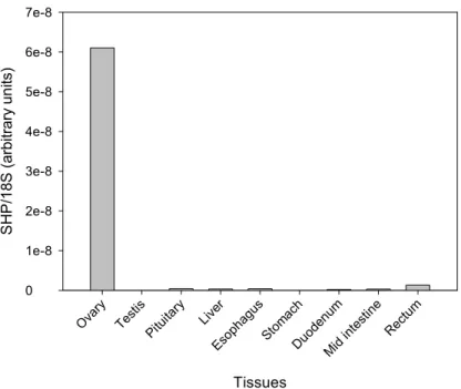
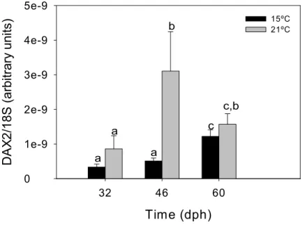
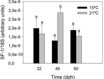
The tissue distribution of the three nuclear receptor members revealed a dimorphic pattern of expression within the hypothalamus-pituitary-gonadal (HPG) axis of male and female fish, suggesting that they may be involved in reproduction. In addition, during the sex determining period, larvae grown at either feminizing (15ºC) or masculinizing (21ºC) temperatures also presented a dimorphic pattern of expression of all three receptors, since larvae grown at low temperature showed repressed gene expression, while at higher temperature they were significantly upregulated. In addition, mRNA expression of other candidate sex determining genes, SOX9.1, SOX9.2 and SF1, was also similar to that obtained for the DAX duplicate genes and SHP. Thus, we have presented data
showing that during the temperature sensitive sex determining period, feminizing or masculinizing temperature regimes affect differently the transcription of different putative sex determining genes, namely of DAX1 and DAX2.
We have also isolated the sea bass DAX1 promoter sequence and compared it with that of other vertebrates. The promoter architecture of vertebrate DAX promoters was remarkably conserved from fish to mammalian species, and we have identified different promoter frameworks that are conserved in fish and mammalians. In addition, we have also identified other gene promoters annotated in the Genebank database that also shared one or more of these frameworks, enabling us to establish a putative role of DAX1, together with the CaM signalling pathway, in the regulation of steroid production within the gonads. To test this hypothesis, we have demonstrated that in male gonads, steroidogenic capacity is highly dependent on the availability of calmodulin within the cells, since inhibition of this protein with an antagonist (W7) blocked steroid production in the gonads of male tilapia, in vitro and in vivo. In addition, we have also studied the DAX1 and DAX2 mRNA expression in testis of tilapias treated with W7, and found that downregulation of the CaM signalling pathway leads to the upregulation of DAX1 and
DAX2 transcription levels.
In conclusion, although we did not obtain any conclusive data supporting a role for fish DAX1 and DAX2 genes in either male specific or female specific sex determination, we did find a link between temperature and DAX1 and 2 transcription levels during the TSD supporting the view of a role of these genes within the early gene cascade involved in TSD. In addition, we have also shown that Ca2+-CaM signalling pathway is not only
involved in the modulation of steroid production within male fish gonads as it is also involved directly or indirectly in the modulation of both DAX1 and DAX2 gene transcription within the gonads.
DAX1, un gène du déterminisme génétique du sexe chez les poissons ? Résumé
La détermination du sexe est un processus essentiel à la survie et l’évolution chez les vertébrés. Bien que la détermination du sexe chez la plupart des vertébrés soit basée principalement sur l’hétérogamétie (mâles XY et femelle XX), les différentes espèces de poissons ne présentent pas forcement un tel system de chromosomes sexuels, mais plutôt divers types de déterminismes sexuels tels que différentes formes d’hermaphrodismes et de déterminismes épigénétiques. Il n’existe donc pas un type général de déterminisme génétique du sexe chez les poissons. Cependant, différentes études ont montré que la plupart des gènes impliqués dans le déterminisme génétique du sexe chez les autres vertébrés, ce dernier étant basé sur un système de chromosomes sexuels, sont aussi présents chez les poissons. Ceci suggère que ces gènes pourraient néanmoins avoir une fonction similaire dans le déterminisme du sexe chez les poissons malgré l’absence d’hétérochromosomes.
DAX1 est un gène essentiel pour la détermination du sexe chez les mammifères.
En effet, des mutations du gène DAX1 chez l’homme et sur des modèles de souris «knockout » aboutissent à une réversion sexuelle mâle. DAX1 à été isolé chez deux espèces de poissons, et étant donné son taux de conservation important nous avons formulé l’hypothèse que ce gène pourrait avoir chez les poissons une fonction similaire à celle qui est connue chez les mammifères. Nous avons choisi le loup ou bar,
Dicentrarchus labrax, comme modèle d’étude, un poisson chez qui il n’existe pas
d’hétérochromosomes. Chez cette espèce, le déterminisme du sexe est fortement dépendant de la température (TSD). Ainsi, nous avons isolé la séquence complète et déterminé la structure du gène DAX1 chez D. labrax. Faisant cela, nous avons également identifié chez notre modèle, un dupliqua de ce gène ou sbDAX2, et un autre gène faisant partie de la famille des récepteurs nucléaires DAX1, appelé SHP (sbSHP). Ces trois gènes, sbDAX1, sbDAX2 et sbSHP, appartenant à la famille des récepteurs nucléaires, présentent une structure conservée et des domaines protéiques putatifs similaires à ceux des gènes mammaliens homologues.
L’étude de l’expression de ces trois gènes dans les différents tissues de D. labrax révèle une expression di-morphique au niveau de l’axe hypothalamus-hypophyse-gonadique chez les mâles et les femelles, suggérant un rôle de ces gènes dans la reproduction. L’expression de ces trois récepteurs nucléaires présente également un
modèle di-morphique au cours de la période de détermination sexuelle puisque l’expression des gènes est réprimée à une température basse féminisante (15°C), alors qu’elle est significativement augmentée à une température élevée masculinisante (21°C). Une modèle d’expression similaire à celui observé pour les gènes dupliqué DAX et le gène SHP a également été observé chez D. labrax pour d’autres gènes candidats à la détermination sexuelle, SOX9.1, SOX9.2 et SF1. Nos résultats montrent donc que pendant la période critique de la détermination du sexe température-dépendante, les températures féminisantes ou masculinisantes agissent différemment sur la transcription des gènes que nous pensons être impliqués dans la détermination sexuelle chez D.
labrax, c'est-à-dire DAX1 et DAX2.
Le promoteur du gène DAX1 a été isolé chez D. labrax et sa séquence comparée à celles d’autres vertébrés. La structure du promoteur des gènes DAX semble remarquablement conservée des poissons jusqu’au mammifères et nous avons identifié différents portions de promoteurs identiques chez les poissons et les mammifères. En utilisant la base de données Genbank, nous avons aussi identifié d’autres gènes comportant une ou plusieurs de ces portions de promoteurs, ceci nous permettant de conclure à un possible rôle de DAX1, ensemble avec la voie de signalisation calmoduline (CaM), dans la régulation de la production de stéroïdes par les gonades. Pour tester cette hypothèse, nous avons démontré que dans la gonade mâle, la capacité stéroïdienne est hautement dépendante de la présence de CaM dans les cellules puisque l’inhibition de cette protéine par l’antagoniste W7 bloque la production de stéroïdes dans la gonade mâle du tilapia, et ceci in vitro et in vivo. Nous avons également étudié l’expression des ARNm de DAX1 et DAX2 dans le testicule du tilapia traité avec W7 et trouvé que l’inhibition de la voie de signalisation CaM entraine une augmentation de la transcription de DAX1 et DAX2.
En conclusion, malgré l’absence de données permettant de conclure avec certitude au rôle des gènes DAX1 et DAX2 dans le déterminisme sexuel mâle ou femelle, nous avons trouvé un lien entre la température et les niveaux de transcription de DAX1 et
DAX2 au cours de la TSD. Ceci conforte l’opinion selon laquelle ces gènes sont liés à la
cascade de gène activés précocement et impliqués dans le TSD. Nous avons de plus montré que la voie de transmission du signal Ca2+-CaM est non seulement impliquée
dans la régulation de production de stéroïdes dans la gonade mâle du poisson mais est également impliquée directement ou indirectement dans la régulation de la transcription des gènes DAX1 et DAX2 dans la gonade.
DAX1, um gene que determina o sexo em peixes? Sumário
O processo de determinação sexual é essêncial à sobrevivência e evolução de todas as espécies de vertebrados. Embora a determinação do sexo na maioria de espécies esteja associada à presença de cromossomas sexuais (e.x., na maioria dos mamíferos, os machos possuem XY e as fêmeas XX), diversas espécies de peixes possuem sistemas de determinação sexual que não estão directamente associados à presença de cromossomas sexuais, uma vez que neste grupo de vertebrados existem diferentes espécies que são hermafroditas ou que utilizam mecanismos dependentes da temperature ambiental. Assim sendo, não é possível criar um modelo genérico que possa explicar o processo de determinação sexual neste grupo de vertebrados. No entanto, estudos efectuados em peixes mostram que, a maioria dos genes envolvidos no processo de determinação sexual em mamíferos que utilizam cromossomas sexuais, estão também presentes em espécies de peixe que não possuem cromossomas sexuais. Estes estudos sugerem que os genes envolvidos no processo de determinação sexual em vertebrados são bastante conservados, e que, independentemente da presença ou ausência de cromossomas sexuais, as funções destes genes nas diversas espécies, deverão ser bastante conservadas.
Um dos genes que está envolvido no processo de determinação sexual em mamíferos é o DAX1. A ocorrência de mutações neste gene em humanos ou a sua ablação do genoma de ratos resulta frequentemente na inversão sexual de machos genéticos. Recentemente, foram identificados genes homólogos do DAX1 em duas espécies de peixe. Dada a elevada conservação ao nível da sequência, nós colocámos como hipótese que provavelmente também deveriam apresentar funções conservadas no processo de determinação sexual dos peixes, independentemente da ausência de cromossomas sexuais. Para testar esta hipótese, decidimos trabalhar com o robalo,
Dicentrarchus labrax, que é uma espécie de peixe cujo processo de determinação sexual
pode ser manipulado através da utilização de diferentes regimes de temperatura nas fases iniciais de desenvolvimento larvar. Assim, começámos por isolar o gene homólogo do DAX1 (designado neste trabalho sbDAX1), caracterizar a estrutura do gene e identificar a sequência complementar do ARN mensageiro que é expressa nesta espécie. Durante este processo, identificámos ainda um gene resultante da duplicação genómica do DAX1 nesta espécie (designado sbDAX2) e de um outro membro da família dos receptores nucleares orfãos à qual o DAX1 pertence, SHP (designado neste trabalho
sbSHP). A identificação destes três genes permitiu-nos verificar que em peixes, os três genes possuem uma elevada conservação ao nível da estrutura dos genes, assim como da sequência e principais domínios das proteínas que codificam e, que a presença de uma duplicação do gene DAX1 é específica nos genomas de peixes.
O estudo da expressão destes três receptores orfãos nucleares em diferentes tecidos mostrou que todos eles apresentam um padrão dimórfico entre machos e fêmeas, nos tecidos pertencentes ao eixo do hipotálamo-pituitária-gónadas (eixo HPG), o que sugere um provável papel destes genes no processo da reprodução em peixes. Durante o periodo no qual existe maior sensibilidade para manipulações dos sexos pela temperatura (nos 60 dias iniciais após eclosão), a expressão dos três receptores nucleares também apresentaram um padrão dimórfico em larvas cultivadas a temperaturas que favorecem o desenvolvimento de fêmeas (15ºC) ou de machos (21ºC). Ou seja, larvas cultivadas a baixas temperaturas apresentam níveis baixos de expressão de sbDAX1, sbDAX2 e sbSHP, enquanto que a altas temperaturas os níveis de expressão destes receptores se apresentaram significativamente estimulados. O estudo da expressão de outros genes que se encontram igualmente envolvidos no processo de determinação sexual em mamíferos, SOX9 e SF1, nestas larvas mostrou ainda que todos eles apresentam também o mesmo padrão de expressão dimórfico. Assim, os dados presentes neste trabalho mostram que durante o periodo de maior sensibilidade à temperatura, a utilização de diferentes regimes de temperatura no cultivo de larvas de robalo afecta significativamente a transcrição de diferentes genes homólogos aos envolvidos no processo de determinação sexual em mamíferos, nomeadamente do DAX1 e DAX2.
No decurso deste trabalho isolámos ainda a sequência do promotor do DAX1 do robalo e comparámo-la com as sequências dos promotores de outros vertebrados. A estrutura e arquitectura do promotor do DAX1 dos peixes apresenta uma elevada conservação quando comparados aos dos mamíferos. Em todos eles, nós identificámos blocos de conservação que se encontram também em diversos promotores de genes anotados na base de dados do Genebank, o que nos permitiu inferir um possível envolvimento do DAX1 de peixes em diversos processos fisiológicos, nomeadamente na sinalização intracelular via Ca2+-Calmodulina (Ca2+-CaM) na produção de esteróides
sexuais nas gónadas. Para testar esta hipótese começámos por demonstrar que, nos testículos de tilápia Mossambicana, a produção basal de esteróides sexuais é fortemente regulada via Ca2+-CaM. Assim, demonstrámos que a administração de um inibidor de
CaM (W7) leva ao bloqueio da produção de testosterona e 11-cetotestosterona, in vitro e
in vivo. De seguida caracterizámos o impacto da administração in vivo deste inibidor na
desta via de sinalização intracelular leva ao aumento significativo dos níveis de RNA mensageiro destes receptores.
Em conclusão, embora não tenhamos obtido dados conclusivos demonstrando que nos peixes o DAX1 e DAX2 são genes de determinação sexual específicos de fêmea ou de macho, nós demonstrámos que a utilização de diferentes regimes de temperatura para manipulação da determinação sexual afecta os níveis de transcrição destes genes, o que sugere que muito provavelmente estarão envolvidos envolvidos na determinação genética do sexo em peixes. Neste trabalho demonstrámos ainda que a produção de androgénios nos machos é regulada via sinalização intracelular de Ca2+-CaM e que a
presença de calmodulina disponivel nas células estimula a produção de esteróides, enquanto que limita directa ou indirectamente a transcrição de DAX1 e DAX2, sugerindo um papel repressor destes receptores no processo da esteroidogenese.
CONTENTS
CHAPTER 1- General Introduction 16
CHAPTER 2- Isolation, characterization and developmental expression of DAX1 in
the European sea bass, Dicentrarchus labrax
34
CHAPTER 3- Characterization of two novel Nr0B family members in the European
sea bass, Dicentrarchus labrax: DAX2 and SHP
57
CHAPTER 4- DAX1 promoter isolation and in silico characterization 86
CHAPTER 5- Ca2+-Calmodulin signalling regulates androgen production in the testis
of adult male Tilapia (Oreochromis mossambicus)
122
CHAPTER 6- Calmodulin inhibitor W7 blocks testis steroid production in adult male
Tilapia (Oreochromis mossambicus) in vivo
141
CHAPTER 7- General Discussion 161
CHAPTER 1
1.1 The quest for a sex determining gene in fish
Sex determination is an integral part of reproduction and an essential process for the survival and evolution of vertebrate species. Most vertebrates rely on a classical system of male heterogamety (males are XY whereas females are XX) or variations thereof, e.g. birds use a female heterogametic system (males are ZZ and females are WZ). However, other vertebrate species have acquired an array of sex determination systems that do not rely exclusively on these classical chromosomal systems. In this respect, fish are one of the most intriguing groups that are presently under study since, aside the classical sex chromosomal XX/XY or WZ/ZZ systems, they can also display a diverse array of sex determining mechanisms that include different forms of hermaphroditism and environmental sex determination (see Devlin and Nagahama, 2002 for review). Thus, to date, no simple genetic sex determining system can be generalized for fish.
In mammalian, the presence of a Y chromosome is sufficient to determine the genetic fate of the gonad. Indeed, the presence of a Y-chromossome specific gene, named SRY, is sufficient to activate male gonadal development. Conversely, the absence of this gene precludes male sexual development and thus, female gonads develop, which has long been seen as a default mechanism. To date, all efforts for identifying an SRY-homolog or functionally equivalent gene in fish have failed. However, recently, a male sex determining gene, named dmY/dmrtY, has been identified in Medaka, Oryzias latipes, which is functionally equivalent to the mammalian SRY (Kobayashi et al., 2004). However, so far, this gene is absent in all other fish genomes, suggesting that Medaka has acquired this male sex determining gene in a recent event and that is species specific (Volff et al., 2003). Nonetheless, although no general sex determining gene has been found in fish, in the last few years different sex-specific probes have been obtained, each one of them shown to be specific to each species and even sometimes to a particular population (Forbes et al., 1994; Nakayama et al., 1994; Reed et al., 1995).
Different sex determining models have been proposed in fish, namely models based on multiple sex chromosomes, polygenic sex determination and autosomal influence (Reinboth, 1983; Chourout, 1994). However, there is still scarce information about the molecular mechanisms involved in these processes. Moreover, an additional level of complexity arises in species that possess a sex determining system that is further modulated by environmental factors. Indeed, sex determination in different fish species (Oreochromis niloticus, Oreochromis aureus, Odonthestes bonariensis, Dicentrarchus
1997; Desprez and Melard, 1998; Pavlidis et al., 2000). Interestingly, the influence of temperature appears to differ amongst species and appears not to be sufficient to induce complete female or male monosex populations. Indeed, there is evidence to suggest that the genetic background is not completely overridden by environmental cues, and that there are individual differences in susceptibility to temperature. Furthermore, different reports for example in the European sea bass, Dicentrarchus labrax, have shown combined effects of temperature and parental influence (Saillant et al., 2002) or genetic strain (Mylonas et al., 2003; Mylonas et al., 2005), but have also failed to produce 100% female or male populations.
Despite the sex determining system used by the different fish species, and lack of a sex determining gene, it is still hypothesized that most factors involved within the sex determination cascade are probably conserved among species. In fact, in the past years, different factors involved in mammalian sex determination, e.g., DMRT-1, SOX9, and
WT-1, have also been found in fish, suggesting a conserved role throughout evolution (Kent et
al., 1995; Kondo et al., 2002; Nakamoto et al., 2005). Nonetheless, the exact functions of these genes are still unclear in fish.
One of the genes that is a strong candidate for a sex determining role in fish is
Ahch or DAX1 (Dosage-sensitive sex-reversal, AHC, on the X chromosome, gene 1). This
gene was originally isolated in the X-chromossome of patients presenting a complex syndrome called adrenal hypoplasia congenital (AHC) (Zanaria et al., 1994). Interestingly, this gene was found to be located within an X-linked locus that overlapped the AHC locus which, when duplicated, results in male sex reversal (Bardoni et al., 1994; Zanaria et al., 1994). After the identification of this gene, different reports have established a direct link between DAX1 mutations and clinical signs of AHC (Yu et al., 1998b). A role of DAX1 in mammalian sex determination was further emphasized when it was established that the gene is repressed by SRY during male sex determination, linking it to ovary formation (Swain et al., 1998). As such, it has been regarded as an anti-testis gene in mammalian. Interestingly, DAX1 homolog genes have been identified in other vertebrate species, namely in bird and reptile embryos and fish larvae during the sex determining period, suggesting that it may also be involved in sex determination of lower vertebrates (Smith et al., 2000; Torres-Maldonado et al., 2002; Wang et al., 2002) .
A key feature of fish sex determination that differentiates it from mammalian sex determination is that steroids are required for box female and male sex differentiation (Baroiller et al., 1999; Devlin and Nagahama, 2002). Indeed, exogenous steroid administration, steroidogenic enzyme inhibitors or steroid receptor antagonists can modify sex during sex differentiation or even in some cases during adulthood (Devlin and Nagahama, 2002). Several of the factors involved in the sex differentiation cascade,
including DAX1, can also target steroidogenic enzymes and/or steroid receptors suggesting possible mechanisms of action (see below). Thus investigations into the structure, regulation and mechanism of action of candidate genes like DAX1 can shed some light on the sex differentiation cascade in fish and how it can be modified by exogenous.
1.2 DAX1 gene and protein structure
The human DAX1 gene has a very simple genomic structure composed of two
exons separated by a single intron (Zanaria et al., 1994). This gene encodes a 470 amino acid protein (Zanaria et al., 1994). Recently, however, a 401 amino acid DAX1 protein, named DAX1α, was identified in humans, as a result of transcription of exon 1 and of the transcription of a cryptic exon that was previously not identified within the intronic sequence of the DAX1 gene (Hossain et al., 2004).
The DAX1 protein has high similarity to other members of the nuclear receptor superfamily. However, due to the lack of known ligands, it has been classified as an orphan member of the nuclear receptor superfamily, and named Nr0B1 (Burris et al., 1995). Nuclear receptors are composed of four modules: the A/B domain, the C domain or DNA-binding domain (DBD), D domain or the hinge region, and E domain or ligand-binding domain (LBD) (Guiguére, 1999). The DAX1 gene has a carboxy-terminal domain composed of a homologous LBD to other nuclear receptors and an AF-2 transactivation domain similar to those found in other nuclear receptor members. However, it lacks the conventional DBD, domain A/B and hinge region. Indeed, the DAX1 amino-terminal domain has an atypical structure composed of 3.5 repeats of a 65–70 amino acids long cysteine-rich motif that has no known homology to any other proteins, with the exception of the small heterodimer partner (SHP), encoded by NR0B2, which is the only known DAX1 family member. Another striking feature of this novel DBD is that it also lacks the classical zinc fingers found in other nuclear receptors and, instead, it possesses 3 LXXLL-like motifs within each of the 65-70 amino acid repeat, that are used for interaction with other receptors (Zhang et al., 2000; Agoulnik et al., 2003; Kawajiri et al., 2003). The C-terminal domain of DAX1 has strongest amino acid similarity to the LBD of the COUP-TF and retinoid X receptor (RXR) (Zhang et al., 2000). Interestingly, this domain has been shown to be responsible for transcriptional silencing activity in mammalian species and is only found in a subset of other members of the nuclear receptor super family: thyroid hormone (TR), the related oncogene product v-erbA, retinoic acid receptor (RAR), and the chicken ovalbumin upstream promoter transcription factor (COUP-TF) (Lalli et al., 1997). The silencing activity of DAX1 is thought to be due to a bipartite domain in the C-terminal
(Lalli et al., 1997) that corresponds to the helix 3 and helixes 11-12, according to the DAX1 protein structure prediction when aligning DAX1 with RAR, TR and v-erbA sequences (Lalli et al., 1997; Altincicek et al., 2000). Nonetheless, overall, the DAX1 structure is more similar to SHP, since the C-terminal region (LBD) of both proteins are well conserved both lack the typical nuclear receptor DBD that is replaced by one of the 3.5 65–70 amino acid repeat in the N-terminal domain (DBD) (Seol et al., 1998).
1.3 DAX1 molecular function
In patients with AHC, all DAX1 mutations localize to the C-terminal LBD, which significantly reduce its repressive effects in cell culture assays (Achermann et al., 2001a), and thus implicating the LBD as a mediator of repression. Indeed, a bipartite transcriptional silencing domain has been shown to exist in the LBD, which interacts directly with co-repressors to mediate repression. Additionally, a direct role for the N-terminal LXXLL motifs and for the LBD AF-2 core have also been shown in mediating repression of different nuclear receptors, namely through direct interaction with target genes: estrogen receptors (Erα and Erβ), androgen receptor (AR), progesterone receptor (PR), NR4A1 encoding Nur77, and NR5A2 encoding liver receptor homologue-1 (LRH1) (Zhang et al., 2000; Holter et al., 2002; Agoulnik et al., 2003; Suzuki et al., 2003; Song et al., 2004). DAX1 transcriptional silencing activity can involve direct protein–protein interactions between DAX1 with known co-repressors, namely Alien, NcoR and MLK2, and RIP140 (Crawford et al., 1998; Altincicek et al., 2000; Sugawara et al., 2001; Eckey et al., 2003). Recruitment of co-repressors has been shown to be a key factor in the regulation of Steroidogenic factor 1 (SF1), a key regulator of steroid hydroxylase enzymes (Lala et al., 1995; Morohashi and Omura, 1996). DAX1 protein interacts with DNA-bound SF1 via the DAX1 N-terminal domain, with the subsequent recruitment of co-repressors to the promoters of target genes, via the DAX1 C-terminal transcriptional silencing domain (Crawford et al., 1998). In cultured cells, DAX1 represses SF1-mediated transcriptional activation of steroidogenic genes, such as Cyp11A, Cyp17 and Cyp19 (Lalli and Sassone-Corsi, 2003). Additionally, when over expressed, DAX1 markedly impairs steroidogenic output and transcriptionaly represses the gene promoters of other steroid enzymes such as StAR and 3β-HSD (Lalli et al., 1997; Zazopoulos et al., 1997; Jo and Stocco, 2004). Moreover, DAX1 is also able to repress SF1-mediated up regulation of the gene encoding anti-Müllerian hormone (AMH, also known as Müllerian inhibiting substance; MIS) during male sexual differentiation, either through repressed synergy between GATA4–SF1 or between SF1-WT1, during activation of the AMH promoter (Nachtigal et al., 1998; Tremblay and Viger, 2001). DAX1 is also able to repress the synergy between SF1-EGR1
and block the GnRH stimulation of the LHβ promoter (Dorn et al., 1999). Thus, collectively, these data suggest that DAX1 is a global negative regulator of genes involved in steroid hormone production and metabolism in endocrine tissues.
Although most of the repressive actions of DAX1 that have been described involve protein: protein interaction with co-repressors, the initial report on DAX1 isolation and characterization has demonstrated that DAX1 could bind directly to the retinoic acid-responsive element (RARE) in vitro (Zanaria et al., 1994). However, other studies support the view that DAX1 binds to particular nucleic acid structures, rather than to particular sequences. Indeed, DAX1 has been shown to bind to DNA loops in target gene promoters. The StAR and DAX1own promoters are two of the examples that have been shown to contain DNA hairpin sequences that are bound by DAX1 (Zazopoulos et al., 1997). The DAX1 protein has not only been shown to regulate its own promoter, as it has also been shown to homodimerize with other DAX1 molecules (Iyer et al., 2006). The motifs involved in DAX1 homodimerization include the LXXLL domains and AF-2 domain. Altogether these data suggest an auto- regulatory feedback loop on its own transcription.
DAX1 is a nucleo-cytoplasmatic shuttling protein that has been shown to be associated with ribonucleoprotein structures and polyribosomes in the cytoplasm, suggesting that DAX1 might also act at a post-transcriptional level, through direct binding to RNA and polyribosomes and thus may be acting as a shuttling protein for export of RNA species into the nucleus (Lalli et al., 2000). Nuclear import requires the integrity of a bipartite transcriptional repression domain in the DAX1 C-terminus. However, in AHC mutations, DAX1 nuclear localization is impaired as all DAX1 mutant proteins are retained within the cytoplasm (Lehmann et al., 2002).
1.4 DAX1 gene expression and regulation
DAX1 gene expression has been detected in the developing adrenal cortex, gonad,
anterior pituitary and hypothalamus, in adult adrenal cortex, Sertoli and Leydig cells in the testis, and in theca, granulosa, and interstitial cells in the ovary (Guo et al., 1996; Ikeda et al., 1996; Swain et al., 1998). Moreover, DAX1 mRNA expression is coincident with that of SF1 in most tissues and developmental stages in mammalian, further suggesting that these two transcription factors are network partners (Ikeda et al., 1996; Ikeda et al., 2001). In addition, different studies have shown that the transcriptional regulation of SF1 target genes during development and in the adult organism appear to be regulated by the intracellular levels of SF1 and DAX1, with the ratio of these two factors determining whether the target genes are activated or repressed. Thus, if higher levels of SF1 than
DAX1 exist, the target genes will be activated, whereas, if more DAX1 is present, the target genes are either not activated or repressed.
In addition to the importance of SF1-DAX1 ratio, both human and rodent DAX1 promoters contain extended AGGTCA-like half sites that are recognized and bound by SF1 (Burris et al., 1995; Yu et al., 1998a). Indeed, SF1 has been shown to use these binding sites to regulate DAX1 transcription in endocrine tissues. Interestingly, the ERR family (estrogen-related receptors, type α, β, γ), which is composed of 3 orphan nuclear receptors, have been shown to bind to the same consensus binding site as SF1. In fact, one of these members, ERRγ, has been demonstrated to bind to the SF1 response element in DAX1 promoter (Park et al., 2005).
Another set of candidate regulators of DAX1 transcription are the GATA transcription factor family genes. Putative GATA binding sites have been found in the mammalian DAX1 promoter (Hoyle et al., 2002), although to date no direct interaction of DAX1 has been reported with GATA elements alone. However, GATA-4 and GATA-6 have been shown to play a role in male sex determination, steroidogenesis, and possibly cell survival (see la Voie, 2003 for review) and to synergize with SF1 in steroidogenic tissue (Nachtigal et al., 1998), thus acting within the same processes as DAX1.
One of the key regulators of DAX1 transcription is Wilms tumor 1 (WT1). WT1 expression precedes that of DAX1 in the urogenital ridge, and at the protein level, both DAX1 and WT1 show an overlapping spatial expression profile in the developing fetal gonad (Kim et al., 1999). Moreover, WT-1 can individually activate DAX1 transcription through direct binding to its promoter or through recruitment of the co-activator FHL2 (Kim et al., 1999; Du et al., 2002). In addition, WT-1 is also able to activate SF1 transcription individually or through recruitment of co-activator Lhx9 (Wilhelm and Englert, 2002). Thus, WT-1 has been proposed as an upstream regulator of SF1 and DAX1 in mammallian sex determination cascade.
Activation of the WNT-beta catenin signaling pathway has been shown to alter
DAX1 transcription. WNT ligands bind to cell-surface Frizzled receptors, initiating a
cytoplasmatic signaling cascade that results in beta-catenin stabilization, cytosolic accumulation, and subsequent translocation to the nucleus. Within the nucleus, beta-catenin classically complexes with a member of the T-cell Factor (TCF) family of transcription factors to activate target-gene transcription (Mulholland et al., 2005). Upon Wnt4 stimulation, beta-catenin activates DAX1 transcription either through direct interaction with the TCF/LEF binding sites on its promoter or through direct interaction with SF1 on the DAX1 promoter, in a TCF/LEF independent process (Mizusaki et al., 2003).
1.5 The role of DAX1 in sex determination 1.5.1 Early embryonic development
Three germ cell layers, the mesoderm, endoderm and ectoderm, are formed during embryonic development. The endoderm develops into the digestive tract; the ectoderm, into skin and the central nervous system and the mesoderm develops into the internal organs. Gonads are formed by cells originating in different locations: Somatic cells originate from proliferation of cells within the genital ridge itself, and primordial germ cells (PGCs) originate in the proximal epiblast. Upon the activation of two genes, c-kit and steel, PGCs start to migrate from the posterior end of the primitive streak into the allantois and then into the adjacent embryonic endoderm, which first forms the gut (Anderson et al., 2000). Here, a cluster of PGC will recover their active migratory capacity and move towards the genital ridges. During migration and early settlement in the genital ridge, PGCs express several markers, e.g., tissue- SF1 (AP) (MacGregor et al., 1995), and the transcription factor Oct3/4 (Yeom et al., 1996). Once in the genital ridge, PGCs start to express different gene markers, within which, the vasa protein will ultimately activate somatic cell development in the genital ridge (Toyooka et al., 2000).
Once PGCs begin to proliferate and establish in the ventral region of the urogenital ridge, they undergo active proliferation (Tam and Snow, 1981; Godin et al., 1990). At this stage, PGCs lose their motile behavior (Wylie et al., 1969) and together with proliferating coelomic and mesenchymal cells, form a cluster of condensed cells that gradually becomes the undifferentiated gonad and differentiate into germ stem cells that divide by mitosis to produce and originate the gametes. In the female, once mitotic proliferation stops, retinoic acid activates the stimulated by retinoic acid 8 (stra8) gene in germ cells, and these initiate meiotic differentiation (Koubova et al., 2006). In males, however, expression of a retinoid-degrading enzyme known as CYP26B1 blocks the retinoic acid stimulation and results in germ cells arrest in the G0/G1 stage of the mitotic cell cycle (McLaren et al., 2004) until after birth. Thus, in the absence of CYP26B1, male germ cells enter meiosis prematurely as in a normal ovary (Bowles et al., 2006).
Embryonic germ (EG) cells, derived from cultured primordial germ cells (PGCs), are the embryonic precursors of the gametes of the adult animal. The pluripotency of EG cells has been demonstrated in widely-used assays and demonstrate that they share many properties with pluripotent embryonic stem (ES) cells (Donovan and Gearhart, 2001). DAX1 was found to be expressed in early pre-implantation embryos as well as in ES cells (Clipsham et al., 2004), together with other genes involved in sex determination, including SF1, and WT-1. In pre-implantation embryos, DAX1, SF1, and WT1 are
expressed in the inner cell mass (Clipsham et al., 2004). In fact, upon implantation, DAX1 is specifically expressed and can function as a marker for lineage determination, separating the extra embryonic visceral endoderm (VE) from the embryonic layers (Niakan et al., 2006). Interestingly, differentiation of ES cells with stimuli in the ES cell media has also been reported to result in loss of DAX1 RNA similar to that observed for
Oct-4. Disruption of the expression of DAX1 by siRNA knockdown as well as a conditional
knockout in ES cells caused their differentiation (Niakan et al., 2006), suggesting that
DAX1 is necessary for the maintenance of pluripotent state of stem cells. Additional proof
for this DAX1 novel role was obtained from the demonstrated interaction with Nanog, a marker of pluripotency, in ES (Wang et al., 2006). Altogether, these results show that DAX1 is determinant for the early mammalian embryonic development and development of embryonic germ cell lineages.
1.5.2 Early gonad development
Genes that are important in the differentiation of the intermediate mesoderm and the urogenital system as a whole will generally have a role in early gonad development. This group of genes is characterized by their mutant phenotypes, which show defects in both early kidney, adrenal and gonad development. A growing list of genes involved in this process has been identified, namely WT-1, SF1, Lim1, Lim-9 and EMX2. WT-1 was found to be determinant for the development of genital ridge, adrenals and kidney in mouse embryos (Pritchard-Jones et al., 1990; Kreidberg et al., 1993). Likewise, SF1 is also expressed in the developing urogenital ridge and undifferentiated gonad while the SF1 knockout in mouse embryos prevents the establishment of undifferentiated gonads and all
SF1 null mutants develop a female phenotype (Luo et al., 1994). Lim-1, Lim-9 and Emx2
are also required for development of the urogenital system (Shawlot and Behringer, 1995; Miyamoto et al., 1997; Birk et al., 2000). Once the indifferent gonad is formed, arrays of autossomal genes are activated in order to establish the genotypic sex of the bipotential gonad either into a female or into a male. In mammalian, the presence of the Y chromosome gene, sex-determining region of the Y-chromosome gene or SRY, is sufficient to trigger differentiation of testes from the indifferent gonad (or genital ridges) that would otherwise develop as ovaries (Gubbay et al., 1990; Sinclair et al., 1990). Upon
SRY activation, pre-sertoli cells start to develop and SOX9 is expressed (Kent et al.,
1995). Yet, although it is not known whether SOX9 responds directly or indirectly to SRY, gain-of-function studies in mice and humans (reviewed in Canning and Lovell-Badge, 2002) indicate that SOX9 may be sufficient for male sex determination even in the absence of SRY.
In mammalian, DAX1 is expressed during early ovarian development but is
suspended during testicular formation (Swain et al., 1996), and has also been shown to be repressed by SRY during testicular development, which implies a critical role for this gene in ovarian formation (Swain et al., 1998). (Swain et al.) (1998) using a transgenic mouse model (Mus musculus origin) carrying extra copies of DAX1 have tested whether
DAX1 function in sex determination was dosage dependent. Surprisingly, when these
mice were bred, they did not observe sex reversal in the offspring, unlike what is seen in human patients with DAX1 duplication in their genome (Bardoni et al., 1994). However, mice with the highest SF1 expression (up to five times that of normal levels) exhibited retarded testis formation, suggesting that DAX1 functioned as a female promoting gene or as an anti-testis gene. The function of DAX1 in ovarian development is still unclear. Although DAX1 expression is detected in the developing ovary at various stages of development homozygous DAX1-defficient female mice are normal and fertile, except for slight ovarian follicular defects, suggesting that DAX1 is not required for ovarian development (Yu et al., 1998b). In addition, female patients with AHC do not present gonadal defects, although later in puberty some patients present compromised endocrine function (Seminara et al., 1999). Thus, these studies suggest that DAX1 is more important for female gonadal endocrine function than for its morphological development.
In contrast, studies with DAX1-deficient male mice provided irrefutable evidence that DAX1 is necessary for proper testicular development and function, in opposition to an anti-testis function. Indeed, the DAX1-deficient mice are hypogonadal and infertile, and have testicular and spermatogenic defects with loss of germ cells and degeneration of the seminiferous epithelium (Yu et al., 1998b). In addition, the DAX1-defcient XY mice develop as phenotypic females when in the presence of a weakened SRY allele, further emphasizing a crucial role in testis determination (Meeks et al., 2003b). Further characterization of the DAX1-deficient mouse model has also shown that this gene is determinant for testis cord organization during development (Jeffs et al., 2001a; Meeks et al., 2003a), and that the lack of DAX1 expression in Sertoli and Leydig cell leads to infertility and low sperm counts, a condition that is only partially rescued when DAX1 expression is allowed in these cells (Jeffs et al., 2001b). However, expression of DAX1 in each of these cell types individually is not sufficient to rescue the testicular pathology, suggesting that DAX1 function in both Sertoli and Leydig cells (in addition to other somatic cell types) is necessary for proper testicular development (Jeffs et al., 2001b; Meeks et al., 2003b). Interestingly, a clear evidence that DAX1 is not an anti-testis gene, but acts instead as ‘pro-testis’ gene was obtained recently, when DAX1-knockout mice were XY sex reversed after they were crossed with the M. d. poschiavianus strain containing the Y chromosome SryPOS allele (Meeks et al., 2003b). These Dax1 -/Y POS mice lacked
testicular cords, Sertoli and Leydig cell markers and were sex reversed. Interestingly,
SOX9 expression was significantly reduced in these mice even in the presence of normal SRY levels. These results imply that DAX1 is determinant for male sex and also that it is
acting downstream the SRY but in concert with SOX9.
Altogether, these studies provide clear evidence for a determinant role of DAX1 in early embryonic development and in male gonadal development. Reduction or increase in
DAX1 expression has no apparent major effect in female development but, in humans and
rodents, DAX1 dosage can influence the final outcome of the phenotypic gonads in genetic males.
1.6 The role of DAX1 in sex differentiation
Once pre-sertoli cells start to develop, anti-müllerian hormone (AMH), a secreted peptide, starts to be produced (Teixeira et al., 2001) upon the activation of SRY, SOX9,
WT1, SF1 and GATA4 (Viger et al., 2005). This peptide causes regression of the
Müllerian ducts, which otherwise wouldgive rise to the female morphological structures. Thus, Sertoli cells organize into the testicular cords, where spermatogenesis later takes place, and are involved in the support of spermatid differentiation. Another cell lineage, the Leydig cells also starts to differentiate with the primary function of initiating steroidogenic capacity and to synthesize testosterone. Testosterone production will support the maintenance of the Wolffian ducts, which develop into the different structural components of the male reproductive tract. Thus, while testicular development is initially independent of steroids, the later proliferation of Sertoli cells and full masculinization requires testosterone (Sharpe, 2006).
The development of the female reproductive tract has received much less attention than the male tract, probably because of the widespread notion that it occurs ‘by default’ in the absence of SRY. Although to date no sex determining gene has been found in females, repressor proteins preventing male differentiation are expressed in the ovary, e.g. WNT-4 (a member of the Wnt/ Wingless family). In early development, WNT-4 suppresses Leydig cell differentiation and the synthesis of testosterone, probably by repressing SF1 function or inhibiting migration of steroidogenic precursor cells into the developing ovary (Fleming and Vilain, 2005). Female WNT- 4 null mice develop virilized gonads and Wolfian derivatives (see Park and Jameson, 2005 for review) , demonstrating an essencial role in female sex differentiation. Moreover, other genes are also essencial for the development of granulosa cells and oocytes within the ovary, namely FOXL 2 and FIG
X (Loffler and Koopman, 2002; Fleming and Vilain, 2005). Interestingly, DAX1 is involved
to its promoter, e.g. SF1 and GATA-4 (Nachtigal et al., 1998; Tremblay and Viger, 2001) . Moreover, DAX1 has been shown to be up regulated by WNT-4 through interaction with SF1 (Jordan et al., 2001). In addition, both DAX1 and WNT-4 are up regulated by WT-1 in female gonads (Sim et al., 2002), suggesting that in concert, DAX1 and WNT-4 are part of a cascade of genes controlling the development of the Müllerian tract and additional morphological structures in the female reproductive system (Rey et al., 2003). Overall, although the activation of AMH secretion is determinant for male sexual differentiation, by itself is not sufficient, since males need to acquire steroidogenic capacity in order to pursue masculinization of the gonads. On the contrary, the inhibition of AMH secretion and steroidogenic function in the early gonad enables the Wolffian Tract regression and the Müllerian tract development into the morphological structures of the female reproductive system. Thus, it is clear that steroid production plays a determinant role in sexual differentiation in mammalian, but also in fish (see below and e.g. Devlin and Nagahama, 2002).
1.6.1 Regulation of steroidogenic development of the gonads
The enzymatic steps and their genetic control in the testicular biosynthesis of testosterone from cholesterol are well documented (reviewed in Payne and Hayes, 2004) The synthesis of steroids from cholesterol involves trafficking between mitochondria and smooth endoplasmic reticulum, mainly via P450 or CYP enzymes. All steps are necessary for androgen production but key points include the rate limiting step controlled by the steroidogenic acute regulatory protein (StAR). In mammalian, the main enzymes involved in the production of testosterone are cytochromes P450 cholesterol side-chain cleavage (P450scc or CYP11A), the 3β-hydroxysteroid dehydrogenases (3β-HSD1-6) types 1–6, and 17β-hydroxylase 17–20 lyase (CYP17). The enzymes 17-hydroxysteroid dehydrogenases, types 1–3 (17β–HSD 1-3) and 5α-reductase function to amplify the androgenic signal through the synthesis of the more potent androgens, testosterone and dihydroxytestosterone (DHT). The expression pattern of CYP genes correlates with that of the SF1 (Honda et al., 1993). SF1 is an orphan receptor that binds to extended AGGTCA-like half sites present in all steroidogenic CYP proximal promoters and stimulates their expression (Lala et al., 1995; Morohashi and Omura, 1996). Interestingly, SF1 and DAX1 are co-expressed in steroidogenic tissues as well as in the hypothalamus and pituitary, early in development (Ikeda et al., 1996). However, when overexpressed, DAX1 markedly impairs steroidogenic output and transcriptionaly represses the promoters of SF1 target genes such as StAR, Cyp11A, 3β-HSD, Cyp17 and Cyp19 (Lalli et al., 1997; Zazopoulos et al., 1997; Sim et al., 2002; Lalli and Sassone-Corsi, 2003; Jo and Stocco, 2004). Thus,
SF1 and DAX1 are suggested to be antagonist regulators of steroid hormone production, and further emphasize the repressive actions on steroid production in endocrine cells.
The initial production of testosterone in Leydig cells is independent of LH secretion, because LH only becomes detectable in the pituitary latter (El-Gehani et al., 2000). In fact, genetic mutant or knock-out male mice deficient in gonadotropin releasing hormone (GnRH), luteinizing hormone (LH) or LH receptors (LHR), all exhibit, although with varying degrees, prenatal masculinization (El-Gehani et al., 2000; Zhang et al., 2001) which corroborates that activation of testosterone production is gonadotropin independent. Upon activation of gonadotropin production in the pituitary, testosterone synthesis in the developing testes is under strict control of chorionic gonadotropin (hCG) and LH (Misrahi et al., 1998). Both hCG and LH stimulate testosterone synthesis via the LH receptor (LHR) in the testis. Mutations in the LHR are associated with gonadotropin unresponsiveness and lead to Leydig cell agenesis and, subsequently, to defective sexual differentiation. More often, inactivation of LHR result in a completely female phenotype, although incomplete virilization due to partial receptor responsiveness with subnormal androgen synthesis has also been described (Misrahi et al., 1998). These molecular abnormalities imply an active role for the LHR in Leydig cell growth and differentiation. DAX1 is expressed in the developing pituitary structure by the time it starts to be expressed in the urogenital ridge (Swain et al., 1998; Ikeda et al., 2001). In addition, DAX1 is expressed throughout development in different structures of the hypothalamus-pituitary-gonadal-adrenal axis (HPGA) (Swain et al., 1998; Ikeda et al., 2001), suggesting an active role in development and early function of this axis. Moreover, patients with DAX1 mutations, presenting hypothalamic hypogonadism (HHG) with absent or delayed puberty, also present impaired or absent production of GnRH from the hypothalamus and/or of FSH and LH from the anterior pituitary (Vaidya et al., 2000; Achermann et al., 2001b) compromising latter steroid production when they reach puberty.
Interestingly, gonadotropins have been shown to regulate DAX1 expression in the gonads (Tamai et al., 1996; Yazawa et al., 2003). In early development of Sertoli cells,
DAX1 expression peaks during the first synchronized spermatogenic wave, and returns to
basal levels thereafter (Tamai et al., 1996). Sertoli cells contain specific G-protein-coupled receptors for the pituitary hormone FSH that upon binding stimulates adenylate cyclase activity and leads to increased concentrations of intracellular cAMP (Heckert and Griswold, 2002). Interestingly, DAX1 expression is significantly down-regulated upon FSH treatment and by treatment with the cAMP analog, (Bu) cAMP, which bypasses the FSH receptor and leads directly to activation of the protein kinase A pathway (Tamai et al., 1996). SF1 is a transcriptional regulator of cAMP-induced genes (e.g. steroid hydroxylases) and is also phosphorylated by cAMP-dependent protein kinase (PKA) (Zhang and Mellon, 1997).
Moreover, PKA stimulation potentiates SF1 function while prevents DAX1 antagonistic interaction with SF1. Moreover, increased cAMP levels have been shown to up-regulate SF1 levels and down-regulate DAX1 levels (Osman et al., 2002). Thus, activation of PKA signaling pathway by gonadotropins stimulate SF1 levels, down regulates DAX1 levels, induces DAX1 and SF1 dissociation, and thus, promotes SF1 interactions with coactivators and activation of SF1 target genes (e.g. P450 steroid hydroxylases) (Tamai et al., 1996; Osman et al., 2002). One of these target genes is the enzyme that is responsible for the aromatization of androgens into estrogens, the cytochrome P450 aromatase (P450arom), the product of the CYP19 gene. The CYP19 promoter activity in male gonads is up-regulated by FSH/LH and through activation of the cyclic AMP-dependent signaling pathway promotes the activation and individual binding of SF1 to promoter response elements, and/or recruitment of beta-catenin (Genissel et al., 2001; Parakh et al., 2006). Interestingly, the expression of the CYP19 gene, is increased significantly in Leydig cells isolated from DAX1 mutant mice (Wang et al., 2001), suggesting a repressive action of DAX1 on the CYP19 promoter. Furthermore, DAX1 was shown to block the cAMP-SF1 pathway-dependent P450arom expression (Gurates et al., 2002). Altogether, these studies show that gonadotropins activate the cAMP-PKA signaling that will lead to SF1 phosphorylation. Subsequently, SF1 increased levels and transcriptional activity supress DAX1 transcription and activate steroid enzymes transcription and activity, leading to increased steroid production. Thus, it is clear that the balance of SF1 and DAX1 levels within steroidogenic tissues is determinant for steroid production, and suggests a determinant role for both transcription factors in the activation and regulation of steroid production in gonads during sexual differentiation.
Interestingly, DAX1 is not only able to modulate steroid production but is also able to regulate androgen and estrogen action, through physical interaction with both androgen receptor (AR) and estrogen receptors (ERs), resulting in repressed activity (Ikonen et al., 1997; Zhang et al., 2000; Holter et al., 2002) . Altogether, it is clear that DAX1 is determinant for gonadal sexual differentiation as it regulates androgen and estrogen production and is also able to regulate their action in the cell through direct protein interaction with both AR and ERs. A general model summarizing DAX-1 interactions is shown in figure 1.
Figure 1– Schematic representation of pathway for the biosynthesis of sexual steroids. DAX1 regulates key steps for androgen production by repressing the
transcriptional activation of SF-1 on the promoters of StAR (Lalli et al., 1997), CYP11A (Lalli et al., 1998) and CYP17 (Hanley et al., 2001). DAX1 binds directly to the CYP19 promoter to downregulate its transcription (Wang et al., 2001). It also regulates estrogen (Zhang et al., 2000) and androgen receptors (Holter et al., 2002) through protein-protein interactions. CYP11A, CYP17 and CYP19 are, respectively, genes that encode for the P450 enzymes cholesterol side chain cleavage, 17- hydroxylase and aromatase. StAR is the mitochondrial cholesterol transporter protein. AR and ER are respectively androgen and estrogen receptors (α and β).
1.7 The role of DAX1 in fish
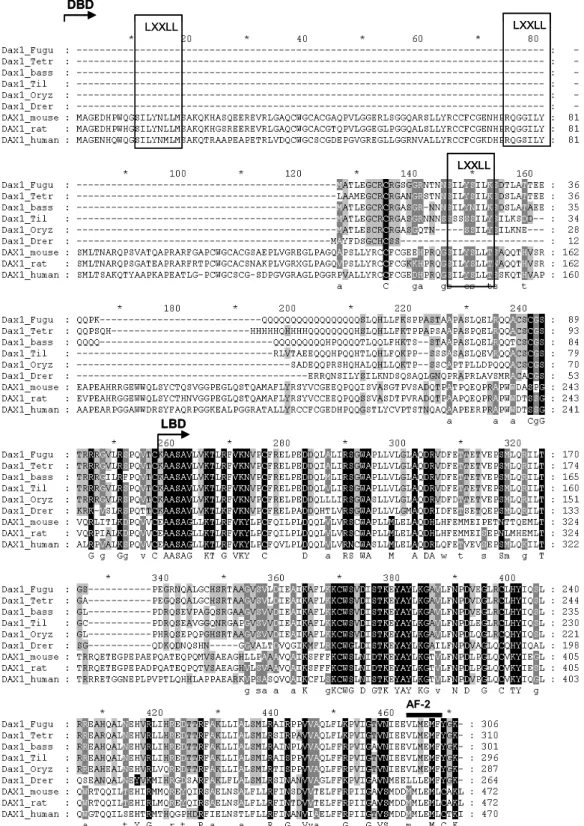
In fish, the DAX1 homologue gene has been recently characterized in zebrafish (Danio rerio) and tilapia (Oreochromis niloticus) (Wang et al., 2002; Zhao et al., 2006). The DAX1 gene structure in both fish species is conserved to that described in mammalian, as they are also composed of 2 exons and 1 intron. The first exon codes for the putative DBD and most of the LBD of the protein, while the second exon, which is a lot smaller, codes for the rest of the LBD and AF-2 core. Thus, the gene structure and main structural features (DBD, LBD and AF-2 core) are also present in fish DAX1 proteins.
Interestingly, the putative DBD of DAX1 fish proteins is also atypical since it does not possess the classical zinc fingers of other nuclear receptor members, but instead, they contain LXXLL-like motifs as identified in mammalian homolog genes. However, unlike mammalian DAX1 proteins, the putative DBD is considerably smaller since it lacks the 3.5 repeat regions. Instead, the DAX1 proteins contains only one of the 3 LXXLL-like motifs found in mammalian DAX1 proteins, resembling the DBD structure of its family member, small heteroprotein (SHP). However, to date, there is no information on whether this motif is sufficient for enabling protein interactions with other receptors as described in mammalian.
Fish DAX1 genes are expressed within the HPAG axis as described in mammalian
(Wang et al., 2002; Zhao et al., 2006). However, unlike mammalian, DAX1 expression is also detected in most of the tissues of adult fish tested so far, contrasting the typical HPAG axis distribution reported earlier in human and rodents (Wang et al., 2002). In addition, tilapia DAX1 is expressed at similar levels in both adult female and male gonads, which contrasts the dimorphic pattern described in mammalian (Wang et al., 2002). Although there is still no data demonstrating a role for DAX1 in fish sex determination, the zebrafish, tilapia and trout (Oncorhyncus mykiss) DAX1 genes are expressed during the sex determining period (Wang et al., 2002; Baron et al., 2005; Zhao et al., 2006). Indeed, a recent report has suggested a role for DAX1 in trout testicular differentiation (Baron et al., 2005). Interestingly, Wang et al. (2002) have identified the fugu (Takifugu rubripes)
DAX1 homolog gene within the genome database, and surprisingly, have also identified a
putative duplicate of the DAX1 gene (which was named DAX2 or Nr0B1b) that is also found in tilapia (Accession number ABB88832). Nonetheless, to date there is still no information on this duplicate gene in either fugu or tilapia.
In zebrafish embryos, DAX1 mRNA has been detected in head kidney, central nervous system, liver, brain and in truck midline region between 4 and 32 days post fertilization (dpf), suggesting that DAX1 is involved in early embryonic development, well before gonads start to be developed (Zhao et al., 2006). Surprisingly, target disruption of
DAX1 in zebrafish resulted in impaired osmoregulation, suggesting that it has a
determinant role in early adrenal function and glucocorticoid production in fish larvae (Zhao et al., 2006).
1.8 Aim of the thesis
The DAX1 gene has been identified in two different fish species. Despite the high conservation in sequence and gene structure, it is still unknown whether this gene is a sex determining gene as described in mammalian. Thus, the aim of this thesis was to study
the DAX1 and DAX2 expression during the sex determining period and also throughout the gonadal sex differentiation period. In addition, to shed some light on the mechanisms underlying the regulation of DAX1 expression in fish, we have aimed to identify the DAX1 promoter in fish and, based on promoter conservation between fish and mammalian, to identify putative regulatory sequences within its promoter that could help to identify possible gene network partners. Finally, we have sought to explore a possible role of DAX1 in male gonadal steroidogenesis as a validation of one of the putative gene networks that have resulted from the promoter analysis.
For this purpose, the organization of this thesis will be as follows:
In Chapter 2, the European sea bass DAX1 (sbDAX1) gene and coding sequence
are described. Protein structure and evolutionary conservation of DAX1 protein was also assessed. The mRNA expression in adult fish was analyzed in both female and male tissues. Within this chapter, the DAX1 mRNA expression was also studied during the temperature sex determining period (TSD) and during the gonadal sex differentiation period (GSD).
In Chapter 3, we describe the isolation of a duplicate DAX1 gene, named DAX2,
and of another DAX1 family member named SHP. For both genes, we have studied the gene structure, sequence and evolutionary conservation. In addition, we have also studied the mRNA expression of these novel members during the GSD and TSD periods described for DAX1 in chapter1. In addition, we have also analyzed the expression of SF1 and of two SOX9 duplicate genes (SOX9.1 and SOX9.2) in order to clarify which genes are involved in the temperature sensitive sex determining cascade in sea bass TSD. In Chapter 4, we describe the isolation of the sea bass DAX1 putative promoter
sequence. We have identified putative transcription factor binding sites (TFBS) that have been shown to regulate the mammalian DAX1 promoter transcription. Moreover, we have compared the promoter conservation of fish DAX1 putative promoters to those described in mammalian and have detected a surprising conservation in promoter architecture, as we have identified different promoter frameworks composed of 3-5 transcription factor binding sites (TFBS) that are present in most of the DAX1 promoters, within the same order and distances. In this chapter we have also identified putative gene networks that share this promoter architecture, allowing the identification of possible roles for DAX1 gene in fish.
In Chapter 5 and Chapter 6, we have studied one of the networks identified in the DAX1 promoter analysis described in chapter 4. Thus, in these two chapters we have
studied the involvement of the DAX1 in male gonad steroid production. In Chapter 5, we have studied the effect of a calmodulin inhibitor on the in vitro androgen production and in
Chapter 6 we have studied the effect of this inhibitor on steroid production, in vivo, and
also its effect on DAX1 and DAX2 mRNA expression, as well as in other steroid enzymes, steroid receptors and other sex determining genes in male gonads.
In Chapter 7, the main results obtained in this thesis are highlighted and
CHAPTER 2
Isolation, characterization and developmental expression of
DAX1 in the European seabass, Dicentrarchus labrax.
With: Laurence A.M. Deloffre, Constantinos C. Mylonas, Deborah M Power and
Adelino V M Canário. Reproductive Biology and Endocrinology (2007) 9: 19.
Abstract
DAX1 (NR0B1), a member of the nuclear receptors super family, has been shown to be
involved in the genetic sex determination and in gonadal differentiation in several vertebrate species. In the aquaculture fish European sea bass, Dicentrarchus labrax, and in the generality of fish species, the mechanisms of sex determination and differentiation have not been elucidated. The present study aimed at characterizing the European sea bass DAX1 gene and its developmental expression at the mRNA level. A full length European sea bass DAX1 cDNA (sbDAX1) was isolated by screening a testis cDNA library. The structure of the DAX1 gene was determined by PCR and Southern blot. Multiple sequence alignments and phylogenetic analysis were used to compare the translated sbDAX1 product to that of other vertebrates. SbDAX1 expression was analysed by Northern blot and relative RT-PCR in adult tissues. Developmental expression of mRNA levels was analysed in groups of larvae grown either at 15ºC (feminizing temperature) or 21ºC (masculinizing temperature) during the first 60 days, or two groups of fish selected for fast (mostly females) and slow growth (mostly males). The sbDAX1 is expressed as a single transcript in testis and ovary encoding a predicted protein of 301 amino acids. A polyglutamine stretch of variable length in different DAX1 proteins is present in the DNA binding domain. The sbDAX1 gene is composed of two exons, separated by a single 283 bp intron with conserved splice sites in same region of the ligand binding domain as other DAX1 genes. sbDAX1 mRNA is not restricted to the BPG axis and is also detected in the gut, heart, gills, muscle and kidney. SbDAX1 mRNA was detected as early as 4 days post hatching (dph) and expression was not affected by the incubation temperature. Throughout gonadal sex differentiation (60-300 dph) no dimorphic pattern of expression was observed. Altogether, we have shown that both sbDAX1 gene and putative protein coding region is highly conserved and has a wide pattern of tissue expression. Although gene expression data suggests sbDAX1 to be important for the development and differentiation of the gonads, it is apparently not sex specific.