*Corresponding author: Cristiana Palmela Pereira, Faculty of Dentistry,
Portuguese National Institute of Legal Medicine and Forensic Sciences, University of Lisbon, Portugal, Tel: +351 218811819; E-mail: cristiana.pereira@fmd.ul.pt
Citation: Abrantes C, Santos R, Pestana D, Pereira CP (2015) Application of
Dental Morphological Characteristics for Medical-Legal Identification: Sexual Diagnosis in a Portuguese Population. J Forensic Leg Investig Sci 1: 001.
Received: June 12, 2015; Accepted: July 11, 2015; Published: July 27, 2015
Introduction
Diagnosis of gender is a significant step in reconstructive postmortem profiling of medico-legal personal identification [1]. Sex classification is a complicated procedure, especially in scenarios of severe mutilation or advanced decomposition, where bodies are damaged beyond recognition and also in cases of non-adult skulls whose sexual characteristics have not yet been developed. In these circumstances, teeth can play an important role as a source of sex information [2,3].
In humans, although only two biological sexes exist, sex differentiation through morphological methods is difficult [2,4,5]. However, the examination of skeleton and dental morphological features represents a method that may help to build the biological profile of an unknown individual, serving as alternative and complementary elements useful in sex discrimination [6-10].
Even though dental sexual dimorphism has been mostly studied by means of odontometric analysis, tooth morphology also represents a valid qualitative method for its study [11-14].
The investigations in dental morphology context seek to observe record, analyze and understand the behavior of the expression (frequency, variability and degree of expression) of the coronary and radicular morphological characteristics of human teeth. These dental traits can take different phenotypic presentations of dental enamel, whose expression is thought to be regulated by the genome of an individual and population during odontogenes is; these can appear in forms of positive structures (such as cusps) or negative (as grooves and depressions). These distinct features or traits have the potential to be present or not in a specific location, occur in one or several individuals within the same population group and, when present, exhibit variable degrees of expression while having both a conservative and a dynamic evolution [15-19].
So far, there are over 100 coronary and radicular dental traits recognized in the human dentition. The excelling analysis method of these features reported in the literature is the ASUDAS method developed at the Department of Anthropology of the Arizona State University. This observational method provides a scoring system to classify the degree of expression of dental morphological variants allowing to evaluate them beyond the dichotomy presence or absence [13,20].
Objectives
The main purpose of this experimental study was to test the possibility of a correct sexual diagnosis using a qualitative method, namely through the analysis of dental morphological traits.
This identification procedure of forensic character was carried out on a sample of the Portuguese population gathered at the School of Dentistry, University of Lisbon.
About 16 Non-metric Dental Crown Traits (NDCT) were investigated on 24 permanent teeth resulting in multiple experimental hypotheses (66) to be tested.
Forensic, Legal & Investigative Sciences
Research Article
Abstract
The main purpose of this work is to investigate the application of dental morphological features as a potential qualitative method of sex classification in medico-legal identification. The classification of 16 non-metric dental traits was performed by ASUDAS (Arizona State University Dental Anthropology System) method. The traits displayed exclusively by the crowns of permanent teeth were analyzed on dental casts of a Portuguese population sample consisting of 53 male and 57 female individuals.
No significant differences (p<0.05) were found between males and females for all investigated traits, except for the dental tubercle of tooth 13, for the metaconule of tooth 27 and the canine distal accessory ridge of teeth 33 and 43. Based on the method applied, tooth 43 has proven to be the most dimorphic among the Portuguese population. Further, a gender classification model using significant different traits between both sexes of the teeth 13, 27 and 43 was developed, and it proved to have a reasonable gender classification reliability (with 76.4 percent of correct classification, Cox & Snell’s pseudo R2 equals to 0.344 and Nagelkerke’s pseudo R2 equals to 0.458) although with a significant probability of misclassification. Keywords: Gender classification model; Gender identification; Medico-legal identification; Non-metric dental crown traits; Permanent dentition; Portuguese population
Catarina Abrantes1, Rui Santos2,5, Dinis Pestana3,5 and
Cristiana Palmela Pereira4,5*
1Faculty of Dentistry, University of Lisbon, Portugal
2Department of Mathematics, School of Technology and Management, Polytechnic Institute of Leiria, Portugal
3Department of Statistics and Operation Research, Faculty of Sciences, University of Lisbon, Portugal
4Faculty of Dentistry, Portuguese National Institute of Legal Medicine and Forensic Sciences, University of Lisbon, Portugal
5Centre of Statistics and Applications, University of Lisbon (CEAUL), Portugal
Application of Dental
Mor-phological Characteristics for
Medical-Legal Identification:
Sexual Diagnosis in a
Portu-guese Population
Volume 1 • Issue 1 • 100001
Materials and Methods
The sample used in this study consisted of dental casts selected from 110 patients of the Orthodontics Post-Graduate Specialization Course, after following the ethical approval by the Ethics Committee for Health of the School of Dentistry, University of Lisbon.
The participants were of Portuguese nationality and Portuguese origin (data obtained from individual records of the clinical process) and had Caucasian population affinity (obtained through the cover photo of the clinical process). Also the presence of permanent teeth (except third molars) as well as the absence of teeth affected by congenital anomalies were inclusion criteria for this study. No restriction of age was applied.
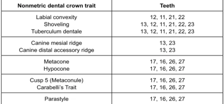
For the observation of 16 NDCT in the permanent dentition (Tables 1 and 2) this study used the Arizona State University Dental Anthropology System (ASUDAS).
Each of the 110 cases was assigned a corresponding code number to the clinical process, not only for the observer to be unaware of the sex of the participants during observations (single-blinded), as well as to facilitate further processing data and in order not to disclose identifying information.
The observation sessions took place in a room with natural light and were macroscopically performed without magnification lens (Figure 1).
The study counted only with the participation of one observer and was performed over approximately 2 months. Inorder to minimize potential eye strain of the viewer that would compromise the following observations, small breaks (10 minutes) were taken between each characteristic assessed during the data collection.
All NDCT were classified by its degree of expression and data were registred in a form developed for this purpose. Permanent teeth were named according to the FDI system.
All data resulting from observations were collected by the same observer. Thus, it was intended to introduce the least possible bias in a
ssessments; simultaneously introducing interobserver variability was excluded. Each observation session of the dental morphological traits followed the same methodological criteria set for its completion. However, it was necessary to estimate the intraobserver error. Thus, the observations were repeated at 10% of the sample after a time interval of more than1 month in order to suppress the memory effect. The data obtained from the NDCT observation from each of the 110 cases were first entered into a template on Excel® sheet and statistical analyses was performed using the IBM® SPSS Statistics® Software, version 22.0. The 16 NDCT combined with the permanent teeth observed (all except third molars, often un erupted or extracted, and upper premolars, frequently extracted in orthodontic cases and due to the fact that the NDCT proposed by the ASUDAS for these teeth were considered irrelevant to include in the study) resulted in 66 independent variables (placed as experimental hypothesis) plus the variable sex.
Descriptive statistics was presented (distribution of the sample population by sex, distribution of all of teeth examined, frequencies of all independent variables and descriptive analysis of each variable by gender) and non-parametric tests were applied (Mann-Whitney U and Wilcoxon). The contingency coefficient was determined and the kappa coefficient was assessed. The conventional 5% level (p<0.05) was considered to be statistically significant.
In addition, abinomial logistic regression allowed modeling the probability of success for the reference category female, based on the development of a model for gender classification with 3 selected variables which showed significant differences among both sexes.
Results
Descriptive statistics
The sample selection consisted of 110 participants, 53 male (48.2%) and 57 female (51.8%). All 66 independent variables were evaluated for each of the 110 individuals giving a total of 7260 observations. From these, 5654 observations (78%) contributed to the analyses of the expression of the dental morphological trait. Scoring of the NDCT was not possible in approximately 1606 observations (22%). In this group, the ASUDAS method could not be applied due to missing orpartially erupted teeth, presence of restorations, dental wear, dental crowding, misshapen teeth, orthodontic appliance, teeth with orthodontic bands and also due to the condition of the dental cast. Intra-observer error: In order to know if the observer was scoring in equal measure, the results from the first time of observation (entire sample cases) and the second time (10% of the sample population) were analysed by a non-parametric test for related samples (Wilcoxon). The level of agreement was done by means of the kappa
Nonmetric dental crown trait Teeth
Labial convexity Shoveling Tuberculum dentale 12, 11, 21, 22 13, 12, 11, 21, 22, 23 13, 12, 11, 21, 22, 23 Canine mesial ridge
Canine distal accessory ridge 13, 2313, 23 Metacone
Hypocone 17, 16, 26, 2717, 16, 26, 27 Cusp 5 (Metaconule)
Carabelli’s Trait 17, 16, 26, 2717, 16, 26, 27 Parastyle 17, 16, 26, 27
Table 1: Dental morphological traits observed in permanent dentition - Upper
arch.
Non-metric dental crown trait Teeth
Shoveling
Canine distal accessory ridge 41, 42, 31, 3243, 33 Premolar lingual cusp variation
Anterior Fovea 45, 44, 34, 3546, 36 Deflecting wrinkle
Cusp 5 (Hypoconulid) 47, 46, 36, 3746, 36 Cusp 6 (Entoconulid) 47, 46, 36, 37 Cusp 7 (Metaconulid) 47, 46, 36, 37
Table 2: Dental morphological traits observed in permanent dentition - Lower
arch.
Figure 1: UI2 shovel plaque for scoring upper lateral incisors and upper
canines. Each of the ASUDAS standard reference plaques gives a physical representation of minimal and maximal characteristic expression and various gradations between these two points.
Volume 1 • Issue 1 • 100001
coefficient (the minimum observed value was 0.083 and it was observed 10 values lower than 0.4).
There were no significant differences observed between the two measurements except for the variables presented in table 3. Although only 3 variables had significant disagreement in scoring the NDCT, their individual values obtained for the kappa coefficient were far from a perfect level of concordance (1).
Sexual dimorphism and bilateral symmetry
The analysis of the expression of dental morphology of the studied sample was performed using a non-parametric test for independent samples (Mann-Whitney U) to determine whether there were differences between sexes. The results of this test did not detect statistically significant differences for all the features identified as independent variables, except for some of those presented in table 4, meaning that those variables presented sexual dimorphism and the null hypothesis was rejected. The lower permanent canines obtained the lowest significance values, with tooth 43 having the most significant statistical value (p<0.01).
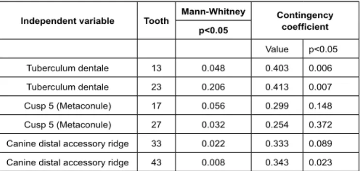
Additionally, in order to understand the degree of association between the dental morphological characteristics and sex, the contingency co-efficient was determined (Table 4). Although the contingency coefficient values were not high, the tuberculum dentale of teeth 13 and 23 and the canine distal accessory ridge of tooth 43 showed an association with statistical significance, thus rejecting the null hypothesis of a non-existing association between NDCT and sex. Let us notice that table 4 contains symmetrical teeth. This may occur because the symmetrical teeth may include the same kind of information about the individual’s gender. Therefore, the analysis of dental bilateral symmetry was executed based on a non-parametric test matched by pairs of antimere teeth (Wilcoxon) for the variables with significant differences between male and female. As evidenced by the results (Table 5), statistically none of the pairs were significantly different at the 5% level. Thus, in the gender classification model discussed in the next subsection it will only be used one tooth of each pair of symmetry (the most significant between the two), in order to obtain a more parsimonious model.
Gender classification analysis
This study intended to analyze the reliability of a gender classification model for dental morphological traits which allowed modeling the probability of success for the female sex.
Through a binomial logistic regression and by taking the variables of the permanent canines (above mentioned in table 4) with significant differences between both sexes (without using symmetric teeth), the developed model uses the information from three teeth: the tuberculum dentale of tooth 13, the cusp 5 (Metaconule) of tooth 27 and the canine distal accessory ridge of tooth 43 (Figure 2).
However, the Pseudo R square proved to below, the Cox and Snell’s pseudo R2 value obtained was 0.334 and the Nagelkerke’s pseudo
R2 equals to 0.458, thus the model did not show great potential to
gender classification although it predicted 76.4 percentage correct. Nevertheless, 24.6 percent of the females and 22.6 percent of the males are misclassified.
In short, even though the model display significant differences between genders, the used information does enable a reliable gender classification model, with low probability of misclassification (the sensitivity and the specificity of the model are around 75 percent) (Table 6).
Independent
variable Tooth Wilcoxonp<0.05 Kappa coefficient value
Strength of agreement (Altman, 1991)
Labial convexity
Anterior Fovea 12*36 0.0340.034 0.1460.314 PoorFair Anterior Fovea 46 0.034 0.302 Fair Table 3: Dental morphological traits withdifference in the twoobservation times.
*Labial convexity of tooth 22 wasclose to be statisticallysignificant (0.059).
Independent variable Tooth Mann-Whitney Contingency coefficient p<0.05 Value p<0.05 Tuberculum dentale 13 0.048 0.403 0.006 Tuberculum dentale 23 0.206 0.413 0.007 Cusp 5 (Metaconule) 17 0.056 0.299 0.148 Cusp 5 (Metaconule) 27 0.032 0.254 0.372 Canine distal accessory ridge 33 0.022 0.333 0.089 Canine distal accessory ridge 43 0.008 0.343 0.023 Table 4: Presence of sexual dimorphism in three dental morphological traits.
NDCT Pair Wilcoxon
p<0.05
Tuberculum dentale 13-23 0.551 Cusp 5 (Metaconule) 17-27 0.961 Canine distal accessory ridge 33-43 0.174 Table 5: Bilateral symmetry of the pairs of antimere teeth.
Figure 2: DAR LC plaque for scoring Canine distal accessory ridge on lowers
canines. Each of the ASUDAS standard reference plaques gives a physical representation of minimal and maximal characteristic expression and various gradations between these two points for each gender.
Predicted
Gender Percentage
Observed Male Female Correct
Male 41 12 77.4
Female 14 43 75.4
Overall Percentage 76.4
Table 6: Classification tablea).
Volume 1 • Issue 1 • 100001
Discussion
The internal value of a laboratory study is related to the statistic validity of the results and the generalization of the findings. The validity of the results depends on the integrity of the experimental methodology. The possibility of intra and inter population generalization depends on population characteristics used and the conditions of implementation of the qualitative analysis.
Integrity of the experimental methodology
This study used a worldwide known method, the ASU Dental Anthropolgy System for the systematic analysis of dental morphological characteristics. Plaster models of the population sample were used to evaluate and classify the degree of expression of NDCT in the permanent dentition.
The experimental procedure consisted of an observational non-destructive qualitative method, easy to perform without leading to tissue damage or using an invasive technique, thus ensuring the physical integrity of the dental pieces [21], in this case the plaster models.
The random sample counted with a total of 110 cases with a minimum of 50 individuals for each group (sex). However, with little time and more limited resources, a hundred individuals are considered a viable sample to provide a good characterization of the population [13].
The cases were selected between the existing plaster models of the orthodontics department of the Faculty of Dentistry from University of Lisbon according to the inclusion criteria, thus absent of a true randomization.
The ideal sample size of a study is determined by the level of significance, the power and the magnitude of the differences to be detected between groups (male and female). By setting up a statistical significance level of 5%, a smaller sample size leads to a reduced statistical power. The main consequence of this reduction is the loss of ability to detect statistically significant differences between groups. For this, the author is aware that data collection should be planed to take sized nM (male) and nF (female) sub samples such that, with a preassigned probability, differences greater than a threshold (bound, “B”) could be detected. Therefore, the half width of the confidence interval (making a Gaussian approximation, legitimate at t(γ) with γ≥30),
(where 1.96=Z0.975, meaning, we are using the confidence coefficient of 0.95, previously declared as normal in preparatory research work) would be enough to solve in order to obtain lower bounds for nM and nF. In this study their calculation was not performed resulting in a convenience sample and the sampling was balanced to
(which means, to seek the same standard error for the two subsets) where in nM = nF = 50, where the variances were identical. So with a prior assessment of the variances, conducted in this pilot study, the sample can be collected with other accuracy for further confirmatory studies.
Plaster models have advantages for the study of dental morphology, they may be obtained from current populations, as was the case in this study, and they allow to define the sample size and age groups depending on the intended goals without being dependent on factors such as post mortem tooth loss. The main disadvantage is clearly the restricted morphological observation of only one part of the teeth, meaning the crowns [22]. Furthermore, a significant fraction of the sample may be lost, for example due to phenomena such as dental tooth wear, carries, congenital anomalies, the presence of dental fillings, dental trauma fractures, dental extractions, dental agenesis and also by influence of environmental factors [23]. Yet the study of dental morphological characteristics in skeletonized remains has the great advantage to include the study of root morphology in the research protocol, even in the absence of dental structures. However, 100 human skeletons do not provide a sample of 100 morphological features to be observed [22].
In the population sample, 22% of the observations failed in classifying the degree of expression of the morphological characteristics determined as variables, mainly due to individuals with absent teeth, the presence of partially erupted teeth and restorations. The first two mentioned reasons refer to the fact that many plaster models belonged to individuals under the age of 12 years with mixed dentition. Accordingly, the selection of morphological characteristics to be assessed in the experimental design could have been more embracing by including also the analysis of dental morphology in primary teeth given the fact that they count as precursors in the development of the permanent ones, representing a possible omen of morphological features later displayed by the permanent dentition [22]. Restorations were observed mostly in teeth with a more complex occlusal anatomy (molars), especially in the first permanent molars probably because these had been for the longest in the oral cavity. Furthermore, the younger population currently requires a greater number of dental treatments and has a higher caries experience than previous generations [24]. Considering that the population of this sample was selected from individuals who attend or attended the orthodontics department, several plaster models showed teeth with orthodontic bands or brackets, fixed orthodontic appliance, fixed containment device and dental crowding. There were also cases where the plaster models were not in perfect condition and, in certain cases, the morphological characteristics were not noticeable as a consequence of dental wear. All these conditions together ultimately reduced the potential of the dental morphology of the sample. The observation in plaster models (the ones included in the study securely placed in an individual box) or directly in teeth require conditions such as an adequate and well-lit environment, preferably using a magnifying lens [22]. This tool was not used and all observations were made macroscopically by a single observer implying a detailed and deep examination of the characteristics from all possible angles, to prevent more subtle features to be unnoticed. Analyzing dental morphological characteristics directly has its drawbacks (as material defects), but the reflection of light by the enamel makes the observation of traits less obvious and more difficult. Scott increases that the analysis of tooth morphology in an intra-oral environment in living individuals is not ideal for two reasons: the reflection of light is exacerbated by the presence of saliva and various observation perspectives become limited in the oral cavity [22]. Data obtained by dental qualitative assessment sin grades have a great inter observer error [25]. In this study the data were all registered by the same investigator-the author. Dental qualitative
2 2
1.96
M
F B
nm
nF
σ
+
σ
≤
2 2 2 2M
F
n M
n F
σ
=
σ
Volume 1 • Issue 1 • 100001
assessments were established by criteria strictly defined before the study, through a careful definition of the variables. Using only one investigator was advantageous in order to limit the introduction of various non quantifiable or controllable factors.
Intra-observer variability: There was a discrepancy between the first and second observations in the assessment of 2 dental morphological characteristics in 3 variables (anterior fovea of teeth 36 and 46 and labial convexity of tooth 12), with low individual kappa values. The reason of this variability may have been due to a lighting difference in both phases of observations, lack of magnification, greater difficulty in classification of these characteristics by an inexperience observer or even plaster models with a less polished surface, with reduced glow offering lower contrast for the analysis of the characteristics. The differences observed may also arise from a possible eyes train from the observer or even due to a combination of all factors mentioned above. In addition to these possibilities, Scott argues that the study of dental morphology requires a certain component of “art”, but still refers to it as a rigorous scientific method [22].
Discussion of the results
Sexual dimorphism: Through analysis of teeth, it is possible to study the sexual dimorphism of an individual from the patterns of dental development and eruption, the expression of the amelogen in protein, dental morphology and dental dimensions [13]. Once formed, teeth do not undergo changes in morphology like bones, but non-metric dental crown traits which are phenotypic forms of the enamel (expressed and regulated by the human genome of each individual) can disappear due to dental wear and as a consequence of certain oral pathologies [15,23,26].
In this study, the characteristics which showed significant differences between male and female were in tooth 13 (tuberculum dentale), tooth 27 (cusp 5/metaconule) and in teeth 33 and 43 (canine distal accessory ridge). However, Rocha et al., [27] achieved significant results for the existence of sexual dimorphism for the shoveling characteristic in the maxillary central incisors, using the ASUDAS method in the permanent dentition in a sample of 42 Colombian women and 42 Colombian men.
Scott and Turner fall within the group of researchers who claim that dental morphological characteristics are equally expressed between both sexes and that the sexual dimorphism exhibited by them is low or nonexistent. In studies of Goyes et al., [8] and Díaz et al., [28], both applied the ASUDAS method in the permanent dentition in different samples of the Colombian population, whose results demonstrated the absence of sexual dimorphism for the tuberculum dentale of the maxillary canines and the distal accessory ridge of the mandibular canines in the first study and, for cusp 5/metaconule for the second study mentioned. Such contradictory facts can reside on the assumption that in both cases the number of the sample was below the population of the present study, or that dental morphological characteristics are specific to a given population, or even because of the existing geographical distance separating population groups. Genetic and environmental factors may also be an explanation for these facts, although it is suggested that tooth morphology, in comparison with tooth size, is less susceptible to external effects [23,29].
As observed in the current study, other researchers concluded that there is no evidence of sexual dimorphism in the meta cone, hypo cone, Carabelli’s trait, parastyle, canine mesial ridge, labial
convexity, shoveling, deflecting wrinkle, cusp 6/entoconulid and cusp 7/metaconulid when applying the ASUDAS method [15,28,30-32]. Reality behind these findings may be, again, due to a low number of individuals in each subpopulation group (gender), including the present study. A possible systematic reduction of sexual dimorphism throughout human evolution, combined with a gradual process of simplification of dental morphology, may have lead to trends increasingly monomorphic [9]. There may be also an association with a line of evolution of the species in the way that currently, a phenomenon related to the gracilization of males phenotypic characteristics, tend to occur [25]. However, according to Moreno-Gómez, contemporary human beings are dimorphic, although less than other hominids, contributing dental morphological traits (especially those of the canines) to this fact.
Dental sexual dimorphism studied by means of odontometric analysis have shown statistically significant differences in the permanent dentition for the canines, especially the mandibular ones [25,33-39]. Although the present study has employed a qualitative method, results are in agreement with this evidence. Based on the results obtained for the canine distal accessory ridge, teeth 33 and 43 were the most dimorphic in the population sample. Also Dumancic et al., [40], who devoted a study exclusively to the canine distal accessory ridge applying the ASUDAS method in a Croatian population, showed that the mandibular canine is a better sexual indicator than the maxillary canine in the permanent dentition. According to Scott [22], the canine distal accessory ridge is considered the NDCT with more dimorphic among the human dentition, displaying frequencies and forms of more pronounced expression in males, as verified by the results of the present study. From a macro evolutionary point of view, it is known that canine teeth are less influenced by environmental factors and have greater stability in their morphogenetic field, hence the possible explanation for the higher degree of sexual dimorphism [7,13]. Nevertheless, further investigations are needed to clarify which genetic factors and sex hormones are directly involved in the process of morphogenesis, in order to better understand the presence/absence of various dental morphological characteristics in each sex, as well as the more pronounced forms of expression observed in males.
Dental symmetry: The pairs of antimere teeth (13-23/tuberculum dentale, 17-27 /metaconule and 33-43/canine distal accessory ridge) did not show bilateral asymmetry in the dental morphological characteristics between the right and the left side of the dental arches. These results corroborate those of other studies in which there was a symmetric expression of the dental morphological characters [6,15,27,41]. In all, the ASUDAS method was employed in the permanent dentition. However, the dental morphological characteristics investigated were not the same, as well as the studied population. Thus, and in spite of the current study did not focus on the same dental morphological traits, it seems that a general symmetry of pairs of homologous teeth exist. The explanation more plausible for the observed bilateral symmetry is related to the gene influence that acts identically on opposite sides of the dental arch [42].
Accordingly, and with regard to the goals of this work, there is no gain in using two symmetrical teeth in the gender classification model since they contain similar information.
ASUDAS method reliability in sex classification: The attempt to model the probability of success for the female sex, by taking dental morphological characteristics of permanent teeth (13, 27 and 43) to
Volume 1 • Issue 1 • 100001
elaborate a model, was innovative. But, despite having obtained more than 75% correct classifications, this did not fully fit the desired goal by presenting a significant probability of misclassification (23.6%).
Conclusion
The results proved the existence, albeit not as significant as it was intended, of sexual dimorphism in dental morphological characteristics of permanent teeth in a sample of the Portuguese population. These not only represent sexually dimorphic phenotypic structures, as may be specific to a particular population. The distal accessory ridge of the mandibular canines, proved to be the NDCT with more evidence of sexual dimorphism, showing frequencies and more pronounced forms of expression in males. Based on the method applied, tooth 43 has proven to be the most dimorphic and best sexual indicator among the Portuguese population through the NDCT canine distal accessory ridge.
However, the ASUDAS method has showed a not significant reliability to be used as a means of sexual diagnosis alone. The classification model elaborated with the dental morphological crown characteristics of teeth 13, 27 and 43 proved to be reasonable (76.4%) to estimate successfully for female (23.6% of misclassification) Future work should include larger samples and comparative studies between distinct population groups to provide a greater reliability of this qualitative method of sex diagnosis, for the purpose of forensic application.
References
1. Zorba E, Moraitis K, Manolis SK (2011) Sexual dimorphism in permanent teeth of modern Greeks. Forensic Sci Int 210: 74-81.
2. Rösing FW, Graw M, Marré B, Ritz-Timme S, Rothschild MA, et al. (2007) Recommendations for the forensic diagnosis of sex and age from skeletons. Homo 58: 75-89.
3. Pereira C (2012) Medicina Dentária Forense. Lidel, Lisbon, Portugal. 4. Iscan MY (2005) Forensic anthropology of sex and body size. Forensic Sci
Int 147: 107-112.
5. Fortes de Oliveira O, Lima Ribeiro Tinoco R, Daruge Júnior E, Silveira Dias Terada AS, Alves da Silva RH, et al. (2012) Sexual dimorphism in Brazilian human skulls: discriminant function analysis. J Forensic Odontostomatol 30: 26-33.
6. Girón G, Gómez P, Morales L, León M, Moreno F (2009) Morphologic and Metric Dental Crown Traits of Upper and Lower Premolars in Scholars of Cali, Colombia. Int J Morphol 27: 913-925.
7. Goyes J, Guerrero L, Narváez N, Moreno F (2011) Non-metric Dental Traits of Deciduous and Permanent Canines in a Group of Mixed Ethnicity Popula-tion in Cali, Colombia. Revista Colombiana de Investigación en Odontología 2: 1-13.
8. Monali C, Pritam P, Tapan M, Kajal D (2011) Gender determination: a View of Forensic Odontologist. Indian Journal of Forensic Medicine and Pathology 4: 147-151.
9. Thapar R, Angadi PV, Hallikerimath S, Kale AD (2012) Sex assessment us-ing odontometry and cranial anthropometry: evaluation in an Indian sample. Forensic Sci Med Pathol 8: 94-100.
10. Rai B, Kaur J (2013) Sex Determination Methods in Forensic Odontology. In: Rai B, Kaur J (eds.). Evidence-Based Forensic Dentistry. Springer Berlin Heidelberg, Germany. Pg no: 73-79.
11. Kondo S, Townsend GC, Yamada H (2005) Sexual dimorphism of cusp di-mensions in human maxillary molars. Am J Phys Anthropol 128: 870-877. 12. Radlanski RJ, Renz H, Hopfenmüller W (2012) Sexual dimorphism in teeth?
Clinical relevance. Clin Oral Investig 16: 395-399.
13. Moreno-Gómez F (2013) Sexual Dimorphism in Human Teeth from Dental Morphology and Dimensions: A Dental Anthropology Viewpoint. In: Hiroshi Moriyama (ed.). Sexual Dimorphism. Intech open science open minds, Cro-atia. Pg no: 97-124.
14. Viciano J, López-Lázaro S, Alemán I (2013) Sex estimation based on decid-uous and permanent dentition in a contemporary Spanish population. Am J Phys Anthropol 152: 31-43.
15. Aguirre L, Castillo D, Solarte D, Moreno F (2006) Frequency and Variability of Five Non-Metric Dental Crown Traits in the Primary and Permanent Den-titions of a Racially Mixed Population from Cali, Colombia. Dental Anthropol-ogy 19: 39-48.
16. Hanihara T (2008) Morphological variation of major human populations based on nonmetric dental traits. Am J Phys Anthropol 136: 169-182.
17. Kimura R, Yamaguchi T, Takeda M, Kondo O, Toma T, et al. (2009) A com-mon variation in EDAR is a genetic determinant of shovel-shaped incisors. Am J Hum Genet 85: 528-535.
18. Pereira C, Antunes MT, Pestana D, Mendonça MCd, Santos JC (2012) An Unidentified Skeletal Assemblage from a Post-1755 Mass Grave of Lisbon: Dental Morphology and Population Affinity. Bulletin of the International Asso-ciation for Paleodontology 6: 12-26.
19. Carter K, Worthington S, Smith TM (2014) News and views: non-metric dental traits and hominin phylogeny. J Hum Evol 69: 123-128.
20. Turner GC, Nichol CR, Scott GR (1991) Scoring Procedures for Key Morpho-logical Traits of the Permanent Dentition: The Arizona State University Dental Anthropology System. In: Kelley MA, Larsen CS (eds.). Advances in Dental Anthropology. Wiley-Liss, New York, USA. Pg no: 13-31.
21. Cunha C (2012) Caracterização da Morfologia Dentária no Médio Guadiana no Neolítico Final-Calcolítico. Fundamentação para o Mapeamento Mor-fológico das Populações Locais na Pré-História Recente. In: António Valera (ed.). Apontamentos de Arqueologia e Património. Núcleo de Investigação Arqueológica-NIA, Lisbon, Portugal. Pg no: 49-55.
22. Scott GR (2008) Dental Morphology. In: Katzenberg MA, Saunders SR (eds.). Biological Anthropology of the Human Skeleton. (2nd edn), New Jersey, John Wiley & Sons. Pg no: 265-298.
23. Scott GR, Turner CG (1988) Dental Anthropology. Annu Rev Anthropol 17: 99-126.
24. Sweet D (2010) Forensic dental identification. Forensic Sci Int 201: 3-4. 25. Pereira C, Bernardo M, Pestana D, Santos JC, Mendonça MC (2010)
Contri-bution of teeth in human forensic identification - discriminant function sexing odontometrical techniques in Portuguese population. J Forensic Leg Med 17: 105-110.
26. Marado L, Campanacho V (2013) Carabelli’s trait: Definition and review of a commonly used dental non-metric variable. Cadernos do GEEvH 2: 24-39. 27. Rocha L, Rivas H, Moreno F (2007) Frequency and variability of the dental
morphology in African-Colombian children of a school institution of Puerto Tejada, Cauca, Colombia. Colombia Médica Julio- 38: 210-221.
28. Díaz E, García L, Hernández M, Palacio L, Ruiz D, et al. (2014) Frequency and variability of dental morphology in deciduous and permanent dentition of a Nasa indigenous group in the municipality of Morales, Cauca, Colombia. Colomb Med (Cali) 45: 15-24.
29. Zorba E, Spiliopoulou C, Moraitis K (2013) Evaluation of the accuracy of dif-ferent molar teeth measurements in assessing sex. Forensic Sci Med Pathol 9: 13-23.
30. Moreno F, Moreno SM, Díaz CA, Bustos EA, Rodríguez JV (2004) Prevalen-cia y variabilidad de ocho rasgos morfológicos dentales en jóvenes de tres colegios de Cali. Colombia Médica 35: 16-23.
31. Delgado-Burbano (2007) Variacíon dental no-métrica y el tráfico de esclavos por el atlántico: la ascendencia biológica y los orígenes geográficos de una población afro-colombiana. Revista Española de Antropología Física 27: 13-32.
Volume 1 • Issue 1 • 100001 32. Aragón N, Bastidas C, Bedón LK, Duque P, Sánchez M, et al. (2008) Rasgos
morfológicos dentales coronales en denticíon temporal y permanente: Dis-tancia biológica entre tres grupos indígenas del Amazonas Colombiano. Re-vista Odontológica Mexicana 12: 13-28.
33. Boaz K, Gupta C (2009) Dimorphism in human maxillary and mandibular ca-nines in establishment of gender. J Forensic Dental Sciences 1: 42-44. 34. Kapila R, Nagesh KS, Iyengar AR, Mehkri S (2011) Sexual Dimorphism in
Human Mandibular Canines: A Radiomorphometric Study in South Indian Population. J Dent Res Dent Clin Dent Prospect 5: 51-54.
35. Sonika V, Harshaminder K, Madhushankari GS, Sri Kennath JA (2011) Sex-ual dimorphism in the permanent maxillary first molar: a study of the Haryana population (India). J Forensic Odontostomatol 29: 37-43.
36. Parekh DH, Patel SV, Zalawadia AZ, Patel SM (2012) Odontometric Study of Maxillary Canine Teeth to Establish Sexual Dimorphism in Gujarat Popula-tion. Int J Biol Med Res 3: 1935-1937.
37. Angadi PV, Hemani S, Prabhu S, Acharya AB (2013) Analyses of odontomet-ric sexual dimorphism and sex assessment accuracy on a large sample. J Forensic Leg Med 20: 673-677.
38. Ayoub F, Shamseddine L, Rifai M, Cassia A, Diab R, et al. (2014) Mandibular Canine Dimorphism in Establishing Sex Identity in the Lebanese Population. Int J Dent 2014: 235204.
39. Chukwujekwu IE, Ezejindu DN, Moses FC (2014) Odontometric Study of Mandibular Canine Teeth Dimorphism in Establishing Sex Identity in South-east Nigeria. Int J Med Med Sci 1: 38-41.
40. Dumancic J, Pavicin IS, Brkic H (2013) Sexual Dimorphism in Canine Mor-phology. J Forensic Odontostomatology 31: 44.
41. Marcovich I, Prado E, Díaz P, Ortiz Y, Martínez C, et al. (2012) Dental Mor-phology Analysis of Afro-Colombian School Children from Villa Rica, Cauca, Colombia. Rev Fac Odontol Univ Antioq 24: 37-61.
42. Wetherell J, Winning T, Townsend G (2004) Localized Asymmetry in Human Dental Crown Form- an Interesting Case. Dental Anthropology 17: 18-23.