Homozygous sequence variants in the
WNT10B
gene underlie split hand/foot
malformation
Asmat Ullah
1*, Ajab Gul
2*, Muhammad Umair
1, Irfanullah
1, Farooq Ahmad
1, Abdul Aziz
3, Abdul Wali
2and
Wasim Ahmad
11
Department of Biochemistry, Faculty of Biological Sciences, Quaid-i-Azam University, Islamabad,
Pakistan.
2
Department of Biotechnology and Informatics, BUITEMS, Quetta, Pakistan.
3Department of Computer Sciences and Bioinformatics, Khushal Khan Khattak University, Karak, Pakistan.
Abstract
Split-hand/split-foot malformation (SHFM), also known as ectrodactyly is a rare genetic disorder. It is a clinically and genetically heterogeneous group of limb malformations characterized by absence/hypoplasia and/or median cleft of hands and/or feet. To date, seven genes underlying SHFM have been identified. This study described four consan-guineous families (A-D) segregating SHFM in an autosomal recessive manner. Linkage in the families was estab-lished to chromosome 12p11.1–q13.13 harboringWNT10B gene. Sequence analysis identified a novel homozygous nonsense variant (p.Gln154*) in exon 4 of theWNT10B gene in two families (A and B). In the other two families (C and D), a previously reported variant (c.300_306dupAGGGCGG; p.Leu103Argfs*53) was detected. This study fur-ther expands the spectrum of the sequence variants reported in theWNT10B gene, which result in the split hand/foot malformation.
Keywords: Split-Hand-Foot Malformation 6,WNT10Bgene, sequence variants.
Received: June 15, 2016; Accepted: April 10, 2017.
Introduction
Split-hand/split-foot malformation (SHFM) is a con-genital limb defect characterized by absence of medial digi-tal rays, median clefts of the hands and feet, aplasia and/or hypoplasia of the phalanges, and syndactyly (McKusick, 1998; Duijfet al., 2003). In severe cases, feet exhibit a lob-ster claw-like appearance with a deep median groove. Asymmetric manifestations, variable syndactyly and non-penetrance of the disorder have also been described (Ozen et al., 1999). Few SHFM patients have been found with signs of intellectual disability, ectodermal and craniofacial defects, and orofacial clefting (Elliott and Evans, 2006). Clinical features of SHFM vary from patient to patient and within the same individual (Duijfet al., 2003; Elliott and Evans 2008; Sowinska-Seidleret al., 2014). Both isolated (non-syndromic) and syndromic forms of SHFM have been reported in the literature.
Seven types of non-syndromic SHFM have been mapped on different human chromosomes. Four of these forms (SHFM-1, SHFM-3, SHFM-4, SHFM-5) inherit in
autosomal dominant pattern, SHFM-2 in X-linked inher-ited, and SHFM-6 in autosomal recessive pattern. The four autosomal dominant forms were mapped on chromosome 7q21 (SHFM-1; MIM183600) (Schereret al., 1994), 10q24 (SHFM-3; MIM246560) (Nuneset al., 1995; Gurrieri et al., 1996; Raas-Rothschildet al., 1996), 3q27 (SHFM-4; MIM605289) (Ianakievet al., 2000) and 2q31 (SHFM-5; MIM606708) (Boleset al., 1995). The autosomal recessive form (SHFM-6; MIM 225300) has been mapped on chro-mosome 12q13.11-q13 (Ugur and Tolun, 2008). The X-linked form was mapped on Xq26, which was later re-fined to a small region of 5.0 cM (Faiyaz-Ul-Haqueet al., 2005). Based on genome-wide linkage analysis in a single family with autosomal recessive inheritance pattern, Gurnettet al.(2006) mapped a novel locus for SHFM on chromosome 8q21.11–q22.3. For these seven loci, only three (SHFM1, SHFM4, SHFM6) have been solved at the gene level. A causative gene of the wingless-type MMTV integration site family, member 10 (WNT10B, MIM 601906) has been reported of causing SHFM-6 (Ugur and Tolun, 2008). Ianakievet al.(2000) reported the geneTP63 (tumor protein p63, MIM 603273), which encodes a homo-log of the tumor suppressor responsible for the SHFM-4 phenotype. Shamseldinet al.(2012) reported a mutation in the geneDLX5responsible for the SHFM-1 phenotype in a
Send correspondence to Wasim Ahmad, Department of Biochemis-try, Faculty of Biological Sciences, Quaid-i-Azam University, Isla-mabad, Pakistan. E-mail: [email protected]
consanguineous family. We reported intragenic variants in theDLX5andDLX6genes that caused the SHFM-1 pheno-type in two different families (Ullahet al., 2016a,b). Good-manet al.(2002) proposed that SHFM-5 and other digit defects may be caused by haploinsufficiency of genes (EVX2,DLX1,DLX2) located at the 5’ end of theHOXD cluster. Recently, Umairet al.(2017) reported a homozy-gous variant in theEPS15L1gene located on chromosome 19p13.11 causing isolated SHFM in a Pakistani family.
In this study, we investigated four consanguineous families of Pakistani origin showing split hand and foot malformation phenotypes. Linkage in the families was es-tablished to SHFM6 on chromosome 12p11.1-q13.13. Se-quence analysis of the WNT10B gene detected a novel nonsense and a previously reported variant in affected indi-viduals.
Materials and Methods
Family history
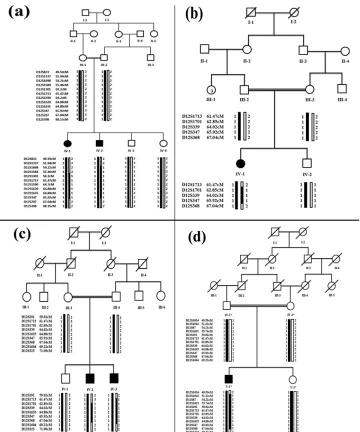
Approval to conduct the study was obtained from the Institutional Review Board (IRB) of Quaid-i-Azam Uni-versity, Islamabad Pakistan. Four consanguineous families with clinical manifestations of split hand and foot malfor-mations (SHFM were recruited from remote areas of Paki-stan. Pedigree reconstruction (Figure 1) provided convinc-ing evidence of an autosomal recessive mode of inheritance of the disorder. Six individuals with SHFM were identified in four families. This included a male (IV-1) and a female (IV-2) in family A, a female (IV-1) in the fourth generation in family B, two males (IV-2, IV-3) in the fourth generation in family C, and one male (V-1) in the fifth generation in family D. All affected and unaffected individuals in the four families were examined by medical specialists at local government hospitals. Blood samples were collected from both affected and unaffected individuals of the families af-ter obtaining written consent.
Isolation of Genomic DNA
Peripheral blood samples were obtained from 19 indi-viduals in EDTA containing vacutainer sets (BD, Franklin Lakes, NJ, USA). Genomic DNA extraction was performed using a standard phenol-chloroform procedure. DNA was quantified using a Nanodrop-1000 spectrophotometer (Thermal Scientific, Wilmington, MA).
Genotyping
Linkage in the families was searched by genotyping microsatellite markers mapped in the flanking regions of autosomal dominant and autosomal recessive forms of SHFM. This included SHFM1 (D7S2537, D7S2481, D7S630, D7S492, D7S627, D7S1813, D7S657, D7S527, D7S479) at chromosome 7q21, SHFM3 (D10S520, D10S91, D10S1736, D10S1726, D10S603, D10S1710, D10S383, D10S1264) at chromosome 10q24, SHFM4
(D3S3570, D3S3600, D3S3596, D3S1661, D3S2747, D3S1662, D3S2311, D3S1305) at chromosome 3q27, SHFM5 (D2S124, D2S2345, D2S294, D2S2302, D2S1274, D2S2257, D2S2173, D2S2978) at chromosome 2q31, SHFM6 (D12S1034, D12S823, D12S1042, D12S1337, D12S1698, D12S87, D12S1584, D12S1621, D12S291, D12S1301, D12S1713, D12S1701, D12S339, D12S1590, D12S1620, D12S1635, D12S347, D12S297, D12S368, D12S398, D12S1604, D12S325) at chromo-some 12q11-q13, and another SHFM locus mapped on chromosome 8q21.11–q22.3 (D8S526, D8S2321, D8S1119, D8S1818, D8S1129, D8S1714, D8S556) (Gurnett et al., 2006). PCR amplification of the micro-satellite markers was performed as previously described (Ullahet al., 2015). The amplified PCR products were re-solved on 8% non-denaturing polyacrylamide gels, stained with ethidium bromide, and genotypes were assigned by vi-sual inspection. DNA ladders of 5, 10 and 20 bp (MBI Fermentas®, Life Sciences, York, UK) were used to deter-mine allele size for respective microsatellite markers. Markers used in the genotyping were arranged according to Rutgers combined linkage-physical map (Build 36.2) of the human genome (Matiseet al.,2007). Haplotypes were ana-lyzed by SIMWALK2 (Sobel and Lange, 1996).
Sequencing of the WNT10B gene
Primers used for PCR amplification, sequencing and coding of intron-exon junctions of theWNT10Bgene were the same as described earlier (Khan et al., 2012). The PCR-amplified products were purified with a commercially available kit (Axygen MD, USA) and sequenced using ABI BigDye Terminator Sequencing Kit v.3.1 (Applied Bio-systems, Foster City, CA, USA). Sequence variants were identified via the BIOEDIT sequence alignment editor, ver-sion 6.0.7 (Ibis Biosciences, CA, USA).
In silicoanalysis
The pathogenicity index of the sequence variants identified here was calculated using the following soft-wares: Mutation Taster (http://www.mutationtaster.org/), Polymorphism Phenotyping V2 (PolyPhen-2) (http://ge-netics.bwh.harvard.edu/pph2/) and Sorting Intolerant From Tolerant (SIFT) (http://sift.bii.a-star.edu.sg/). The fre-quency of the variants in the general population was deter-mined using the Exome Variant Server (EVS) (http://evs.gs.washington.edu/EVS/), and 1000 genomes.
Results
Clinical features
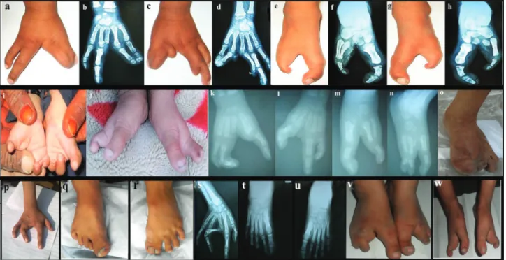
showed cleft hand and cleft foot deformities associated with mesoaxial syndactyly (Figure 2).
In family A, the affected female (IV-1) showed cleft hand deformity with absence of the middle finger in the
right hand. Radiographs showed pre-axial syndactyly of the index finger and thumb at the distal phalanx as well as in the 3rd and 4th metacarpals (Figure 2a,b). The same patient showed cleft hand deformity with central syndactyly in her left hand with an additional bud on the 2nd metacarpal. The distal phalanx of the middle finger was also missing (Figure 2c,d). Classical cleft feet deformities, characterized by cen-tral deficiency, were present. Radiographs showed hallux valgus deformities of the big toe and missing central toes associated with postaxial syndactyly in the metatarsal bones. Some of the tarsal bones were absent (Figure 2e-h). The male individual of the same family (IV-2) showed the same SHFM phenotypes as the female (IV-1). Abnormal-ities such as syndactyly, radial ray malformation, dysplastic hands, and cleft feet were noted (Figure 2i-j). The radiological study revealed bilateral cleft hand/feet de-formities associated with pre-axial and postaxial syndac-tyly in hands and feet, respectively (Figure 2k-n).
In family B, an affected girl (IV-1) was missing the big toe and toes 2 and 3 in the right foot. The left foot and the hands were apparently normal (Figure 2o).
One of the affected members (IV-2) of family C showed aplasia of the middle phalanx of the 3rd finger in the right hand, severe bilateral hypoplasia and fusion of the 3rd and 4th finger (Figure 2p). Fusion of the big toe and 2nd toe in both feet, and an additional rudimentary bud were ob-served in the 3rd toe of the right foot. Radial ray malforma-tion including hypoplasia of the 1st metacarpal, as well as complex fusion of the middle and ring fingers. Fingers with contractures and deviations were observed in radiographs of the right hand of patient IV-2 (Figure 2q-u). The other af-fected member (IV-3) of family C showed bilateral cleft foot with missing 2nd and 3rd toes, characterized by central deficiency with rudimentary bud of lesser toe, classical cleft foot, and basal syndactyly formation (Figure 2v).
In family D, the affected person (V-1) showed cleft foot deformity, i.e. longitudinal deficiency of the digital ray
of the foot except rays 1 and 5, central deficiency and varus deformity of the lesser toe, claw toe deformity, classical cleft foot and fusion of middle and distal phalanx of the 5th toe (Figure 2w). Abnormalities in skin, teeth, face, nails, and eyes were not observed in affected members of the four families. Parents of the affected people were normal and healthy.
Linkage and mutation analysis
Linkage in the families was tested by genotyping microsatellite markers. Haplotypes revealed that affected individuals were homozygous for markers linked to the WNT10Bgene on chromosome 12q11-q13 (Figure 1).
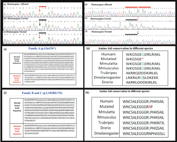
Sub-sequently, theWNT10B gene was sequenced in affected and unaffected members of the families. Sequence analysis detected a novel homozygous nonsense variant (c.460C > G, p.Gln154*) in exon 4 of theWNT10B gene in all the three affected members of the families A and B. In the two other families (C and D) a 7 bp duplication mutation (c.300_306dupAGGGCGG; p.Leu103Alafs*53) was de-tected in the affected members (Figure 3). The two se-quence variants were present in heterozygous state in the obligate carriers in the families and were not identified in 150 ethnically matched control individuals. This absence and the available databases of genetic variations (ExAc,
Figure 3- Sequence analysis ofWNT10Bgene showing a novel nonsense variant (c.460C > G, p.Gln154*) in affected individuals in family A and B. The upper panel (a) represents nucleotide sequences in an affected individual, the middle panel (b) in a heterozygous carrier and lower panel (c) in a normal in-dividual; arrows indicate position of the sequence variant. (d) 7-bp duplication mutation (c.300_306dupAGGGCGG; L103Rfs*53) in the geneWNT10B
1000 genome, EVS or dbSNP) confirm that these were not polymorphisms.
Discussion
To date, five sequence variants in theWNT10Bgene causing SHFM have been reported. Clinical findings result-ing from these variants are summarized in Table 1. Ugur and Tolun (2008) reported a large consanguineous family of Turkish origin in which affected individuals were homo-zygous for p.Arg332Trp in theWNT10Bgene. Blattneret al.(2010) found a 4 bp duplication (c.458_461dupAGCA) in a Swiss woman affected with sporadic SHFM6. Previ-ously, four families of Pakistani origin carrying variants in the WNT10B gene have been reported. This included a missense variant (p.Thr329Arg) (Khanet al., 2012), 4 bp deletion (c.1165_1168delAAGT), and 7 bp duplication (c.300_306dupAGGGCGG) (Azizet al., 2014).A family reported by Khanet al.(2012) showed absence of fingers and thumbs, which were not observed in the families stud-ied by Ugur and Tolun (2008) and Blattneret al.(2010).
In the present study, we have reported four additional Pakistani families with SHFM6 in autosomal recessive pat-tern. The SHFM features observed in affected members of the families included syndactyly, cleft hand/foot malforma-tion, hallux valgus deformities, aplasia, hypoplasia, radial ray malformation, presence of extra rudimentary bone, hypoplastic finger and missing of phalanges. Features ob-served in affected members of the four families were simi-lar to those reported previously (Ugur and Tolun, 2008; Khanet al., 2012; Azizet al., 2014). However, polydactyly in few affected members of the families reported by Khan
et al.(2012) and Azizet al.(2014) were not observed in our study.
Microsatellite-based genotyping established linkage in the four families to SHFM6 on chromosome 12q11-q13, harboring theWNT10Bgene. The geneWNT10Bcontains five exons spanning 6.5 kb of genomic DNA and gives rise to a 389 amino acids protein. WNT10B is a 45 kDa glyco-protein that plays a role in fetal limb bud development and adult hematopoiesis. Sequence analysis of the gene led to the identification of a novel nonsense variant (p.Gln154*) in two families (A and B) and a previously reported dupli-cation (c.300_306dupAGGGCGG; p.Leu103Argfs*53) in the other families (C and D). The sequence variant (p.Gln154*) identified in family A and B is the first non-sense and sixth variant detected in theWNT10Bgene. The premature stop codon at position 154 (p.Gln154*) results in a truncated transcript, which is probably degraded by non-sense mediated mRNA decay (Maquat, 1996, 2005). The 7 bp duplication (c.300_306dupAGGGCGG; Leu103Argfs*53) found in the other two families (C, D) changes the downstream nucleotide sequence, resulting in a premature stop codon (TGA). Thus, the mutant transcript produced is probably also degraded by nonsense-mediated mRNA decay.
The geneWNT10Bbelongs to the WNT gene family. Proteins encoded by the WNT gene family are involved in the activation of a canonical signaling pathway, acting as ligands for cell surface receptors complexes composed of frizzled (FZ) and low-density lipoprotein receptor-related protein 5/6 (LRP5/6) family members. Gordon and Nusse (2006) reported that binding of WNT to FZ and LRP5/6
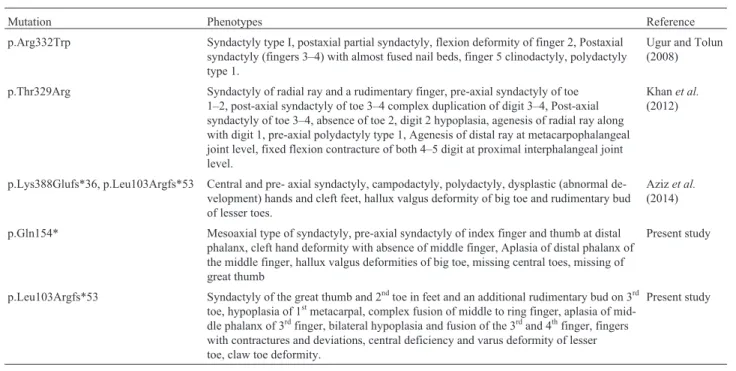
re-Table 1- Comparison of the clinical features, due to sequence variants in theWNT10Bgene, observed in participants of the present study and those re-ported previously.
Mutation Phenotypes Reference
p.Arg332Trp Syndactyly type I, postaxial partial syndactyly, flexion deformity of finger 2, Postaxial syndactyly (fingers 3–4) with almost fused nail beds, finger 5 clinodactyly, polydactyly type 1.
Ugur and Tolun (2008)
p.Thr329Arg Syndactyly of radial ray and a rudimentary finger, pre-axial syndactyly of toe 1–2, post-axial syndactyly of toe 3–4 complex duplication of digit 3–4, Post-axial syndactyly of toe 3–4, absence of toe 2, digit 2 hypoplasia, agenesis of radial ray along with digit 1, pre-axial polydactyly type 1, Agenesis of distal ray at metacarpophalangeal joint level, fixed flexion contracture of both 4–5 digit at proximal interphalangeal joint level.
Khanet al.
(2012)
p.Lys388Glufs*36, p.Leu103Argfs*53 Central and pre- axial syndactyly, campodactyly, polydactyly, dysplastic (abnormal de-velopment) hands and cleft feet, hallux valgus deformity of big toe and rudimentary bud of lesser toes.
Azizet al.
(2014)
p.Gln154* Mesoaxial type of syndactyly, pre-axial syndactyly of index finger and thumb at distal phalanx, cleft hand deformity with absence of middle finger, Aplasia of distal phalanx of the middle finger, hallux valgus deformities of big toe, missing central toes, missing of great thumb
Present study
p.Leu103Argfs*53 Syndactyly of the great thumb and 2ndtoe in feet and an additional rudimentary bud on 3rd toe, hypoplasia of 1stmetacarpal, complex fusion of middle to ring finger, aplasia of mid-dle phalanx of 3rdfinger, bilateral hypoplasia and fusion of the 3rdand 4thfinger, fingers with contractures and deviations, central deficiency and varus deformity of lesser toe, claw toe deformity.
sults in the disruption of the APC/Axin/GSK3 complex that is required for the targeted degradation ofb-catenin.
In conclusion, we have reported a novel and a previ-ously known sequence variant in theWNT10Bgene in four consanguineous families affected by SHFM. This further extended the spectrum of mutations in theWNT10Bgene that result in split hand/foot malformation. These findings can improve diagnosis and genetic counseling, and form the basis of prenatal testing.
Acknowledgments
We are highly indebted to the family members for their invaluable cooperation and participation in this study. Muhammad Umair was supported by HEC Indigenous Ph.D. Fellowship.
References
Aziz A, Khan S, Zimri FK, Muhammad N, Rashid S and Ahmad W (2014) Novel homozygous mutations in the WNT10B gene underlying autosomal recessive split hand/foot malfor-mation in three consanguineous families. Gene 534:265-271.
Blattner A, Huber AR and Rothlisberger B (2010) Homozygous nonsense mutation inWNT10Band sporadic split-hand/foot malformation (SHFM) with autosomal recessive inheri-tance. Am J Med Genet A 152:2053-2056.
Boles RG, Pober BR, Gibson LH, Willis CR, McGrath J, Roberts DJ and Yang-Feng TL (1995) Deletion of chromosome 2q24-q31 causes characteristic digital anomalies: case re-port and review. Am J Med Genet 55:155-160.
Duijf PH, van Bokhoven H and Brunner HG (2003) Pathogenesis of split-hand/split-foot malformation. Hum Mol Genet 12:51-60.
Elliott AM and Evans JA (2006) Genotype-phenotype correla-tions in mapped split hand foot malformation (SHFM) pa-tients. Am J Med Genet 140A:1419-1427.
Elliott AM and Evans JA (2008) The association of split hand foot malformation (SHFM) and congenital heart defects. Birth Defects Res 82:425-434.
Faiyaz-Ul-Haque M, Zaidi SH, King LM, Haque S, Patel M, Ahmad M, Siddique T, Ahmad W, Tsui LC and Cohn DH (2005) Fine mapping of the X-linked split-hand/split-foot malformation (SHFM2) locus to a 5.1-Mb region on Xq26.3 and analysis of candidate genes. Clin Genet 67:93-97. Goodman FR, Majewski F, Collins AL and Scambler PJ (2002) A
117 kb microdeletion removing HOXD9, HOXD13 and EVX2 causes synpolydactyly. Am J Hum Genet 70:547-555.
Gordon MD and Nusse R (2006) Wnt signaling: Multiple path-ways, multiple receptors, and multiple transcription factors. J Biol Chem 281:22429-22433.
Gurnett CA, Dobbs MB, Nordsieck EJ, Keppel C, Goldfarb CA, Morcuende JA and Bowcock AM (2006) Evidence for an additional locus for split hand/foot malformation in chromo-some region 8q21.11-q22.3. Am J Med Genet A 140:1744-1748.
Gurrieri F, Prinos P, Tackels D, Kilpatrick MW, Allanson J, Genuardi M, Vuckov A, Nanni L, Sangiorgi E, Garofalo G,
et al.(1996) A split hand-split foot (SHFM3) gene is located at 10q24-25. Am J Med Genet 62:427-436.
Ianakiev P, Kilpatrick MW, Toudjarska I, Basel D, Beighton P and Tsipouras P (2000) Split-hand/ split-foot malformation is caused by mutations in the p63 gene on 3q27. Am J Hum Genet 67:59-66.
Khan S, Basit S, Zimri FK, Ali N, Ali G, Ansar M and Ahmad W (2012) A novel homozygous missense mutation inWNT10B in familial split-hand/foot malformation. Clin Genet 82:48-55.
Maquat LE (1996) Defects in RNA splicing and the consequence of shortened translational reading frames. Am J Hum Genet 59:279-286.
Maquat LE (2005) Nonsense-mediated mRNA decay in mam-mals. J Cell Sci 118:1773-1776.
Matise TC, Chen F and Chen W (2007) A second-generation com-bined linkage physical map of the human genome. Genome Res 17:1783-1786.
McKusick VA (1998) Mendelian inheritance in man. A catalog of human genes and geneticdisorders. 12th edition. Johns Hopkins University Press, Baltimore, 3,972 p.
Nunes ME, Schutt G, Kapur RP, Luthardt F, Kukolich M, Byers P and Evans JP (1995) A second autosomal split hand/split foot locus maps to chromosome 10q24-q25. Hum Mol Genet 4:2165-2170.
Ozen RS, Baysal BE, Devlin B, Farr JE, Gorry M, Ehrlich GD and Richard CW (1999) Fine mapping of the split-hand/split-foot locus (SHFM3) at 10q24: Evidence for anticipation and segregation distortion. Am J Hum Genet 64:1646-1654. Raas-Rothschild A, Manouvrier S, Gonzales M, Farriaux JP,
Lyonnet S and Munnich A (1996) Refined mapping of a gene for split hand-split foot malformation (SHFM3) on chromosome 10q25. J Med Genet 33:996-1001.
Scherer SW, Poorkaj P, Allen T, Kim J, Geshuri D, Nunes M, Soder S, Stephens K, Pagon RA, Patton MA,et al.(1994) Fine mapping of the autosomal dominant split hand/split foot locus on chromosome 7 band q21.3-q22.1. Am J Hum Genet 55:12-20.
Shamseldin HE, Faden MA, Alashram W and Alkuraya FS (2012) Identification of a novel DLX5 mutation in a family with autosomal recessive split hand and foot malformation. J Med Genet 49:16-20.
Sobel E and Lange K (1996) Descent graphs in pedigree analysis: applications to haplotyping, location scores, and marker-sharing statistics. Am J Hum Genet 58:1323-1337. Sowinska-Seidler A, Badura-Stronka M, Latos-Bielenska A,
Stronka M and Jamsheer A (2014) HeterozygousDLX5 non-sense mutation associated with isolated split hand/foot mal-formation with reduced penetrance and variable expressivity in two unrelated families. Birth Def Res A Clin Mol Teratol 100:764-771.
Umair M, Ullah A, Abbas S, Ahmad F, Basit S and Ahmad W (2017) First direct evidence of involvement of a homozy-gous loss-of-function variant in the EPS15L1 gene underly-ing split-hand/split-foot malformation. Clin Genet doi: 10.1111/cge.13152.
Ugur SA and Tolun A (2008) HomozygousWNT10Bmutation and complex inheritance in split hand/foot malformation. Hum Mol Genet 17:2644-2653.
DSG4 gene underlies autosomal recessive hypotrichosis with variable phenotype in two unrelated consanguineous families. Clin Exp Dermatol 40:78-84.
Ullah A, Ullah MF, Khalid ZM and Ahmad W (2016a) A novel heterozygous frameshift mutation inDLX5gene underlies isolated split hand foot malformation type 1. Pediatr Int 58:1348-1350.
Ullah A, Hammid A, Umair M and Ahmad W (2016b) A novel heterozygous intragenic sequence variant in DLX6 probably underlies first case of autosomal dominant split-hand/foot malformation type 1. Mol Syndromol 8:79-84.
Associate Editor: Maria Rita Passos-Bueno