Recebido em 05.12.2001. / Received on December 05, 2001.
Aprovado pelo Conselho Consultivo e aceito para publicação em 10.09.2003. / A p p roved by the Consultive Council and accepted for publication on September 10, 2003. * Trabalho realizado no Instituto de Dermatologia R D Azulay; Centro de Estudo da Unha - Céu; Santa Casa da Misericórdia do Rio de Janeiro. / Work done at R D Azulay Dermatology
Institute; “Centro de Estudo da Unha- Ceu” (Nail Study Center); “Santa Casa da Misericórdia do Rio de Janeiro”, Rio de Janeiro State.
1Especialista em Dermatologia. Chefe do Centro de Estudo da Unha do Instituto de Dermatologia da Santa Casa- RJ. / Dermatology specialist. Head of the Santa Casa Dermatology
Institute nail Study Center, Rio de Janeiro state.
2Chefe do Setor de Histopatologia do Instituto de Dermatologia da Santa Casa- RJ. / Head of the histopathology sector of the Santa Casa Dermatology institute, Rio de Janeiro state. 3Alunos do Curso de Pós-Graduação em Dermatologia. / Students of the Postgraduate program in Dermatology.
©2 0 0 4 by Anais Brasileiros de Dermatologia
Melanoma do aparelho ungueal
*Nail apparatus melanoma
*Ignez Regina dos Santos Muri Mendonça
1Bernard Kawa Kac
2Renata Teixeira da Silva
3Letícia Pereira Spinelli
3Renata Rodrigues Orofino
3Janine Ribeiro França
3Resumo: O melanoma do aparelho ungueal é apresentação relativamente rara dessa neoplasia, muitas vezes diagnosticada como nevo juncional, hematoma subungueal ou mesmo onicomicose. Esse fato leva a um atraso no diagnóstico e, conseqüentemente, na instituição da terapêutica específica, contribuindo para agravar o prognóstico de uma doença que por si só já é muito agressiva. Os autores relatam um caso de melanoma no primeiro quirodáctilo esquerdo de uma paciente negra com evolução de um ano, ressaltando a importância de avaliar certos critérios clínicos para obter o diagnóstico em fases mais pre-coces da doença.
Palavras-chave: doenças da unha; melanoma; neoplasias.
Abstr a ct: Na il a ppa ra tus mela noma is a ra re presenta tion of mela noma a nd ma y be misdia gnosed
a s junctiona l nevus, subungua l hema toma or onychomycosis. This fa ct often lea ds initia lly to ina p -propria te trea tment a nd significa nt dela ys in a p-propria tely ma na ging such a n a ggressive disea se. The a uthors report a ca se of mela noma on the left thumb of a bla ck pa tient evolving for a yea r. Empha sis wa s pla ced on the importa nce of a ssessing certa in clinica l cha ra cteristics in order to rea ch a n ea rly dia gnosis.
Key words: na il disea ses; mela noma ; neopla sms.
INTRODUCTION
Nail Apparatus Melanoma (N A M) is a rare pre s e n t a t i o n of this neoplasm. It is considered a variant of acral lentiginous melanoma. Its incidence is estimated to vary from 0.7% to 3.5%
of all cases of melanoma.1It is frequently diagnosed in aged
patients between the fifth and seventh decade of life, and no p redilection for sex has been noticed. It is common among black and Asian persons. Nonetheless, many authors prefer not to associate the epidemiology of the tumor in this topography with race, skin type or sun exposure .1Pain and discomfort sel
-dom occur as symptoms. A deformation of the ungual blade may be noticed when originating in the nail bed. However, most cases arise gradually in the form of a pigmented lesion on the thumb or the hallux (great toe).2The most common clinical pre
-sentation is a brown or black macule of short duration. Amelanotic forms might be mistaken for pyogenic granuloma. Onychomycosis, subungual hematoma, striated melanonychia
and junctional nevus might simulate N A Mand must be included
INTRODUÇÃO
O melanoma do aparelho ungueal (M A U) é apresenta-ção rara dessa neoplasia, sendo considerada uma variante do melanoma lentiginoso acral. Estima-se que sua incidência
varie entre 0,7% e 3,5% de todos os casos de melanoma.1
Freqüentemente diagnosticado nos idosos, entre a quinta e a sétima décadas , sem predomínio entre os sexos. É forma comum em negros e asiáticos, muito embora alguns autores prefiram não associar a epidemiologia do tumor nessa topo-grafia com a raça, tipo de pele ou exposição ao sol.1Dor e
desconforto são sintomas pouco freqüentes. Pode determinar deformidade da lâmina ungueal quando tiver origem no leito, porém grande parte surge casualmente, como uma lesão pig-mentada no polegar ou no hálux.2Aapresentação clínica mais
in the differential diagnosis.3The spread of the melanoma pig
-ment into the proximal and lateral edges of the blade (Hutchinson's sign) is indicative of the advanced stage of the d i s e a s e .1 , 1 3In addition to the clinical diagnosis, which is often
assisted by dermatoscopy, the histopathological analysis is of capital importance for the diagnosis. Treatment depends on sta -ging, and surg e ry is a frequent option. The prognosis is subject to a re s e rvation owing to the disease's aggressive behavior and, above all, a delayed diagnosis in most cases.4
CASE REPORT
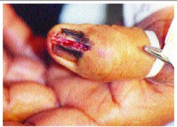
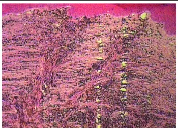
The re p o rt discusses the case of a 65-year-old black female patient. A year after experiencing trauma on the left thumb, the patient noticed a dark lesion close to the cuticle extending to the ungual, asymptomatic blade. The patient had no previous alterations on the fingernail. The examination of the finger demonstrated a clear case of melanonychia vir -tually occupying the entire surface of the ungual blade, distal onycholysis, and median nail dystrophy with a distal fracture of the ungual blade, in addition to periungual pigmentation ( F i g u res 1, 2 and 3). The patient presented with left axillary lymphadenomegaly that was mobile and painless, ro u g h l y measuring 1.5 cm. The clinical diagnosis was melanoma. The patient was re f e rred for an incisional biopsy. Histopathology exposed atypical melanocytes infiltrating the reticular dermis, with oval hyperc h romatic nuclei, and accentuated cytoplas -mic melanin pigmentation (Figures 4 and 5) in addition to an infiltration of the blade itself and the surrounding epidermis. The distal phalanx was amputated and the axillary ganglions removed. It was carried out at the oncology service, which also took charge of following up the case.
DISCUSSION
N A M was first described by Boyer in 1834,5and in
English by Hutchinson in 1886.6
o M A Ue devem entrar no diagnóstico diferencial.3O
derra-me de pigderra-mento derra-melânico nas bordas proximal e lateral da lâmina (sinal de Hutchinson) é indicativo de doença avança-d a .1 , 1 3 Além do diagnóstico clínico, muitas vezes auxiliado
pela dermatoscopia, a análise histopatológica é de fundamen-tal importância para o diagnóstico. O tratamento depende do estadiamento e tem a cirurgia como grande opção. O prog-nóstico é reservado devido a seu comportamento agressivo e, sobretudo, pelo diagnóstico tardio na maioria dos casos.4
RELATO DO CASO
Paciente do sexo feminino, 65 anos, negra, que, um ano após traumatismo no polegar esquerdo, notou lesão enegrecida próximo da cutícula estendendo-se pela lâmina ungueal, assintomática. Não apresentava alterações prévias na unha. O exame do dígito demons-trava evidente melanoníquia ocupando quase toda a superfície da lâmina ungueal, onicólise distal, distrofia mediana com fratura distal da lâmina ungueal, além de pigmentação periungueal (Figuras 1, 2 e 3). A p r e s e n t a v a linfoadenomegalia axilar esquerda, móvel, indolor, com cerca de 1,5cm. O diagnóstico clínico foi melanoma, e a paciente, submetida à biópsia incisional. A h i s t o p a t o l o-gia evidenciou melanócitos atípicos infiltrando a derme r e t i c u l a r, com núcleos ovalados, hipercromáticos e
acentuada pigmentação melânica citoplasmática
(Figuras 4 e 5 ) além da infiltração da lâmina e da epi-derme circunjacente. Foi feita a amputação da falange distal e retirada dos gânglios axilares no serviço de oncologia que se responsabilizou pelo acompanhamento do caso.
DISCUSSÃO
O MAU foi primeiramente descrito por Boyer, em
1834,5e, na língua inglesa, por Hutchinson, em 1886.6
Figura 1: Lesão melanocítica envolvendo matriz, lâmina, leito e dobra ungueal proximal (Sinal de Hutchinson). Figure 1: Mela no cytic lesio n with invo lvement o f the na il ma trix,
pla te, bed a nd pro xima l na il fo ld (Hutchinso n` s sign).
Figure 3: Lesio n pro file. Figura 3:
Perfil da lesão.
It is estimated that there is a 23% variation in melano mas localized on the nail appa -ratus in whites and 15-20% in black persons. Never t h e l e s s , there is no significant differen -ce in the number of cases of nail melanoma between white
and black persons.7 Incidence
in children is rare despite the increase in the overall number
of melanoma cases.8
This disease must be suspected anytime a (brown or black) hyperchromic macule appears on the nail matrix, bed or blade. Also suggestive of melanoma is a black longi tudinal streak of short duration in white persons, and a cli nical change of a preexisting lesion. The spread of the mela -nin pigment is always suggestive of a tumor. The literature refers to how individuals casually perceive the appearance of a previously inexistent melanocytic lesion, with only
two-thirds of patients seeking medical care as a result.7A biopsy
is recommended for every longitudinal melanonychia acquired after puberty in fair-skinned individuals or in the presence of longitudinal melanonychia showing rapid and progressive growth.7,9
Congenital or acquired melanocytic nevi may stem from the nail matrix and clinically present as a longitudinal melanonychia. They are rare and usually of the junctional type. The histopathologic architecture is similar to a nevus localized on the skin. Simple nevic cells may be found in the basal and supra basal layers of the onychocytes. Dendritic
Estima-se que varia-ções de 2 a 3 % dos melano-mas em brancos e de 15 a 20 % em negros estejam locali-zadas no aparelho ungueal. Entretanto, não há diferença significativa do número de casos de melanoma ungueal entre indivíduos brancos e n e g r o s .7 A incidência em
crianças é rara, apesar do
aumento do número de casos de melanoma.8
Deve ser suspeitado toda vez que surgir mácula hiper-crômica (marrom ou negra) na matriz, leito ou lâmina ungueal. Também sugere melanoma uma faixa longitudinal enegrecida com pouco tempo de evolução em caucasianos ou a mudança clínica de uma lesão preexistente. O derrame de pigmento melânico é sempre sugestivo do tumor. A l i t e r a t u r a faz referência ao fato de, casualmente, o indivíduo perceber uma lesão melanocítica antes ausente, e apenas dois terços dos pacientes procurarem o médico em função do surg i m e n-to da lesão.7A biópsia está indicada em toda melanoníquia
longitudinal adquirida após a puberdade em indivíduos de pele clara ou na presença de melanoníquia longitudinal que apresente crescimento rápido e progressivo.7 , 9
Os nevos melanocíticos congênitos ou adquiridos podem ter origem na matriz e apresentar-se clinicamente como uma melanoníquia longitudinal. São raros e, geralmente, do tipo juncional. Aarquitetura histopatológica é semelhante à de um nevo de localização na pele. Células névicas simples podem ser encontradas na camada basal e suprabasal dos
oni-Figura 4: A histopatologia evidenciou melanócitos atípicos infiltrando a derme reticular. / Figure 4: Histo pa tho lo gy sho wed
a typica l mela no cytes infiltra ting the reticula r dermis.
Figura 5: Melanócitos atípicos constituídos por núcleos ovalados e hipercromáticos, e acentuada pigmentação melânica citoplasmática /
melanocytes are only present occasionally on the ungual matrix. Benign melanocytic hyperplasia, which is characte -rized by an increase in the number of melanocytes among the keratinocytes of the matrix without forming nests, will
have to be equally distinguished from nevus and NAM,
which is at times difficult. Suspected cases of longitudinal melanonychia must be completely excised and analyzed from a histopathologic and immunohistochemical point of view. The frequency of a nevus-to-melanoma progression of the nail apparatus is not known. The literature informs us
that it may disappear spontaneously.7Dermatoscopy might
assist the differential diagnosis of the melanocytic lesions.10
Occasionally, a subungual hematoma might require a differential diagnosis with melanoma. The bluish-red color, lesion irregularity and absence of pigment on the blade point to hematoma. Moreover, the blood stored bet -ween the ungual blade and bed is displaced toward the front as the appendage grows. In addition, subungual hemoglo -bin is not degraded in hemosiderin by the macrophage. Staining with Prussia blue favors a diagnosis of hematoma. However, the difference between hemosiderin and melanin pigment in histopathology sometimes requires an ultra-structural analysis: iron is intercellular whereas melanin is intracellular. In dermatoscopy the formation of pseudopods is usually the outcome of erythrocytes penetrating into the layers of the ungual blade. They can act as criteria to dis -tinguish a hematoma from melanoma.
Agents such as Candida guilliermondii,
Trichophyton rubrum nigricans, Scytalidium sp,
Trichosporum beigelli, among others, might cause mela
-nonychia. The clinical suspicion of mycosis, in association with the positive mycological examinations and histopatho -logy, are decisive for clarifying the diagnosis.
Unlike cutaneous melanoma in which 80% of cases
are MNT-1,11only 20% of NAMcases are at stage I of MNT
(a classification for the primary tumor, regional lymphno -des and metastases) at the moment of diagnosis. In an attempt to reverse this situation, Levit and col.11idealized an
easy-to-memorize application system. A few characteristics were grouped together according to the letters of the alpha
-bet and designated as the ABC of NAM:
A (age) - Incidence peak between the fifth and seventh decade of life:
B (nail band) - Examine the streak: color usually brown or black, breath and border. The presence of color variations, breath greater than 3 mm, or an irregular bor -der should increase the suspicion of melanoma;
C (change) - Speed and recent increase in size of the streak, compatible with the phase of radial growth, or chan -ge in morphology of the nail sug-gest malignity;
D (digit involved) - The most common sites to be affected, in order of fre q u e n c y, are assumed to be the thumb, then the hallux and index, and the dominant hand. A pigmented streak on one digit is more indicative of a tumor than its presence on several digits. This is especially
cócitos. Melanócitos dendríticos só estão presentes ocasional-mente na matriz ungueal. Ahiperplasia melanocítica benigna, que se caracteriza pelo aumento do número de melanócitos entre os queratinócitos da matriz sem formarem ninhos, deve-rá ser igualmente distinguida do nevo e do M A U, o que é por vezes difícil. As melanoníquias longitudinais suspeitas deverão ser completamente excisadas e analisadas do ponto de vista histopatológico e imuno-histoquímico. A freqüência da pro-gressão de um nevo para melanoma do aparelho ungueal não é conhecida, e a literatura informa que podem desaparecer e s p o n t a n e a m e n t e .7A dermatoscopia poderá auxiliar no
diag-nóstico diferencial das lesões melanocíticas.1 0
Ocasionalmente, um hematoma subungueal pode exi-gir diagnóstico diferencial com melanoma. A cor azul-aver-melhada, a irregularidade da lesão e a ausência do pigmento na lâmina sugerem hematoma. Além disso, o sangue armaze-nado entre a lâmina e o leito ungueal é deslocado para frente com o crescimento do fânero. Além disso, a hemoglobina subungueal não é degradada em hemossiderina pelo macró-fago. A coloração pelo azul da Prússia favorece o diagnósti-co de hematoma. A diferença entre a hemossiderina e o pig-mento melânico na histopatologia, porém, algumas vezes exige análise ultra-estrutural: o ferro é intercelular, e a mela-nina, intracelular.7Na dermatoscopia a formação de
pseudó-podes é em geral resultado da penetração de eritrócitos nas camadas da lâmina ungueal e pode servir de critério para dis-tinguir um hematoma de um melanoma
Agentes como Candida guilliermondii, Tr i c h o
-phyton rubrum nigricans, Scytalidium sp, Trichosporum beigelli, entre outros, podem determinar melanoníquia. A
suspeita clínica de micose, associada aos exames micológi -cos positivos e a histopatologia são definitivas para esclare-cer o diagnóstico.
Ao contrário do melanoma cutâneo em que 80% dos casos são TNMI,11apenas 20% dos casos de MAUestão no
estágio I do TNM(classificação para tumor primário, linfo-nodos regionais e metástases) no momento do diagnóstico. Visando reverter esta situação, Levit e col.11idealizaram um
sistema de fáceis memorização e aplicação. Algumas carac-terísticas foram agrupadas segundo as letras do alfabeto e
designadas como o ABCDEF do MAU:
A( age) - Pico de incidência entre a quinta e a sétima décadas de vida;
B (nail band) - Examinar a faixa: cor geralmente marrom (brown) ou preta (black), espessura (breadth) e bordas (border). A presença de variação de cores, espessu-ra maior do que 3mm ou borda irregular devem aumentar a suspeição de melanoma;
C (change) - Rápido ou recente aumento no tamanho da faixa, compatível com a fase de crescimento radial, ou mudança na morfologia da unha sugerem malignidade;
i m p o rtant, given that striated melanonychia may be found in virtually all Africans over 50 years in age as an ethnic f e a t u re ;
E (extension) - This is the Hutchinson's sign itself,13
which consists of the pigment extending to the periungual region;
F (family) - Family and personal background for dysplastic nevus syndrome and previous melanomas.
Even though each letter has its own importance, the sum of them leads to the possibility of a more accurate diag nosis. Of the six letters of the alphabet utilized for the clini -cal diagnosis of melanoma, five were present in the case reported.
A presumed diagnosis of subungual melanoma when the Hutchinson's sign is present has proved to be the
rule.11,12,13 However, Baran et al.12focused on three excep
tions: benign diseases, nonmelanomous tumors and the illu -sory condition (pseudo Hutchinson's sign). Accordingly, it might be present in Peutz-Jeghers and Laugier-Hunziquer s y n d romes, subungual hematoma, ethnic pigmentation,
AIDS, drug use (minocycline and zidovudine), as well as
Bowen's disease.
A histopathologic examination continues to be the golden standard for confirming melanoma. The lesions in
situ may simulate a benign pattern, especially along the
contour, with an increase in basal melanocytes and hyper -pigmentation with only focal atypia of the melanocytes. Nevertheless, in the middle of the lesion, intense and uni -form cytologic atypia is found. Pigmentation is pronounced, resulting in the presence of melanophages in the upper der -mis and large melanin aggregates in the corneal layer. Also, lichenoid lymphocytic infiltrate could plausibly blur the dermoepidermal junction. These histopathologic findings were present in the case described. The biopsy can be either incisional or excisional.
Based on the clinical and histopathologic criteria,
the prognosis of N A M is worse than cutaneous melanoma,
with survival rates over five years varying from 16% to 87%. M o re than 50% of patients die prior to completing the five y e a r s .11The traditional treatment has been amputation of the
affected digit, at the height of the proximal joint, once the m a rgins are freed of the disease. Some studies have assessed the effectiveness of Mohs micrographic surg e ry for tre a t i n g the melanoma in situ. Its advantage is to pre s e rve noble tis
-sue for the affected segment to function adequately.1 3
Detecting the first N A Mlesions is fundamental not only for improving the patient's chances of survival, but also for p re s e rving the digit. The physician must be alert and corre c t l y identify any nail alterations that might be suggestive of mela -noma, in addition to being aware of the epidemiological data associated with the disease. q sua presença em vários dígitos. Isso é especialmente
impor-tante, visto que a melanoníquia estriada pode ser encontra-da em quase todos os africanos com mais de 50 anos de idade como uma expressão étnica;
E (extension) - É o próprio sinal de Hutchinson,13
que consiste na extensão do pigmento na região periun-gueal;
F (family) - História familiar e pessoal para síndro-me do nevo displástico e síndro-melanomas prévios.
Apesar de cada letra ter sua importância, o somató-rio delas implica a possibilidade de diagnóstico mais preci-so. Das seis letras do alfabeto para diagnóstico clínico do melanoma, cinco estavam presentes no caso relatado.
O diagnóstico presuntivo de melanoma subungueal na presença do sinal de Hutchinson11 , 1 2 , 1 3é regra, porém, Baran e
c o l .1 2enfocam três exceções: doenças benignas, tumores não
melanomas e condição ilusória (pseudo sinal de Hutchinson). Dessa feita, poderá estar presente nas síndromes de Peutz-Jeghers, Laugier- H u n z i q u e r, hematoma subungueal, pigmen-tação étnica, S I D A, uso de drogas (minociclina e zidovudina), assim como na doença de Bowen.
O exame histopatológico continua sendo o padrão ouro para a confirmação do melanoma. As lesões in situ podem simular um padrão benigno, especialmente na peri-feria, com aumento nos melanócitos basais e hiperpigmen-tação com atipia apenas focal dos melanócitos. Entretanto, no centro da lesão, atipia citológica intensa e uniforme é encontrada. A pigmentação é pronunciada, resultando da presença de melanófagos na derme superior e de grandes agregados de melanina na camada córnea. Há possibilidade de infiltrado linfocítico liquenóide que pode obscurecer a junção dermoepidérmica. Esses achados histopatológicos estavam presentes no caso descrito. A biópsia poderá ser incisional ou excisional.
Com base nos critérios clínicos e histopatológicos o
prognóstico do MAUé pior do que o do melanoma cutâneo,
com taxas de sobrevida em cinco anos variando entre 16% e 87%, sendo que mais de 50% dos pacientes morrem antes de completar cinco anos.11
O tratamento tradicional tem sido a amputação do dígi-to acometido, na altura da articulação proximal, desde que as m a rgens estejam livres de doença. Alguns estudos têm avalia-do a eficácia da cirurgia micrográfica de Mohs para o trata-mento do melanoma in situ, com a vantagem de preservar os tecidos nobres para a função adequada do segmento afetado.1 3
A detecção das lesões iniciais do MAU é
two patients. J Am Acad Dermatol. 1996; 34:765-71. 10. KawabataY, Ohara K, Hino H, Tamaki K. Two kinds of Hutchinson's sign, benign and malignant. J Am A c a d Dermatol. 2001; 44:305-7.
11. Levit EK, Kagen MH, Scher RK, Grossman M, Altman E. The ABC rule for clinical detection of subungual melanoma. J Am Acad Dermatol. 2000; 42:269-74. 12. Baran R, Kechijian P. Hutchinson's sign: a reappraisal. J Am Acad Dermatol. 1996; 34:87-90.
13. Banfield CC, Dawber RP, Walker NP, Stables GI, Zeina B, Schomberg K. Mohs Micrographic surgery for the treat-ment of in situ nail apparatus melanoma: a case report. J Am Acad Dermatol. 1999; 40:98-9.
REFERÊNCIAS / REFERENCES
1. Langley RG, Barnhill RL, Fitzpatrick TB. Fitzpatrick's Dermatology in general medicine. 5nd ed vol I. New York: Mc Graw Hill; 1999. p. 1080-116.
2. O'Toole E, Stephens R, Young M, Tanner A, Barnes L. Subungual Melanoma: a relation to direct injury? J Am Acad Dermatol. 1995; 33:525-8.
3. Glat PM, Spector JA, Roses DF. The management of pig-mented lesions of the nail bed. Ann Plast Surg. 1996; 37: 25-34.
4. Paul E, Kleiner H, Bodeker RH. Epidemiology and prog-nosis of subungual melanoma. Hautarzt. 1992; 43: 286-90 5. Boyer A. Fungus Hematide du petit digit. Ganz Med Paris. 1834: 212.
6. Hutchinson J. Melanosis often not black: melanotic whit-low. Br Med J. 1886; 1: 491.
7. Baran R, Dawber RPR. Diseases of the nail and their management. 2nd ed. Oxford: Blackwell Science;1994. p. 483-497.
8. Goettman-Bonvallot S, Andre J, Belaich S. Longitudinal melanonychia in children: a clinical and histopathologic study of 40 cases. J Am Acad Dermatol. 1999; 41:17-22. 9. Tosti A, Baran R, Piraccini BM, Cameli N, Fanti PA. Nail matrix nevi: a clinical and histophatologic study of
twenty-ENDEREÇO PARA CORRESPONDÊNCIA: / MAILINGADDRESS:
Ignez Regina do s Sa nto s Muri Mendo nça Rua Do na Ma ria na , 136/ 204 - Bo ta fo go 22280-020 Rio de Ja neiro RJ
Tel./Fa x: (21) 2538-0049