Clinical impact of
Achromobacter xylosoxidans
colonization/infection in patients with cystic
fi
brosis
M.C. Firmida
1, R.H.V. Pereira
2, E.A.S.R. Silva
3, E.A. Marques
1,2,3and A.J. Lopes
11Programa de Pós-Graduac
¸ão em Ciências Médicas, Universidade do Estado do Rio de Janeiro, Rio de Janeiro, RJ, Brasil 2Departamento de Microbiologia, Imunologia e Parasitologia, Faculdade de Ciências Médicas,
Universidade do Estado do Rio de Janeiro, Rio de Janeiro, RJ, Brasil 3Laboratório de Bacteriologia, Hospital Universitário Pedro Ernesto, Universidade do Estado do Rio de Janeiro,
Rio de Janeiro, RJ, Brasil
Abstract
The rate of diagnosis of colonization/infection of the airways withAchromobacter xylosoxidanshas increased in cysticfibrosis patients, but its clinical significance is still controversial. This retrospective, case-control study aimed to evaluate the clinical impact ofA. xylosoxidanscolonization/infection in cysticfibrosis patients. Individuals who were chronically colonized/infected (n=10), intermittently colonized/infected (n=15), and never colonized/infected withA.xylosoxidans(n=18) were retrospectively evaluated during two periods that were 2 years apart. Demographic characteristics, clinical data, lung function, and chronic bacterial co-colonization data were evaluated. Of the total study population, 87% were pediatric patients and 65.1% were female. Individuals chronically colonized/infected with A. xylosoxidanshad decreased forced expiratory volume in 1 s (51.7% in the chronic colonization/infection group vs82.7% in the intermittent colonization/infection groupvs76% in the never colonized/ infected group). Compared with the other two groups, the rate of co-colonization with methicillin-resistantStaphylococcus aureus
was higher in individuals chronically colonized/infected withA.xylosoxidans(P=0.002). Changes in lung function over 2 years in the three groups were not significant, although a trend toward a greater decrease in lung function was observed in the chronically colonized/infected group. Compared with the other two groups, there was a greater number of annual hospitalizations in patients chronically colonized/infected withA. xylosoxidans(P=0.033). In cysticfibrosis patients, there was an increased frequency of
A. xylosoxidanscolonization/infection in children, and lung function was reduced in patients who were chronically colonized/ infected withA. xylosoxidans. Additionally, there were no differences in clinical outcomes during the 2-year period, except for an increased number of hospitalizations in patients withA. xylosoxidans.
Key words: Cysticfibrosis;Achromobacterspp.;Achromobacter xylosoxidans; Microbiology
Introduction
The genus Achromobactercontains genetically distinct species and subspecies, and has not been fully character-ized (1–5).Achromobacterspp. are Gram-negative, aerobic,
nonfermenters of glucose bacilli that are widely distributed in the environment. Achromobacter xylosoxidans is the most common bacillus in clinical samples and is recognized as an emerging and multidrug-resistant microorganism that causes various opportunistic infections and nosocomial outbreaks (3,6). Most knowledge onA. xylosoxidanshas been obtained from studies on populations living in regions where cystic
fibrosis (CF) is prevalent (3,6).
The rate of colonization/infection withA. xylosoxidans
in individuals with CF varies between 2% and 17.9% (7,8) and is increasing worldwide. However, this frequency may be underestimated because this organism can be con-fused with Pseudomonas aeruginosa, bacteria from the
Burkholderia cepacia complex (BCC), and Stenotropho-monas maltophilia, particularly in laboratories that are not specialized for evaluation of CF (9).
The factors that predispose patients to colonization/ infection have not been fully determined. Frequent exposure to antibiotics, particularly during treatment for chronic colonization withP. aeruginosa, may favor the emergence of this and other Gram-negative, multidrug-resistant bacteria (10,11). The possibility of person-to-person transmission, the association of A. xylosoxidans colonization/infection with pulmonary inflammation, and an increased frequency of exacerbations have been demonstrated. However, the clinical impact of colonization/infection of A. xylosoxidans
in CF patients is still controversial (6,11–15). Therefore, the
present study aimed to evaluate the clinical impact of
A. xylosoxidanscolonization/infection in patients with CF.
Correspondence: A.J. Lopes:<agnaldolopes.uerj@gmail.com>
Material and Methods
Study design
This retrospective, case-control study evaluated patients with a confirmed diagnosis of CF (16). These patients were regularly monitored at the Instituto Fernandes Figueira, Fundac¸ão Oswaldo Cruz and Policlínica Piquet Carneiro,
Universidade do Estado do Rio de Janeiro (Brazil). Patients’
respiratory secretion culture results were obtained between January 2003 and December 2011 at the Laboratório de Bacteriologia, Hospital Universitário Pedro Ernesto (LBACT-UERJ).
The protocol conformed to the World Medical Associa-tion DeclaraAssocia-tion of Helsinki and was approved by the Research Ethics Committee of the Universidade do Estado do Rio de Janeiro (No. CAAE: 00716512.0.3001.5269).
Patients
A total of 238 individuals (155 females and 83 males) with CF were regularly monitored in these referral centers, of whom 25% were adults (X18 years). The routine follow-up period consisted of quarterly consultations, except for infants, who were monitored monthly. The interval between consultations was shortened depending on clinical need. At each visit, the general medical condition, weight, height, and lung function of patients were evaluated; and res-piratory secretions were obtained for culture (sputum or oropharyngeal swab for non-expectorating children). All material obtained at these centers was sent to the LBACT-UERJ. In this institution, cultures of respiratory secretions were carried out according to standardized protocols established for CF patients. Cultures were performed every 3 months throughout the study (17).
Identification ofAchromobacter
Phenotypic methods. Isolates that were identified as
Achromobacterspp. by the Vitek 2 Compact system using Gram-negative cards (reference no. 21341; bioMérieux, France) were subjected to further identification via a large panel of phenotypic tests, as previously described (18,19).
Molecular methods. To identify each isolate, DNA was extracted by the boiling lysis method, and the entire 16S
rRNA gene was amplified by PCR, sequenced, and used for BLAST searches against the GenBank database (20). The presence of the A. xylosoxidans species-specific marker
blaOXA-114 was investigated by PCR amplification as described by Barrado et al. (6). After amplification, the PCR products were sequenced and compared with sequences in the GenBank database at the NCBI using BLAST.
Inclusion and exclusion criteria
The respiratory secretion culture results of patients with CF were evaluated using the LBACT-UERJ database. The inclusion criteria were as follows: 1) patients with one or more cultures that were positive for A. xylosoxidans (the term‘‘colonization/infection’’is used in reference to positive
cultures), and 2) patients who were colonized/infected with
A. xylosoxidans and chronically colonized with P. aerugi-nosa, defined as more than 50% of cultures positive for the latter agent during 1 year (21). The exclusion criteria consisted of colonization with BCC bacteria and/or the absence of chronic colonization withP. aeruginosa.
Definition of the groups
Patients were subdivided according to theirA. xylosox-idanscolonization/infection status into a chronically colo-nized/infected group and an intermittently colonized/ infected group. The criterion for chronic colonization/in-fection byA. xylosoxidanswas the same as that adopted for P. aeruginosa (21). Any shorter frequency was considered to be intermittent colonization/infection. The control group consisted of individuals who never had a positive culture for A. xylosoxidans, and subjects were matched with those in the case groups according to age (±1 year), sex, and chronic colonization with P. aerugi-nosa. All of the patients were chronically colonized by
P. aeruginosa, and the status ofA. xylosoxidans(chronic, intermittent, and never) was defined as described pre-viously. Therefore, the three study groups were as follows: group I, chronic colonization/infection withA. xylosoxidans
and chronic colonization with P. aeruginosa; group II, intermittent colonization/infection withA. xylosoxidansand chronic colonization with P. aeruginosa; and group III, never colonized/infected withA. xylosoxidans,but chroni-cally colonized byP. aeruginosa.
Clinical outcomes
The general population was described according to the demographic characteristics, diagnostic criteria for CF, and the presence of exocrine pancreatic insufficiency, cystic fibrosis-related diabetes, and liver disease. The frequency of the F508delmutation was described when available. In addition, other chronic bacterial co-coloniza-tions were recorded by adopting the same criteria for chronic colonization as those used forP. aeruginosa(21). Cross-sectional registration of clinical data was per-formed on two occasions: when thefirst positive culture for
A. xylosoxidansoccurred (moment 1 [M1]) and as close as possible to 24 months after the first positive culture (moment 2 [M2]). In the control group, data from M1 were paired with those of subjects in the case groups (groups II and III), and the same criteria were followed for M2.
With regard to lung function, the values of forced expiratory volume in 1 s (FEV1) and forced vital capacity
results are reported as a percentage of the predicted values for the Brazilian population (23). The weight and height of patients were used to calculate body mass index (BMI). FEV1, FVC, and BMI were compared for the two
time periods within and between groups. The median number of annual admissions was also compared between the groups.
Statistical analysis
Numerical data are reported as means±SD or
medians and ranges (minimum–maximum). Categorical
data are reported as frequencies (%). The variables had a non-normal distribution according to the Kolmogorov-Smirnov test. Therefore, a non-parametric test was applied. Kruskal-Wallis ANOVA, with the corresponding Dunn’s multiple comparison test, was used to compare numerical variables between the three groups. Fisher’s exact test was used to compare categorical variables. When the association between categorical variables within the group was significant at 5%, Fisher’s exact test, set for each peer group separately, was used. Therefore, we aimed to identify which groups differed from each other at a level of 1.7%. A level of 1.7% (5% divided by the number comparisons: 0.05/3=0.017) was used to control for type I error. To determine the existence of significant variations in FEV1, FVC, and BMI values between M1 and M2, the
Wilcoxon signed rank test was used. Data analysis was performed using SAS software version 6.11 (SAS Institute, Inc., USA). The level of statistical significance was set at Po0.05.
Results
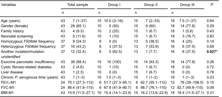
Of the 238 individuals with culture results, 47 (19.7%) had at least one positive culture for A. xylosoxidans, among whom 25 met the inclusion criteria for the study. Ten patients were classified as chronically colonized/ infected and 15 were classified as intermittently colonized/ infected. The control group consisted of 18 patients. No participants died during the study period. The general characteristics of the study population and comparison between groups at baseline are reported in Table 1.
The median period of chronic colonization with
P. aeruginosawas 1 year, and this ranged from 1 to 3 years. The baseline values for age, sex, F508del mutation frequency, exocrine pancreatic insufficiency, diabetes, liver disease, length of colonization withP. aeruginosa, and BMI were similar among the three groups. FEV1and FVC values
were lower in the chronically colonized/infected group, but this difference was not significant compared with the other groups (Table 1).
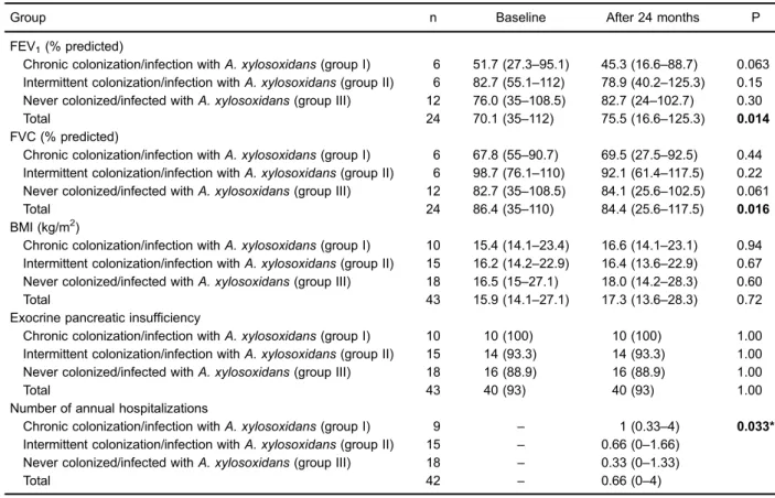
When the two periods (M1 and M2) were compared, there was a significant increase in FEV1(P=0.014) and
significant reduction in FVC (P=0.016) for the total sample. However, no significant changes were observed for these parameters for the patient groups. The median number of annual admissions during the study period was significantly different between the groups (P=0.033). There was a higher number of annual admissions in the chronically colonized/infected group compared to the never colonized/infected group. Information regarding
Table 1.General characteristics of the study population and comparison between groups at baseline.
Variables Total sample Group I Group II Group III P
n n n n
Age (years) 43 7 (1–37) 10 10.5 (3–18) 15 7 (2–33) 18 7.5 (1–37) 0.64
Gender (female) 43 28 (65.1) 10 5 (50) 15 9 (60) 18 14 (77.8) 0.29
Family history 43 4 (9.3) 10 2 (20) 15 1 (6.7) 18 1 (5.6) 0.43
Neonatal screening 43 5 (11.6) 10 1 (10) 15 1 (6.7) 18 3 (16.7) 0.83
HomozygousF508del frequency 37 9 (24.3) 8 0 (0) 13 5 (38.5) 16 4 (25) 0.14 HeterozygousF508delfrequency 37 16 (43.2) 8 3 (37.5) 13 7 (53.9) 16 6 (37.5) 0.69 Another mutation/mutation
unidentified
37 12 (32.4) 8 5 (62.5) 13 1 (7.7) 16 6 (37.5) 0.027*
Exocrine pancreatic insufficiency 43 38 (88.4) 10 10 (100) 15 14 (93.3) 18 14 (77.8) 0.26 Cysticfibrosis-related diabetes 43 2 (4.6) 10 1 (10) 15 1 (6.7) 18 0 (0) 0.72
Liver disease 43 1 (2.3) 10 0 (0) 15 1 (6.7) 18 0 (0) 0.78
ChronicP. aeruginosatime (years) 43 1 (1–3) 10 1.5 (1–3) 15 1 (1–2) 18 1 (1–3) 0.23 FEV1-M1 24 70.1 (27.3–112) 6 51.7 (27.3–95.1) 6 82.7 (55.1–112) 12 76 (35–108.5) 0.15 FVC-M1 24 86.4 (41.9–115) 6 67.8 (41.9–90.7) 6 98.7 (76.1–110) 12 82.7 (49.9–115) 0.09 BMI-M1 43 15.9 (11.5–27.1) 10 15.4 (14.1–23.4) 15 16.2 (13.6–22.9) 18 16.4 (11.5–27.1) 0.31
pulmonary function, BMI, and clinical data for each group at M1 and M2 is reported in Table 2.
Chronic co-colonization in each group is shown in Figure 1. A significant difference (P=0.002) in chronic co-colonization with methicillin-resistant S. aureus (MRSA) among the three groups was observed. Chronic co-colonization with MRSA was observed in 50% and 26.7% of the patients who were chronically colonized/ infected withA. xylosoxidansand intermittently colonized/ infected with A. xylosoxidans, respectively. No patients without colonization/infection with A. xylosoxidans had chronic co-colonization with MRSA. No significant differ-ence was observed for other types of chronic colonization.
Discussion
The mainfindings of this study were as follows: in CF patients, a relatively high frequency of A. xylosoxidans
colonization/infection was present among children, and reduced lung function in patients who were chronically colonized/infected withA. xylosoxidanswas observed. In addition, we did not observe any differences in clinical
endpoints over 2 years when we compared patients who were chronically colonized with P. aeruginosa, with or withoutA. xylosoxidans,except for an increased number of hospital admissions for patients withA. xylosoxidans.
In the present study, the frequency of colonization/infection withA. xylosoxidans(19.7%) was similar to the upper limit of the range reported in other studies (2% to 17.9%) (7,8). This reported frequency in our study was cumulative, which explains the higher percentage than other studies. The large range inA. xylosoxidanscolonization/infection frequency may be partly attributed to methodological differences between the studies (6–8,11,12). The prevalence of colonization/infection
withA. xylosoxidansin pediatric patients in our study (median, 7 years) was different from that reported in other studies, which showed that it was predominantly observed during late adolescence or early adulthood (10,12–14). The most similar
results to our study are those of a French study that reported a median age of 10.3 years (variation of 6 to 14 years) for the
first positive culture among patients with CF who became chronically colonized/infected with A. xylosoxidans (24). However, notably, this French study only included children and adolescents.
Table 2.Lung function, body mass index, and clinical data according to the groups at baseline and after 24 months.
Group n Baseline After 24 months P
FEV1(% predicted)
Chronic colonization/infection withA. xylosoxidans(group I) 6 51.7 (27.3–95.1) 45.3 (16.6–88.7) 0.063 Intermittent colonization/infection withA. xylosoxidans(group II) 6 82.7 (55.1–112) 78.9 (40.2–125.3) 0.15 Never colonized/infected withA. xylosoxidans(group III) 12 76.0 (35–108.5) 82.7 (24–102.7) 0.30
Total 24 70.1 (35–112) 75.5 (16.6–125.3) 0.014
FVC (% predicted)
Chronic colonization/infection withA. xylosoxidans(group I) 6 67.8 (55–90.7) 69.5 (27.5–92.5) 0.44 Intermittent colonization/infection withA. xylosoxidans(group II) 6 98.7 (76.1–110) 92.1 (61.4–117.5) 0.22 Never colonized/infected withA. xylosoxidans(group III) 12 82.7 (35–108.5) 84.1 (25.6–102.5) 0.061
Total 24 86.4 (35–110) 84.4 (25.6–117.5) 0.016
BMI (kg/m2)
Chronic colonization/infection withA. xylosoxidans(group I) 10 15.4 (14.1–23.4) 16.6 (14.1–23.1) 0.94 Intermittent colonization/infection withA. xylosoxidans(group II) 15 16.2 (14.2–22.9) 16.4 (13.6–22.9) 0.67 Never colonized/infected withA. xylosoxidans(group III) 18 16.5 (15–27.1) 18.0 (14.2–28.3) 0.60
Total 43 15.9 (14.1–27.1) 17.3 (13.6–28.3) 0.72
Exocrine pancreatic insufficiency
Chronic colonization/infection withA. xylosoxidans(group I) 10 10 (100) 10 (100) 1.00 Intermittent colonization/infection withA. xylosoxidans(group II) 15 14 (93.3) 14 (93.3) 1.00 Never colonized/infected withA. xylosoxidans(group III) 18 16 (88.9) 16 (88.9) 1.00
Total 43 40 (93) 40 (93) 1.00
Number of annual hospitalizations
Chronic colonization/infection withA. xylosoxidans(group I) 9 – 1 (0.33–4) 0.033* Intermittent colonization/infection withA. xylosoxidans(group II) 15 – 0.66 (0–1.66)
Never colonized/infected withA. xylosoxidans(group III) 18 – 0.33 (0–1.33)
Total 42 – 0.66 (0–4)
There is no universal criterion for the definition of chronic colonization with A. xylosoxidans (6–8,11–13).
The criterion of Pereira et al. (25) is more consistent for ensuring chronicity. However, the criterion of chronicity that was adopted in the present study (21) included an assumption that patient care needs were satisfied. There-fore, clinical measures must be adopted during the short period in which colonization can harm the patient. One of the suspected risk factors for colonization/infection with
A. xylosoxidansis treatment forP. aeruginosa(11). In our study, of the 47 patients who had at least one positive culture for A. xylosoxidans, 41 were colonized/infected withP. aeruginosa. However, only 25 met the criteria for chronic colonization/infection with P. aeruginosa without BCC colonization. All six patients in whomP. aeruginosa
was not identified were colonized with BCC. Despite the restriction of the sample size by the selection criteria, chronic colonization with P. aeruginosa was considered important for subject pairing because it decreased the chance of bias in the outcomes of interest.
Interestingly, none of the patients in the group of patients who were chronically colonized/infected with A. xylosox-idanswere homozygous for theF508delmutation, and its frequency was smaller than that found in the other groups. Therefore, other serious mutations may be more frequent in this population, as suggested by Cabello et al. (26). With regard to lung function, FEV1values in the group of patients
who were chronically colonized/infected with A. xylosox-idanswere lower than those found in the other two groups. Although this finding was not statistically significant, clinically, this difference suggests a more advanced stage
of lung damage among individuals who became chronically colonized/infected with A. xylosoxidans. These data are consistent with the hypothesis proposed by De Baets et al. (8), in which individuals with increased lung impairment appear to be more prone to chronic colonization/infection withA.xylosoxidans.
A higher frequency of hospitalizations and chronic co-colonization with MRSA was observed in the group of patients who were colonized/infected withA.xylosoxidans,
and this frequency was highest in the chronically colonized/ infected group. Zemanick et al. (27) found a higher number of exacerbations requiring intravenous treatment and a higher relative risk of isolation of MRSA,S. maltophilia, and
A. xylosoxidans after the first isolation of P. aeruginosa. A recent multicenter study showed that the frequency of colonization with MRSA has increased in recent years (28). Additionally, colonization with P. aeruginosa and more intensive therapeutic interventions may be risk factors for chronic colonization with MRSA, particularly for healthcare-associated MRSA (HA-MRSA) (28). In our study, although chronic colonization withP. aeruginosawas a criterion for pairing, MRSA was not found in the group that was not colonized/infected withA. xylosoxidans.
Compared with the other two groups, there was higher number of hospitalizations in the chronically colonized/ infected group. Thisfinding may be explained by the fact that the condition of this group of patients was more severe at the beginning of the study or because other conditions contributed to this outcome. Notably, we were unable to determine whether the association between chronic colo-nization with MRSA andA. xylosoxidanswas the result of
increased hospitalization and more intensive antimicrobial therapy, or whether any real association existed between these two agents or between these agents and mutations, as previously reported forP. aeruginosa(29).
The chronically colonized/infected group showed much smaller FEV1 values than the intermittently colonized/
infected and non-colonized/infected groups, at the time of colonization/infection and approximately 2 years later. Similarly, other studies have shown a higher frequency of colonization/infection withA. xylosoxidansin individuals with CF with more severe lung disease (13). In the present study, no significant difference in intra- or inter-group variation was observed for these parameters. Nevertheless, over 2 years, FEV1values decreased in the chronically colonized/infected
group by 6.4% of the predicted value and by 3.8% in the intermittently colonized/infected group (Table 2). Interest-ingly, Llorca Otero et al. (30) observed a mean annual decline in FEV1of 2.49% in patients who were chronically
colonized/infected withA. xylosoxidans.
A strength of the current study is that it is thefirst Brazilian study to determine a relationship between clinical data and colonization/infection with A. xylosoxidans. However, the present study has major limitations. First, our study was
limited by the broad age range and small sample size. These factors can, at least in part, be explained by the study’s retrospective design and the fact thatA. xylosoxidanshas a low incidence/prevalence in CF patients. Second, our population was exclusively composed of patients who were chronically colonized withP. aeruginosa. However, treatment for chronic colonization with P. aeruginosa might favor the emergence ofA. xylosoxidans(10,11). Notwithstanding these limitations, this study can serve as a starting point for future clinical trials to evaluate intervention protocols in CF patients who are colonized/infected withA. xylosoxidans.
In conclusion, a relatively high frequency ofA. xylosox-idans colonization/infection was present in children; and reduced lung function was observed in patients who were chronically colonized/infected with A. xylosoxidans. This colonized/infected group also showed an increased frequency of chronic colonization with MRSA. In addition, no significant differences in clinical endpoints were observed over 2 years, except for an increased number of hospitalizations in patients with A. xylosoxidans. With regard to the change in lung function over 2 years, a trend toward a decrease in FEV1
values of patients who were chronically colonized/infected withA. xylosoxidanswas observed.
References
1. Li X, Hu Y, Gong J, Zhang L, Wang G. Comparative genome characterization of Achromobacter members reveals poten-tial genetic determinants facilitating the adaptation to a pathogenic lifestyle. Appl Microbiol Biotechnol 2013; 97: 6413–6425, doi: 10.1007/s00253-013-5018-3.
2. Vandamme P, Moore ER, Cnockaert M, Peeters C, Svensson-Stadler L, Houf K, et al. Classification of Achro-mobacter genogroups 2, 5, 7 and 14 as Achromobacter insuavis sp. nov., Achromobacter aegrifaciens sp. nov., Achromobacter anxifersp. nov. andAchromobacter dolens sp. nov., respectively.Syst Appl Microbiol2013; 36: 474–482, doi: 10.1016/j.syapm.2013.06.005.
3. Swenson CE, Sadikot RT. Achromobacter respiratory infections. Ann Am Thorac Soc 2015; 12: 252–258, doi: 10.1513/AnnalsATS.201406-288FR.
4. Spilker T, Vandamme P, Lipuma JJ. Identification and distribution of Achromobacter species in cysticfibrosis.J Cyst Fibros2013; 12: 298–301, doi: 10.1016/j.jcf.2012.10.002. 5. Gomila M, Prince-Manzano C, Svensson-Stadler L, Busquets
A, Erhard M, Martinez DL, et al. Genotypic and phenotypic applications for the differentiation and species-level identifi ca-tion of achromobacter for clinical diagnoses.PLoS One2014; 9: e114356, doi: 10.1371/journal.pone.0114356.
6. Barrado L, Branas P, Orellana MA, Martinez MT, Garcia G, Otero JR, et al. Molecular characterization of Achromobacter isolates from cysticfibrosis and non-cysticfibrosis patients in Madrid, Spain.J Clin Microbiol 2013; 51: 1927–1930, doi: 10.1128/JCM.00494-13.
7. Tan K, Conway SP, Brownlee KG, Etherington C, Peckham DG. Alcaligenes infection in cysticfibrosis.Pediatr Pulmonol 2002; 34: 101–104, doi: 10.1002/ppul.10143.
8. De Baets F, Schelstraete P, Van Daele S, Haerynck F, Vaneechoutte M. Achromobacter xylosoxidans in cystic
fibrosis: prevalence and clinical relevance. J Cyst Fibros 2007; 6: 75–78, doi: 10.1016/j.jcf.2006.05.011.
9. Saiman L, Chen Y, Tabibi S, San GP, Zhou J, Liu Z, et al. Identification and antimicrobial susceptibility ofAlcaligenes xylosoxidans isolated from patients with cystic fibrosis. J Clin Microbiol2001; 39: 3942–3945, doi: 10.1128/JCM.39. 11.3942-3945.2001.
10. Kanellopoulou M, Pournaras S, Iglezos H, Skarmoutsou N, Papafrangas E, Maniatis AN. Persistent colonization of nine cystic fibrosis patients with an Achromobacter (Alcaligenes) xylosoxidans clone. Eur J Clin Microbiol Infect Dis2004; 23: 336–339, doi: 10.1007/s10096-004-1105-9.
11. Lambiase A, Catania MR, Del Pezzo M, Rossano F, Terlizzi V, Sepe A, et al.Achromobacter xylosoxidansrespiratory tract infection in cysticfibrosis patients.Eur J Clin Microbiol Infect Dis2011; 30: 973–980, doi: 10.1007/s10096-011-1182-5. 12. Ronne Hansen C, Pressler T, Hoiby N, Gormsen M. Chronic
infection withAchromobacter xylosoxidansin cysticfibrosis patients; a retrospective case control study.J Cyst Fibros 2006; 5: 245–251, doi: 10.1016/j.jcf.2006.04.002.
13. Raso T, Bianco O, Grosso B, Zucca M, Savoia D. Achromobacter xylosoxidansrespiratory tract infections in cystic fibrosis patients. APMIS 2008; 116: 837–841, doi: 10.1111/j.1600-0463.2008.00995.x.
15. Mantovani RP, Levy CE, Yano T. A heat-stable cytotoxic factor produced by Achromobacter xylosoxidans isolated from Brazilian patients with CF is associated within vitro increased proinflammatory cytokines.J Cyst Fibros2012; 11: 305–311, doi: 10.1016/j.jcf.2012.02.002.
16. Farrell PM, Rosenstein BJ, White TB, Accurso FJ, Castellani C, Cutting GR, et al. Guidelines for diagnosis of cystic
fibrosis in newborns through older adults: Cystic Fibrosis Foundation consensus report.J Pediatr2008; 153: S4–S14, doi: 10.1016/j.jpeds.2008.05.005.
17. Gilligan PH, Kiska DL, Appleman MD.Cumitech 43: Cystic fibrosis microbiology. Washington: ASM Press; 2006. 18. Barth AL, de Abreu E Silva FA, Hoffmann A, Vieira MI,
Zavascki AP, Ferreira AG, et al. Cysticfibrosis patient with Burkholderia pseudomalleiinfection acquired in Brazil.J Clin Microbiol2007; 45: 4077–4080, doi: 10.1128/JCM.01386-07. 19. Leao RS, Carvalho-Assef AP, Ferreira AG, Folescu TW, Barth AL, Pitt TL, et al. Comparison of the worldwide transmissiblePseudomonas aeruginosawith isolates from brazilian cysticfibrosis patients.Braz J Microbiol2010; 41: 1079–1081, doi: 10.1590/S1517-838220100004000028. 20. Hiraishi A. Direct automated sequencing of 16S rDNA
amplified by polymerase chain reaction from bacterial cultures without DNA purification.Lett Appl Microbiol1992; 15: 210–213, doi: 10.1111/j.1472-765X.1992.tb00765.x. 21. Lee TW, Brownlee KG, Conway SP, Denton M, Littlewood
JM. Evaluation of a new definition for chronicPseudomonas aeruginosainfection in cysticfibrosis patients.J Cyst Fibros 2003; 2: 29–34, doi: 10.1016/S1569-1993(02)00141-8. 22. Miller MR, Hankinson J, Brusasco V, Burgos F, Casaburi R,
Coates A, et al. Standardisation of spirometry.Eur Respir J 2005; 26: 319–338, doi: 10.1183/09031936.05.00034805. 23. Pereira CA, Sato T, Rodrigues SC. New reference values for
forced spirometry in white adults in Brazil. J Bras Pneumol 2007; 33: 397–406, doi: 10.1590/S1806-37132007000400008.
24. Moissenet D, Baculard A, Valcin M, Marchand V, Tournier G, Garbarg-Chenon A, et al. Colonization by Alcaligenes xylosoxidansin children with cysticfibrosis: a retrospective clinical study conducted by means of molecular epidemio-logical investigation.Clin Infect Dis1997; 24: 274–275, doi: 10.1093/clinids/24.2.274.
25. Pereira RH, Carvalho-Assef AP, Albano RM, Folescu TW, Jones MC, Leao RS, et al. Achromobacter xylosoxidans: characterization of strains in Brazilian cysticfibrosis patients. J Clin Microbiol 2011; 49: 3649–3651, doi: 10.1128/JCM. 05283-11.
26. Cabello GM, Cabello PH, Otsuki K, Gombarovits ME, Llerena JC Jr, Fernandes O. Molecular analysis of 23 exons of the CFTR gene in Brazilian patients leads to thefinding of rare cysticfibrosis mutations.Hum Biol2005; 77: 125–135, doi: 10.1353/hub.2005.0027.
27. Zemanick ET, Emerson J, Thompson V, McNamara S, Morgan W, Gibson RL, et al. Clinical outcomes after initial pseudomonas acquisition in cysticfibrosis.Pediatr Pulmo-nol2015; 50: 42–48, doi: 10.1002/ppul.23036.