wmm,
^-TESE DE DOUTORAMENTO
CARMEN DE LURDES FONSECA JERÓNIMO
M O L E C U L A R DETECTION O F PROSTATE CANCER
D E T E C Ç Ã O MOLECULAR DO CARCINOMA DA PRÓSTATA
C A R M E N D E L U R D E S F O N S E C A J E R Ó N I M O
Dissertation for applying to a Doctor degree in Biomedical Sciences, presented to the
Institute of Biomedical Sciences Abel Salazar of the University of Oporto
Dissertação de candidatura ao grau de Doutor em Ciências Biomédicas apresentada ao
Instituto de Ciências Biomédicas de Abel Salazar da Universidade do Porto
To Rui,
This is only the first project together....
INTRODUCTION
II CHAPTER 1
PAPER I
PAPER II
PAPER III
PAPER IV
Quantitation of GSTP1 hypermethylation distinguishes between
non-neoplastic prostatic tissue and organ confined prostate adenocarcinoma.
(J. Natl. Cancer Inst, accepted for publication)
Detection of prostate cancer in urine by GSTP1 hypermethylation.
(Clin. Cancer Res., in press)
Quantitative GSTP1 hypermethylation in bodily fluids of prostate
cancer patients (J. Urol, submitted)
1105V polymorphism and promoter methylation of GSTP1 gene in
prostate adenocarcinoma. (Cancer Epidemiol. Biomark. Prev.,
submitted)
III CHAPTER 2
PAPER V Detection of endothelin receptor B hypermethylation in early stage
prostate adenocarcinoma. (Cancer Letters, submitted)
13
32
45
62
80
IV CHAPTER 3
PAPER VI Mitochondrial mutations in early stage prostate cancer and bodily
fluids (Oncogene, in press)
V MAIN CONCLUSIONS AND FUTURE STUDIES
94
109
V I S U M M A R Y - R É S U M É - R E S U M O 114
Carmen Jerónimo - Doctoral Thesis
I. INTRODUCTION
BACKGROUND
Prostate cancer has become a major health issue. Over the last decade the incidence of prostate cancer in Western world has been sharply increasing, and is nowadays the most common cancer in men from those countries, as well as the second leading cause of cancer-related mortality (1). Since adequate prophylactic measures are not available, the role of early detection has been emphasized, as organ confined disease is still the best chance for the patient to benefit from curative treatment (2).
The utilization of serum PSA level as screening tool, in addition to digital rectal examination and transrectal ultrasonography, results in an increased ability to detect prostate cancer while still organ-confined disease (3, 4). However, although elevated blood levels of PSA (greater than 4 ng/ml) correlate with the presence of prostate cancer, false positive results do occur and efforts have been made to accurately distinguish benign from malignant disease, even for slightly elevated PSA levels that can be indicative of either (4). Thus new approaches are needed that may allow a confident detection of malignancy, namely through DNA-based techniques (5).
Indeed, it is acknowledged that a series of genetic alterations are associated with the transformation of normal glandular epithelium to prostate intraepithelial neoplasia, and from this in situ lesion to invasive carcinoma as depicted in the figure (6). Moreover, increasing efforts have been undertaken to characterize the molecular events implicated in the
Carmen Jerónimo - Doctoral Thesis
suggest that certain genetic alterations may becommon in theevolution tomalignancy. Thus,
a potentially useful application of these studies would be the identification of molecular
markers already present in early stage prostate cancer that could provide the means for more
effective screening and diagnosis ofthedisease.
Multistep
carcinogenesis
model
for
Prostate
Adenocarcinoma
Prostatic intraepithelial neoplasia Normal epithelium _^, Histological prostate adenocarcinoma ->■ Metastatic adenocarcinoma -Hereditary factors: HPC1,BRCA1 -AR,vitDreceptor
HypermethylationGSTP1,HIC Telomcraseactivity
-GrowthFactorsReceptors -Human papillomavirus?
AR
refractory adenocarcinoma
PS3,Rbl,PTEN
CD44 KAI-1 mutation/amplification E-cadherin/a-catenin Growth Factors Rasoncogene Receptors activation
Montironi et «/.,2000
Genetic alterations
Tumor suppressor gene (TSG) alterations have a major role in prostate cancer
development, which fits perfectly well with the biology of a chronic, slowly growing
neoplastic disease (8). According to Knudson's Two-hit hypothesis, TSG fuction is lost by
independent inactivation events of both parental alleles (9). In sporadic tumors, bi-allelic
inactivation is required to silence gene function in TSG and this is frequently accomplished
byasomatic mutation inone alleleandloss ofheterozigozity (LOH)in theother (9).
Several studies, using LOH analysis, indicated that the most common chromosomic
losses (located at 8p, lOq, 7q, and 16q, and firstly described by cytogenetic studies) are
chromosome 8p abnormalities were found to be a preferential site of LOH in prostate cancer, occurring in the majority of adenocarcinoma cases examined (11). Additionally, Emmert-Buck et al. identified frequent loss of portions at 8p21-pl2 in a high percentage of prostatic intraepithelial neoplasia (PIN) lesions, thus becoming an early event in prostate tumorigenesis (12). It has also been suggested that loss of 8p could be related to the development of androgen independence (13). Furthermore, deletions of portions of both lOp and lOq have been associated with advanced stage and rapidly progressive disease (14). Another tumor suppressor gene located on chromosome 10q23 (PTEN) was found to be the main inactivation target of lOq loss inactivated in three human prostate cancer cell lines derived from metastatic tissues (15, 16). Concerning 16q, the available data is not consistent. Carter et al. reported LOH in 30% of clinically localized tumor, and Bergerheim et al. found a higher rate (11, 17). Moreover, it is worthwhile noting that E-cadherin gene (a cell adhesion molecule critical for normal differentiation) was mapped at 16q22.1, which is a frequent site of LOH (18). Additionally, abnormalities in chromosome 7 have been observed in several studies. Trisomy 7 was found to be common in both PIN and cancer lesions, and, on the contrary, aneusomy of chromosome 7 and loss of 7q31.1 were found to be associated with advanced stage and poor
prognosis (19, 20).
Other chromosomal losses have also been reported in prostate carcinoma: 3p, 5q, 9q, 1 lp, 13q, 17p, and 18q. Interestingly, the p53 gene maps to 17p, the retinoblastoma gene (RB)
to 13q, and DCC (deleted in colon cancer) to 18q (10, 21). These findings add further support to the importance of TSG alteration in the genesis of prostate cancer.
Moreover, a number of these TSG were found to be mutated in prostate cancer, including P53, RB, pl6, and PTEN (10, 21). However, P53 mutations are uncommon in
Carmen Jerónimo - Doctoral Thesis
cancer, but point mutations are present in less than 20% of tumor samples (23). Furthermore, although pl6 is one of the most frequently altered genes in prostate cancer, it is rarely found to be mutated (24, 25). Finally, sequence analysis revealed that a second mutational event at the PTEN locus occurs in as much as 43% of the tumors with LOH of this gene (15). Additionally, it has been suggested that PTEN point mutations and homozygous deletion are late events, associated with advanced cancers (15, 16). Cairns et al. observed a higher frequency of these gene alterations in pelvic lymph node metastases than in clinically localized cancers, indicating that PTEN inactivation contributes to the acquisition of metastatic potential of prostate cancer (16).
Epigenetic alterations
Alongside with sequence loss, chromosomal deletions and point mutations, gene promoter hypermethylation is an alternative inactivation mechanism for TSG. Indeed, hypermethylation of CpG islands (areas of the genome rich in the sequence CpG, associated with the 5' regulatory regions of genes) has been found to correlate with gene transcriptional inactivation in several human cancers, including those of prostate (26, 27). Most of the initial studies focused on gene methylation patterns in prostate cancer cell lines. In this regard, Jarrard et al. showed that CpG methylation of pl6 occurred in three of five cell lines analyzed, although this alteration was found to be less common in prostate primary tumors (13%) (28). Concerning E-cadherin, a similar result was found: the promoter region was commonly methylated in prostate cancer cell lines, but low detectable rates of methylation were present in primary prostatic tumors (29).
Interestingly, in prostate cancer, there are also genes inactivated by CpG methylation but not recognized as tumor suppressor genes at present time. Glutathione-S-transferase PI gene (GSTP1) is the prototype of this class of genes. GSTP1 codes for a phase II
detoxification enzyme, which has an important role in preventing DNA damage due to cell exposure to a wide range of carcinogens (30). This gene was found to be hypermethylated in the vast majority of prostate carcinomas (-90%), and even in precursor lesions such as PIN (31, 32). However, GSTP1 promoter hypermethylation has also been reported in a non-negligible number of benign prostate tissue samples (32).
Another frequent (-70%) site of hypermethylation in prostate cancer is within the transcriptional region of the endothelin B receptor gene (ENDRB) (33), which mediates the clearance and secretion of the vasoconstrictor endothelin-1, another protein associated with prostate cancer progression. Hypermethylation is also an alternative mechanism for transcriptional repression of CD44, a metastasis suppressor gene, which seems to be involved in cancer progression (34). Finally, a recent report showed that inactivation of androgen receptor gene expression by hypermethylation was linked with advanced hormone
independent prostate cancer (35).
Clearly, the elevated frequency of hypermethylation of several of the above mentioned
genes, together with its apparent specificity, led us to envision its use as molecular markers for the detection of prostate carcinoma cells.
Mitochondrial mutations
Besides the alterations in nuclear DNA found in cancer cells, recent studies have associated mitochondrial DNA (mtDNA) mutations with neoplasia. The mtDNA mutation rate is at least 10 times higher than nuclear DNA (36), which is most likely due to the production of reactive oxygen species (ROS) during oxidative phosphorylation, and to an
inefficient mtDNA repair system (37, 38).
Recently, several somatic mutations in mtDNA have been described in human cancer (39-42). These mutations were scattered throughout the mtDNA genome in the various
Carmen Jerónimo - Doctoral Thesis
studied tumors (40-42). The non-coding displacement-loop region (D-loop), an area considered to be a replication start site of the closed circular mitochondrial genome (36), was found to be a mutational hotspot (41). Regarding the coding mutations, most of them were confined to the respiratory complex I (39, 40). Additionally, because mtDNA has a higher copy number than nuclear DNA, and they were found to be homoplasmic in tumor cells, they are easier to detect, even in diluted clinical samples such as bodily fluids (41).
Although the functional significance of these alterations is still largely unknown, these results could prove to be very useful in other neoplasms, particularly prostate cancer. To the best of our knowledge, an analysis of mtDNA mutations in prostate cancer cells, either in tumor tissue or in urine, has not been previously reported.
AIMS
Based on the previous section that summarizes very briefly (and for sure incompletely) the contemporary knowledge about prostate cancer molecular genetics and cytogenetics, one realizes that there is still a vast field of research. It would be unreasonable and unrealistic that this Doctoral Thesis would aim at the exhaustive characterization of prostate cancer at the molecular level. Moreover, we are limited by time and logistic means, and thus, a specific area of research was chosen. Since the biological and clinical material was provided by The Portuguese Institute of Oncology - Porto Regional Center, an institution mainly devoted to cancer diagnosis and treatment, we felt that basic and clinical research
should allied, to provide the basis for future clinical trials, eventually including screening in a population setting. Thus, the main goal of this study was to identify genetic abnormalities and to define methodologies that may contribute for the early detection of prostate cancer.
Specifically, the aims of this Doctoral Thesis were:
1. To investigate the potential of GSTP1 methylation level quantitation as a prostate cancer specific marker, using real-time quantitative MSP.
2. To assess whether detection of GSTP1 hypermethylation in voided urine may be used as a
prostate cancer specific marker.
3. To exam the clinical usefulness of GSTP1 hypermethylation quantitation as a prostate cancer specific marker in bodily fluids (voided urine and plasma), comparing conventional MSP with real-time quantitative MSP.
4. To study the relation between GSTP1 polymorphism and epigenetic alterations linked to
GSTTT expression in prostate cancer.
5. To test the usefulness of the detection of ENDRB somatic methylation as a prostate cancer
marker.
6. To determine the frequency of mitochondrial mutations in prostatic adenocarcinoma, paired PIN lesions, voided urine specimens, and plasma samples and whether these changes can be used as tumor molecular markers.
The results of this Doctoral Thesis are presented in 3 chapters, each of them dealing with a potential molecular marker of prostatic malignancy (GSTP1, ENDRB, and mitochondrial mutations). For each of the above mentioned aims a research project was
Carmen Jerónimo - Doctoral Thesis
designed and the resultant paper is presented in the accepted or submitted form for
publication.
Finally, in the last part of this Thesis, the more relevant conclusions of our studies and
References
1. Landis, S.H. et al. Cancer Statistics. Ca. Cancer Clin. 49: 8-31, 1999.
2. Isaacs, W.B. & Bova, G.B. Prostate cancer. In: B. Vogelstein and K.W. Kinzler (eds.), The Genetic Basis of Human Cancer pp. 653-660. New York, NY: McGraw-Hill, 1998.
3. Brawer MK. Prostate-specific antigen. Semin Surg Oncol 18: 3-9, 2000.
4. Zlotta, A.R. & Schulman, C.C. Clinical evolution of prostatic intraepithelial neoplasia.
Eur Urol 35: 498-503, 1999.
5. Sidransky, D. Nucleic Acid-Based Methods for Detection of Cancer. Science 278: 1054-1058,1997.
6. Montironi, R. et al. Expression of pi-class glutathione S-transferase: two populations of
high grade prostatic intraepithelial neoplasia with different relations to carcinoma. Mol
Pathol. 53: 122-128,2000.
7. Bostwick, D.G. et al. Independent origin of multiple foci of prostatic intraepithelial
neoplasia. Cancer 83: 1995-2002, 1998.
8. Gao, X., et al.Involvement of multiple tumor suppressor genes and 12-lipoxygenase in human prostate cancer: therapeutic implications. Adv Exp Med Biol 407: 41-53, 1997. 9. Fearon, E.R. Tumor Suppressor Genes. In: B. Vogelstein and K.W. Kinzler (eds.), The
Genetic Basis of Human Cancer pp. 653-660. New York, NY: McGraw-Hill, 1998.
10. Isaacs, W.B. Molecular genetics of prostate cancer, in. Ponder, B.A., Cavenee, W.K., Solomon, E. (eds.), Genetics and Cancer: a second look, pp. 403-408. Plainview, NY: Cold Spring Harbor Laboratory Press, 1995.
ll.Bergerheim U S et al. Deletion Mapping of chromosomes 8, 10, and 16 in prostatic
carcinoma. Genes Chrom.Cancer 3: 215-220, 1991.
Carmen Jerónimo - Doctoral Thesis
12. Emmert-Buck M et al. Allelic loss chromosome 8pl2-21 in microdissected Prostatic
Intraepithelial Neoplasia. Cancer Res 55: 2959-2962, 1995.
13. Cher M L et al. Genetic alterations in untreated prostate cancer metastases and androgen
independent prostate cancer detected by comparative genomic hybridization and allelotyping. Cancer Res 56: 3091-3102, 1996.
14. Ittman, M. et al Allelic loss of chromosome 10 in prostate adenocarcinoma. Cancer Res
56: 2143-2147, 1996.
15. Cairns, P. et al. Frequent inactivation of PTEN/MMAC1 in primary prostate cancer.
Cancer Res 57: 4997-5000, 1997.
16. Suzuki, H. et al. Interfocal heterogeneity of PTEN/MMAC1 gene alterations in multiple metastatic prostate cancer tissues. Cancer Res 58: 204-209, 1998.
17. Carter, B.S. et al. Allelic loss on chromosomes 16p and lOq in human prostate cancer.
Proc Natl acad Sci USA 87: 8751-73, 1993.
18. Suzuki, H. et al. Three distinct commonly deleted regions of chromosome arm 16q in human primary and metastatic prostate cancers. Genes Chrom.Cancer 17: 225-233, 1996
19. Qian, J.Q. et al. Chromosomal abnormalities in prostatic intraepithelial neoplasia and
carcinoma detected by fluorescence in situ hybridization. Cancer Res 55: 5408-5412,
1995.
20. Oakahashi S. et al. Frequent loss of heterozygosity at 7q31.1 in primary prostate cancer is associated with tumor aggressiveness and progression. Cancer Res 55: 4114-4119, 1995.
21. Isaacs, W.B. Molecular genetics of prostate cancer. Cancer Surv 25: 357-79, 1995.
22. Dinjens, W.N. et al. Frequency and characterization of p53 mutations in primary and metastatic human prostate cancer. Int J Cancer 56: 630-634, 1994.
23. Kubota, Y. et al. Retinoblastoma gene mutations in primary human prostate cancer.
Prostate 27: 314-320, 1995.
24. Cairns, P. et al. Frequency of homozygous deletion at pl6/CDKN2 in primary human
tumours. Nat Genet 11: 210-212, 1995.
25. Tamimi, Y. et al pl6 mutations/deletions are not frequent events in prostate cancer. Br J
Cancer 1 A: 120-122,1996.
26.Esteller, M. Epigenetic lesions causing genetic lesions in human cancer: promoter hypermethylation of DNA repair genes. Eur J Cancer 36: 2294-2300, 2000.
27. Malik, K. Epigenetic gene deregulation in cancer. Br J Cancer 83: 1583-1588, 2000.
28.Jarrard, D.F. et al. Deletional, Mutational, and Methylation Analyses of CDKN2 (pl6/MSTl) in primary and Metastatic Prostate Cancer. Genes Chrom.Cancer 19: 90-96,
1997.
29. Graff, J.R. et al. E-Cadherin expression is silenced by DNA hypermethylation in human
breast and prostate carcinomas. Cancer Res 55: 5195-5199, 1995.
30. Henderson, C.J. et al. Pi-class glutathione S-transferase: regulation and function. Chem.
Biol. Interact. 111-112: 69-82, 1998.
31. Lee, W-H. et al. Cytidine methylation of regulatory sequences near the pi-class glutathione S-transferase gene accompanies human prostatic carcinogenesis. Proc Natl
AcadSci USA 91: 11733-11737, 1994.
32. Lee, W-H. et al. CG island methylation changes near the GSTP1 gene in prostatic carcinoma cells detected using the polymerase chain reaction: a new prostate cancer biomarker. Cancer Epidemiol Biomark Prev 6: 443-50, 1997.
Carmen Jerónimo - Doctoral Thesis
34. Lou, W. et al. Methylation of the CD44 metastasis suppressor gene in human prostate
cancer. Cancer Res 59: 2329-2331, 1999.
35. Hidefumi, K. Methylation of the androgen receptor minimal promoter silences transcription in human prostate cancer. Cancer Res 60:3630-3630, 2000.
36. Wallace, D.C. Diseases of the mitochondrial DNA. Annu.Rev.Biochem. 61: 1175-1212,
1992.
37. Cadet, J. et al. Oxidative damage to DNA: formation, measurement, and biological
significance. Rev. Physiol. Biochem. Pharmacol 131: 1-87, 1997.
38. Croteau, D.L. & Bohr, V.A. Repair of oxidative damage to nuclear and mitochondrial
DNA in mammalian cells. J. Biol. Chem. 272: 25409-25412, 1997.
39. Horton, T.M. et al. Novel mitochondrial DNA deletion found in a renal cell carcinoma.
Genes, Chromosomes Cancer 15: 95-101, 1996.
40. Polyak, K. et al. Somatic mutations of the mitochondrial genome in human colorectal
tumours. Nat. Genet. 20: 291-293, 1998.
41. Fliss, U.S. et al. Facile detection of mitochondrial DNA mutations in tumors and bodily
fluid. Science 287: 2017-2019, 2000.
42. Habano, W. et al. Microsatellite instability and mutation of mitochondrial and nuclear
DNA in gastric carcinoma. Gastroenterol. 118: 835-841, 2000.
PAPER I: Quantitation of GSTP1 Hypermethylation Distinguishes Between Non-Neoplastic Prostatic Tissue and Organ Confined Prostate Adenocarcinoma
QUANTITATION OF GSTP1 HYPERMETHYLATION
DISTINGUISHES BETWEEN NONNEOPLASTIC PROSTATIC
TISSUE AND ORGAN CONFINED PROSTATE ADENOCARCINOMA
Carmen Jerónimo11", Henning Usadel1'1', Rui Henrique2, Jorge Oliveira3, Carlos Lopes2, William G. Nelson4 & David Sidransky1'4
Department of 'Otolaryngology-Head and Neck Surgery, Head and Neck Cancer Research
Division, 4Department of Urology, Johns Hopkins University School of Medicine, 818 Ross,
720 Rutland Avenue, Baltimore, Maryland 21205, USA
2
Unit of Molecular Pathology-Department of Pathology and department of Urology,
Instituto Português de Oncologia de Francisco Gentil - Centro Regional do Porto, Portugal
Correspondence should be addressed to D. S.; email: [email protected]
Running Title: GSTP1 HYPERMETHYLATION IN PROSTATE CANCER Key words: real-time quantitative MSP, prostate cancer, early detection, GSTP1 hypermethylation
Footnotes:
'These authors contributed equally to this work.
This work was supported by NIH grants ROI DE 012488, ROI CA 77664, POl CA 58184, and UOl CA 84986. C.J. and H.U. are supported by grants from the Fundação para a Ciência e Tecnologia, Portugal (Program PRAXIS XXI - BD 13398/97), and the Dr. Mildred Scheel-Stiftung fur Krebsforschung, Deutsche Krebshilfe, respectively.
Funding for the study described in this article was provided by Virco, Inc. Under a licensing agreement between The Johns Hopkins University and Virco, Dr. Sidransky is entitled to a share of royalty received by the University on sales or products described in this article. The University and Dr. Sidransky own Virco stock, which is subject to certain restrictions under University policy. Dr. Sidransky is a paid consultant to Virco. The terms of this arrangement are being managed by The Johns Hopkins University in accordance with its conflict of interest policies.
Dr. Nelson has a patent (U.S. Patent 5,552,277) entitled "Genetic Diagnosis of Prostate Cancer".
Carmen Jerónimo - Doctoral Thesis
ABSTRACT
Background - Methylation of regulatory sequences near the pi-class glutathione S-transferase (GSTP1) gene is the single most common (>90%) reported epigenetic alteration found in prostate cancer. We compared quantitative GSTP1 methylation to standard histopathologic
assessment of prostate tissues.
Methods - Tissue samples from 69 patients with early stage prostatic adenocarcinoma, 28 prostatic intraepithelial neoplasia lesions, and 31 patients with benign prostatic hyperplasia were tested for GSTP1 hypermethylation by quantitative fluorogenic real-time methylation specific PCR. To further verify the clinical applicability of this assay we performed a blinded investigation of prospectively collected prostate sextant biopsies of 21 patients with raised serum prostate-specific antigen (PSA) levels (11 with histologically identified adenocarcinoma, and 10 with no morphological evidence of adenocarcinoma).
Results - The median ratios (methylated GSTP1/MYOD1) found in resected hyperplastic prostatic tissue, intraepithelial neoplasia, and adenocarcinoma were 0.0, 1.4, and 250.8, respectively (P < 0.00001). The median GSTP1 methylation ratios found in adenocarcinomas and normal prostate tissue in sextant biopsies from the 21 prospective patients with high PSA levels also differed significantly (410.6 and 0.0, respectively; P = 0.0007).
Conclusion - Quantitation of GSTP1 hypermethylation may augment standard pathology by accurately discriminating between normal hyperplastic tissue and prostatic carcinoma within a
small tissue sample.
INTRODUCTION
Prostate adenocarcinoma is the most commonly diagnosed cancer among men in Western countries, and the second leading cause of cancer related deaths in the United States . Treatment of the advanced stages of this disease has met with limited success. Hence, the development of reliable methods for early detection of the tumor while still organ-confined elevates the likelihood of cure after radical therapy2. Serum level measurement of prostate-specific antigen (PSA) is the most powerful screening test available, but a large proportion of false positive elevations still limit this approach. Newer molecular tests may be able to more precisely identify the presence of neoplastic cells, namely in biopsies and/or bodily fluids.
It is known that several genes are mutated in prostate adenocarcinoma. Notwithstanding, these alterations have been consistently found either just in a small number of cases, including ras oncogenic activation and TP53 inactivation3, or mainly in advanced disease, like PTEN inactivation4. Thus, the identification of more common genetic alterations in earlier stages of the disease, and in premalignant lesions like prostate intraepithelial neoplasia (PIN) is a major challenge, and may allow DNA-based detection of this major tumor type'.
In this setting, loss of expression of the DNA detoxification enzyme glutathione S-transferase (GST-rc) is associated with the methylation of the 5'-regulatory region of the GSTP1 gene and is the most common event described so far in prostate adenocarcinoma " . This alteration seems to be present even in precursor lesions such as PIN, but is rare in benign prostatic hyperplasia (BPH)9. Thus, this epigenetic alteration represents a new and potentially powerful molecular marker for the detection of prostatic tumor cells in an early stage of this disease.
Carmen Jerónimo - Doctoral Thesis
lavage, sputum, and serum from lung cancer patients , and serum from head and neck cancer patients". In these studies, a highly sensitive (1:1000) methylation specific PCR (MSP) method was introduced with high reproducibility12. However, this method does not permit a quantification of the extent of the gene methylation status.
More recently, a specific real-time quantitative MSP method, allowing the performance of non-isotopic, rapid, and highly accurate quantitative amplification analysis via the continuous optical monitoring of a fluorogenic PCR assay was developed13. The application of this method to evaluate the methylation status of the pi6 gene in bone marrow aspirates from patients with multiple myeloma, revealed complete concordance with conventional MSP analysis14. In this same study, it was shown that real-time quantitative MSP was sensitive enough to detect up to 10 genome equivalents of a methylated pi6 sequence.
Here, we investigated the potential of quantitation of GSTP1 hypermethylation levels as a prostate cancer specific marker, using real-time quantitative MSP. We performed this study in prospectively collected tissue samples from patients harboring clinically localized prostate cancer, and a control group of patients with BPH. We then tested 21 patients with high (> 4.0 ng/ml) PSA values who underwent prostate sextant biopsies (11 with prostate cancer and 10 with histologically normal tissue). Our results suggest that quantitation of GSTP1 methylation-levels may accurately distinguish between benign and neoplastic prostate cells.
MATERIAL AND METHODS
Patients and Sample Collection:
Sixty-nine patients with clinically localized prostate adenocarcinoma (TRP) [Tic, according to the TNM staging system15], consecutively diagnosed and primarily treated with radical prostatectomy at the Portuguese Cancer Institute - Porto, were selected for this study. In addition, 31 patients with BPH that submitted to transurethral resection of the prostate (TURP), were included as controls. To further test the accuracy of GSTP1 methylation quantitation in small biopsy samples, prospectively collected sextant prostate biopsies from 21 patients with raised serum PSA levels were analyzed in a blinded fashion. These cases consisted of 10 patients with no morphological evidence of adenocarcinoma (BWT) in the biopsy fragments, and 11 patients with histologically proven adenocarcinoma within the biopsy (CPB).
Carmen Jerónimo - Doctoral Thesis
extracted using the method described by Ahrent et al. Briefly, DNA was digested overnight at 48 °C in 1% SDS/Proteinase K (0.5 mg/ml), extracted with phenol-chloroform, and ethanol precipitated17.
Bisulfite Treatment:
Sodium bisulfite conversion of 2 |ig of genomic DNA was performed by a modification of a previously described method18. Briefly, NaOH was added to denature DNA (final concentration 0.2 M) and incubated for 20 minutes at 50 °C. A volume of 500 (il freshly
made bisulfite solution (2.5 M sodium metabisulfite and 125 mM hydroquinone, pH = 5.0) was added to each sample and incubation was continued at 50° C for 3 hours in the dark. Modified DNA was purified using the Wizard DNA purification resin according to the manufacturer (Promega Corp., Madison, WI) and eluted in 45 ul of water at 80 °C. After treatment with NaOH (final concentration, 0.3 M) for 10 minutes at 37 °C, isolation was
continued with 75 |il 7.5 M ammonium acetate followed by an incubation step of 5 minutes at room temperature. Finally, the modified DNA was precipitated by adding 2.5 volumes of 100% ethanol and 2 |il glycogen (5 mg/ml). The pellet was washed with 70% ethanol, dried, and eluted in 30 |il 5 mM Tris (pH 8.0).
Real-Time Quantitative MSP:
Templates were amplified by fluorescence based real-time MSP (TaqMan ®-technology) as previously described19 using a 96-well reaction plate format in a PE Applied Biosystems 7700 Sequence Detector (Perkin Elmer, Foster City, CA). In brief, primers and probes were designed to specifically amplify either bisulfite-converted DNA within the 3'-end of the promoter for the gene of interest, GSTP1, as well as for the internal reference gene, MYOD1.
thus amplifying this gene independently of the methylation status of CpG nucleotides. The ratio between the values of GSTP1 versus MYOD1 obtained by the TaqMan® analysis were used as a measure for representing the relative level of methylated GSTP1 DNA, in the particular sample. These ratios were multiplied by 1000 for easier tabulation. The specificity of the reaction for methylated DNA was confirmed separately using LNCaP cell line DNA, which is methylated for the GSTP1 gene6. The primer and probe sequences were as follows:
(a) GSTP1 methylation specific: 5'-AGTTGCGCGGCGATTTC-3' (sense); 6FAM-5'-CGGTCGACGTTCGGGGTGTAGCG-(TAMRA)-3'-TAMRA
(TaqMan®probe);
5'-GCCCCAATACTAAATCACGACG-3'(antisense).
(b) MYOD1: 5'-CCAACTCCAAATCCCCTCTCTAT-'3 (sense);
6FAM-5'-TCCCTTCCTATTCCTAAATCCAACCTAAATACCTCC-3'-TAMRA
(TaqMan® probe);
5'-TGATTAATTTAGATTGGGTTTAGAGAAGGA-'3(antisense).
Fluorogenic PCRs were setup in a reaction volume of 25 [il using components supplied in a TagMan® PCR Core Reagent Kit (Perkin-Elmer, Foster City, CA). Fluorogenic probes were custom-synthesized by PE Applied Biosystems, primers by Life Technologies (Gaithersburg, MD). The final PCR reaction mixture consisted of 600 nM of each primer, 200 nM of probe 200 |iM of each dATP, dCTP, dGTP, 400 nM dUTP, 5.5 mM MgCl2, 1 X TaqMan® Buffer A
and 3 |il bisulfite-converted DNA. PCRs were performed using the following conditions: 50 °C for 2 min, 95 °C for 10 min, followed by 50 cycles at 95 °C for 15 s and 60 °C for 1 min.
Carmen Jerónimo - Doctoral Thesis
dilutions of the positive control (bisulfite-converted LNCaP cell line DNA) considering a
conversion factor of 6.6 pg of DNA per diploid cell .
Statistical Analysis:
The median and interquartile range of the methylation ratios was determined for each group of tissue samples (BPH, PIN, TRP, BWT, and CPB). These values were analyzed with the Kruskal-Wallis one-way analysis of variance, followed by the Bonferroni-adjusted Mann-Whitney U test (non-adjusted statistical significance was considered for P values < 0.05). The same procedure was used to compare the age and PSA distributions among the patients with BPH, TRP, BWT, and CPB. Correlation analyses between the tumor methylation ratio and the PSA, on the one hand, and the Gleason score on the other hand, were performed, using Spearman's correlation analysis. Analyses were carried out using a computer-assisted program (Statistica for Windows, version 6.0). A cut-off level for GSTP1 methylation level at 10.0, to distinguish benign from malignant tissue in prospectively tested biopsies, was chosen after analysis of the GSTP1 methylation levels in the initial resected samples (BPH, PIN and TRP).
RESULTS
We initially studied GSTP1 promoter hypermethylation in prospectively collected samples from 69 patients with clinically localized prostate adenocarcinoma who underwent radical prostatectomy. As a control group, 31 patients with BPH documented by TURP and histologic analysis and were also included.
Quantitative real-time MSP identified 63 of 69 (91.3%) adenocarcinomas (TRP) and 15 of 28 (53.6%) paired high grade PIN lesions as positive for GSTP1 methylation. Moreover, 9 of 31 (29%) patients with BPH also displayed GSTP1 methylation. The distribution of the ratios of methylated GSTP1/MYOD1 in BPH, PIN and TRP was however markedly different (Fig. 1). The medians and interquartile ranges (IQR) were, respectively, 0 (IQR: 0 - 0 . 1 ) , 1.4 (IQR: 0 - 45.9), and 210.8 (IQR: 53.5 - 697.5). The Kruskal-Wallis test revealed a statistically significant difference among the methylation ratios of these 3 groups of tissue samples (P = 0.00001). Using the Bonferroni-corrected Mann-Whitney U test, significant differences between BPH and PIN (P = 0.014), and between BPH and adenocarcinoma (P <
6) were found (Fig. 1). Likewise, a significant difference between PIN and TRP (P = 1E-5) was observed. After reviewing the initial data, we set a cut-off level of 10.0 for GSTP1/MYOD1 ratios to distinguish benign (i.e., hyperplastic) from malignant tissue (TRP). Using this cut-off value, the sensitivity of the test was 85.5% and the positive predictive value was 100%. Specificity could not be determined since all prostatectomy specimens harbored adenocarcinoma.
Based on our initial observations, we sought to investigate the potential of quantitative methylation to detect prostate cancer in small prostate biopsies. Twenty-one patients with raised serum PSA levels, submitted for a prostate sextant biopsy, were prospectively collected and tested in blinded fashion. Eleven patients harbored histologically proven prostatic adenocarcinoma (CPB, median PSA levels: 21.4 ng/ml), while 10 patients had no evidence of
Carmen Jerónimo - Doctoral Thesis
malignant disease within their biopsies (BWT, median PSA levels: 10.8 ng/ml). Ten of 11 (90.9%) CPB patients as well as 4 of 10 (40%) BWT patients harbored some level of GSTP1 methylation. We were able to predict the histological diagnosis of prostate cancer in 10 out of 11 sextant biopsies from patients with prostate cancer and excluded all 10 patients with BWT, (Fig. 2). The BWT methylation ratios were significantly different from those determined in adenocarcinomas, i.e., CPB (P = 0.0007, Fig. 2) as well as from TRP samples (P = 0.00001). Using the same cut-off value of 10.0, the sensitivity of the test when applied to this group of biopsies was 90.9%, the specificity was 100%, and the positive predictive value was 100%.
Using the Bonferroni-corrected Mann-Whitney U-test there were no significant differences in serum PSA levels between BWT and CPB patients (P = 0.014). In contrast, significant differences in serum PSA levels were observed between BPH patients on the one hand, and CPB and TRP patients on the other hand (P = 0.00002, and P < 1E-6). In TRP patients, no correlation was found between the tumor methylation ratio and the Gleason score (r = 0.13, P = 0.36), nor between the tumor methylation ratio and the Gleason score (r = 0.13, p = 0.36). No correlation was observed between the tumor methylation ratios (TRP and CPB) and PSA levels (r = 0.04, P = 0.74; and r = 0.41, P = 0.20, respectively). The age distribution among all groups of patients also did not differ significantly (median age: 64 yrs, range: 52 -82; P = 0.087).
DISCUSSION
Our study demonstrated that GSTP1 promoter methylation is present in most prostate adenocarcinoma cases (91.3%, and 90.9% in TRP and CPB cases, respectively). Other studies using conventional non quantitative MSP also reported GSTP1 hypermethylation in over 90% of the cases6,7. Conventional MSP for GSTP1 is severely limited for specific cancer detection because many BPH lesions are also positive and cannot be distinguished from cancer cases. By using a robust quantitative assay, we demonstrated a clear difference in GSTP1 methylation levels between benign (both BPH and BWT) and neoplastic prostate tissues.
In agreement with previous studies, GSTP1 hypermethylation was found in a large proportion of PIN lesions9. In our cases, the paired adenocarcinomas also displayed the same alteration, but the methylation ratio was significantly lower in PIN lesions when compared with the tumor sample. These findings add further support to the precursor role of at least some PIN lesions in prostate adenocarcinoma. Furthermore, a loss or decreased expression of
GSTTI protein in PIN lesions has been reported, although the basal cells present in these lesions retained the normal pattern of expression21. Thus, the lower levels of GSTP1 hypermethylation detected in PIN lesions could also be related to the presence of non-methylated alleles in basal cells. However, a significant number of these PIN lesions were negative for GSTP1 hypermethylation. This result could be associated with the well-known multi-focality of PIN lesions by other types of genetic analysis22. Future studies need to be done to see if the level of methylation in PIN lesions predicts progression to invasive cancer.
Fourty-one patients without clinical or pathological evidence of prostate adenocarcinoma, showing either BPH or BWT were tested. Thirteen of these patients displayed methylation of the GSTP1 promoter in the normal (BWT: 4 cases) or hyperplastic (BPH: 9 cases) prostatic tissue. The former patients are good clinical controls because they represent the bulk of patients who present with high PSA values and a need to rule out
Carmen Jerónimo - Doctoral Thesis
prostate cancer. Importantly, the median ratio of hypermethylated GSTP1 was significantly lower in non-neoplastic (i.e., BPH and BWT) tissue samples, when compared with PIN and adenocarcinoma (both CPB and TRP). Indeed, since GSTP1 hypermethylation appears to be an early genetic alteration9, it could also occur in morphologically normal tissue, as an early step in prostatic carcinogenesis. Although recent evidence suggests that CpG island methylation of the promoter region of certain genes in normal-appearing tissues may be associated with aging 23'24, we did not see age related methylation differences in this set of
patients.
The distinct segregation (Fig. 1 and 2) in GSTP1 methylation levels between non-neoplastic lesions and prostate cancer suggests that this test could be quite useful in distinguishing men with a very low risk of prostate cancer, which cannot be discriminated by PSA measurement alone (Fig. 2). Indeed, the accuracy of the GSTP1 methylation test is excellent, with a positive predictive value of 100% (a cut-off value of 10.0). Moreover, since no correlation was found between PSA levels and GSTP1 methylation levels in prostate cancer patients, the latter potentially represents an independent marker for this disease. Indeed, BWT patients did not differ significantly from prostate cancer patients as far as serum PSA is concerned, but the methylation ratios confirmed that these biopsies were non-neoplastic.
additional patients with cancer for further during clinical evaluation. Our blinded preliminary analysis of small biopsy samples from 21 individuals suggests important clinical utility, but further follow-up of these patients is required, including clinical, serological, pathological and molecular evaluation.
The present study demonstrates that quantitation of GSTP1 hypermethylation is promising as an exceptionally useful marker for prostate cancer in clinically localized disease. The use of the real time PCR technology further enhances this approach as a powerful ancillary tool in molecular detection of prostate cancer. Intriguingly, methylated DNA was also recently detected in urine and plasma samples from patients with prostate cancer27, . Thus, this approach could be useful in patient monitoring and detection of minimal residual disease, once the GSTP1 methylation status of the primary tumor is established. Because so many patients at risk for prostate cancer present with a high serum PSA, quantitation of GSTP1 hypermethylation in tissue biopsies could augment cytologic techniques and triage patients into appropriate risk categories for further intervention .
Carmen Jerónimo - Doctoral Thesis
10000
1000
100
10
0,1
0,01
B P H ( n = 3 1 ) P I N ( n = 2 8 ) TRP
(n
= 69)
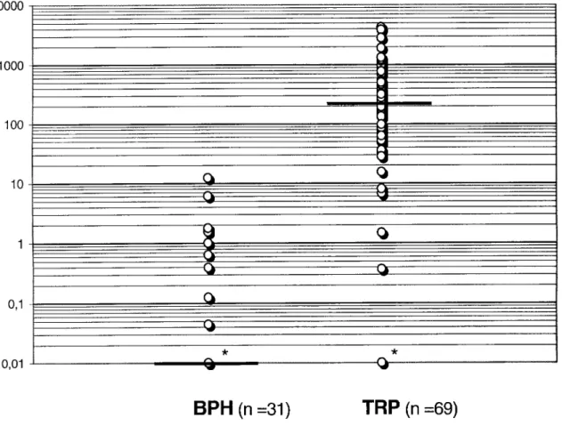
Fig. 1 Distribution of GSTP1 methylation levels in prostate tissues from benign prostatic hyperplasia (BPH), prostate intraepithelial neoplasia (PIN) and clinically localized prostate adenocarcinoma (TRP). Twenty-nine % of patients with BPH, 91.3% of patients with TRP and 53.6% paired PIN lesions were positive for GSTP1 methylation by real-time MSP. As indicated, the GSTP1/MYOD1 methylation ratios differed significantly. Solid bars indicate the median within a group of patients. Asterisks indicate the samples with 0-values which can not be plotted on a log scale (BPH: n = 22; PIN: n = 13; TRP: n = 6).
10000
1000
100
0) 10
o
0,1
0,01
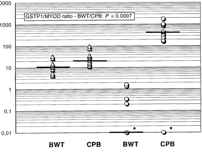
GSTP1/MYOD ratio - BWT/CPB: P = 0.0007
3
BWT
CPB
BWT
CPB
Fig. 2 Distribution of serum PSA and GSTP1 methylation levels in biopsy samples of patients without (BWT; n = 10) and with a histological diagnosis of prostate cancer (CPB; n = 11). The range of GSTP1/MYOD1 methylation ratios (O) between BWT and CPB was
significantly different as well as serum PSA levels (A). Solid bars indicate the median within a group of patients. Asterisks indicate the samples with 0-values which can not be plotted on a log scale (BWT: n = 6 ; C P B : n = l ) .
Carmen Jerónimo - Doctoral Thesis
REFERENCES
1. Landis, S.H., Murray, T., Bolden, S. & Wingo, P.A. Cancer Statistics. Ca. Cancer Clin.
49,8-31 (1999).
2. Andriole, G.L. & Catalona, W.J. in Principles and Practice of Genitourinary Oncology. 1st edn. (Ed. Raghavan, D., Scher, H.I., Leibel, S.A. & Lange, P.H.) 457-464 (Lippincott-Raven, Philadelphia, PA, 1996).
3. Isaacs, W.B. & Isaacs, J.T. in Principles and Practice of Genitourinary Oncology. 1st edn. (Ed. Raghavan, D., Scher, H.I., Leibel, S.A. & Lange, P.H.) 403-408 (Lippincott-Raven, Philadelphia, PA, 1996).
4. Cairns, P. et al. Frequent inactivation of PTEN/MMAC 1 in primary prostate cancer.
Cancer Res. 57, 4997-5000 (1997).
5. Sidransky, D. Nucleic acid-based methods for the detection of cancer. Science. 278, 1054-1058(1997).
6. Lee, W.-H. et al. Cytidine methylation of regulatory sequences near the pi-class glutathione S-transferase gene accompanies human prostatic carcinogenesis. Proc Natl AcadSci USA. 91, 11733-11737 (1994).
7. Lee, W.-H., Isaacs, W.B., Bova, G.S. & Nelson WG. CG island methylation changes near the GSTP1 gene in prostatic carcinoma cells detected using the polymerase chain reaction: a new prostate cancer biomarker. Cancer Epidemiol Biomark Prev. 6, 443-450 ( 1997). 8. Esteller, M. et al. Inactivation of glutathione S-transferase PI gene by promoter
hypermethylation in human neoplasia. Cancer Res. 58, 4515-4518 (1999).
9. Brooks, J.D. et al. CG island methylation changes near the GSTP1 gene in prostatic intraepithelial neoplasia. Cancer Epidemiol Biomark Prev. 7, 531-536 (1998).
10. Esteller, M. et al. Detection of aberrant promoter hypermethylation of tumor suppressor genes in serum DNA from non-small cell lung cancer patients. Cancer Res. 59, 67-70 (1999).
11. Sanchez-Cespedes, M. et al. Gene promoter hypermethylation in tumors and serum of head and neck patients. Cancer Res. 3, 1229-1235 (2000).
12. Herman, J.G., Graff, J.R., Myõhánen, S., Nelkin, B.D. & Baylin SB. Methylation-specific PCR: a novel PCR assay for methylation status of CpG islands. Proc Natl Acad Sci USA.
93, 9821-9826 (1996).
13. Heid, C.A., Stevens, J., Livak, K.J. & Williams, P.M. Real time quantitative PCR.
Genome Res. 6, 986-994 (1996).
14. Lo, Y.M.D. et al. Quantitative analysis of aberrant pl6 methylation using real-time quantitative methylation-specific polymerase chain reaction. Cancer Res. 59, 3899-3903 (1999).
15. Hermanek, P., Hutter, R.V.P., Sobin, L.H., Wagner, G. & Wittekind, C. in Illustrated Guide to the TNM/pTNM Classification of malignant tumors. 4th edn. (eds. Hermanek, P., Hutter, R.V.P., Sobin, L.H., Wagner, G. & Wittekind, C.) 272-280 (Springer-Verlag, Heidelberg, Germany, 1997).
16. Gleason, D.F., Mellinger, G.T. & Veterans Administration Cooperative Urological Research group. Prediction of prognosis for prostatic adenocarcinoma by combined histologic grading and clinical staging. J Urol. I l l , 58-64 (1974).
17. Ahrendt, S.A., et al. Molecular detection of tumor cells in bronchoalveolar lavage fluid from patients with early stage lung cancer. J Natl Cancer Inst. 91, 332-339 (1999).
18. Olek, A., Oswald, J. & Walter, J.A. A modified and improved method of bisulfite based cytosine methylation analysis. Nucleic Acids Res. 24, 5064-5066 (1996).
Carmen Jerónimo - Doctoral Thesis
19. Eads, C.A. et al. CpG island hypermethylation in human colorectal tumors is not associated with DNA methyl transferase overexpression. Cancer Res. 59, 2302-2306 (1999).
20. Saiki, R.K. et al. Primer - directed enzymatic amplification of DNA with a thermostable DNA polymerase. Science. 239, 47-491 (1988).
21. Montironi, R. Mazzucchelli. R., Stramazzotti, D., Pomante, R., Thompson, D. & Bartels, P.H. Expression of pi-class glutathione S-transferase: two populations of high grade prostatic intraepithelial neoplasia with different relations to carcinoma. Mol Pathol. 53,
122-128 (2000).
22. Bostwick, D.G. et al. Independent origin of multiple foci of prostatic intraepithelial neoplasia. Cancer. 83, 1995-2002 (1998).
23. Toyota, M. & Issa, J.P. CpG island methylator phenotypes in aging and cancer. Semin Cancer Biol. 9, 349-357 (1999).
24. Ahuja, N., Li, Q., Mohan, A.L., Baylin, S.B. & Issa, J.P. Aging, DNA methylation in colorectal mucosa and cancer. Cancer Res. 58, 5489-5494 (1998).
25. Keetch, D.W., Catalona, W.J. & Smith, D.S. Serial prostatic biopsies in men with persistently elevated serum prostate specific antigen values. J Urol. 151, 1571-1574 (1994).
26. Epstein, J.I., Walsh, P.C., CarMichael, M. & Brendler, C.B. Pathological and clinical findings to predict tumor extent of non-palpable (stage Tic) prostate cancer. JAMA. 271, 368-374(1994).
27. Goessl, C. et al. Fluorescent methylation-specific polymerase chain reaction for DNA-based detection of prostate cancer in bodily fluids. Cancer Res. 60, 5941-5945 (2000). 28. Cairns, P. et al, Molecular Detection of Early Stage Prostate Cancer in Urine. Clin
Cancer Res. In press, (2001).
29. Sharifi, R. et al. Evaluation of cytologic techniques for diagnosis of prostate cancer.
PAPER II: Detection of Prostate Cancer in Urine by GSTP1 Hypermethylation
DETECTION OF PROSTATE CANCER IN URINE
BY
GSTP1
HYPERMETHYLATION
1Paul Cairns, Manel Esteller, James G. Herman, Mark Schoemberg, Carmen Jerónimo, Montserrat Sanchez-Cespedes, Nan-Haw Chow, Marc Grasso, Li Wu, William B. Westra, and
David Sidransky2
Fox Chase Cancer Center, 7701 Burholme Avenue, Philadelphia, PA 19111, USA (P.C.) And Tumor Biology, Oncology Center (M.E., J.G.H., D.S.), Department of Urology (M.S., D.S.), Department of Otolaryngology, Head & Neck Surgery, Division of Head and Neck Cancer Research (C.J., M.S-C, N-H. C , M.G., L.W., D.S.), and Department of Pathology (W.B.W., D.S.), Johns Hopkins University School of Medicine, 818 Ross Research Building, 720 Rutland Avenue, Baltimore, Maryland 21205-2195, USA
'Supported by ROl CA77664-01 (P.C., M.S-C, M.G, L.W., & D.S.) and PRAXIS XXI - BD 13398/97 Fundação Para a Ciência e Tecnologia, Portugal (C.J.)
2
To whom requests for reprints should be addressed, at Head and Neck Cancer Research, 820 Ross Research Building, Johns Hopkins University School of Medicine, 720 Rutland Avenue, Baltimore, Maryland 21205-2195, USA. Phone: (410) 502-5153; Fax: (410) 614- 1411.
Carmen Jerónimo - Doctoral Thesis
ABSTRACT
Novel approaches for the early detection and management of prostate cancer is urgently needed. Clonal genetic alterations have been used as targets for the detection of neoplastic cells in bodily fluids from many cancer types. A similar strategy for molecular diagnosis of prostate cancer requires a common and/or early genetic alteration as a specific target for neoplastic prostate cells. Hypermethylation of regulatory sequences at the
INTRODUCTION
Prostate cancer is the most commonly detected male cancer and the second leading cause of male cancer deaths in the US.1 Diagnosis and management are confound by the lack of symptoms and the lack of cancer specific diagnostic techniques during early stages of the disease. Prostate cancer is indeed curable if detected early while still localized within the capsule2. Novel approaches for the detection and control of this cancer is therefore extremely important. Adult sporadic cancers are known to arise through the accumulation of multiple genetic events,3 and these clonal genetic alterations can be used as targets for the detection of neoplastic cells in clinical samples.4 To develop such targets, a common and early genetic event unique to neoplastic cells must be identified and combined with a sensitive molecular assay able to detect this genetic event, among a high background of normal wild type cells. Several specific genetic alterations have been identified in prostate cancer5 including ras
oncogenic activation, and inactivation of the tumor suppressor genes, Rb, p53, CDKN2a and
PTEN. However, RAS or p53 mutations are infrequent5 and PTEN inactivation generally occurs relatively late in prostate cancer progression.6 Loss of heterozygosity (LOH) at critical suppressor loci, such as 8p and 16q, occurs frequently,5 but successful LOH detection requires a high proportion of tumor cells for robust analysis of a diagnostic sample.
Hypermethylation of normally unmethylated CpG islands in the promoter regions of tumor suppressor genes correlates with loss of gene expression in human tumors. " Hypermethylation of regulatory sequences at the detoxifying glutathione S-transferase
(GSTP1) gene locus is found in the majority (>90%) of primary prostate carcinomas but not in normal prostatic tissue or other normal tissues nor in benign hyperplasia of the prostate.
GSTP1 methylation is thus the most common genetic alteration so far described in prostate cancer. The initial studies of GSTP1 methylation status in prostate tumors and cell lines were
Carmen Jerónimo - Doctoral Thesis
performed using Southern blot analysis.10 A new method, methylation specific PCR (MSP), has since been described" which is more sensitive and requires less DNA. MSP utilizes a DNA modification step before PCR to determine the presence or absence of methylation of a gene locus at a sensitive level of up to 1 methylated allele in 1000 unmethylated alleles.
Bodily fluids from several types of cancer have been successfully utilized for the molecular detection of neoplasia including stool in colon and pancreas, urine in bladder, and sputum and bronchial lavage fluid (BAL) in lung cancer.4 Recently promoter hypermethylation has been successfully used to detect neoplastic DNA in sputum, BAL
and serum14 from lung cancer patients and serum from liver cancer,15 head and neck cancer and breast cancer patients.17 Most prostate tumors occur in the peripheral zone which contains 3/4 of the glands, the minilobes of which form secretory ducts that empty their contents into the urethra. We hypothesized that urine from prostate cancer patients might therefore contain shed neoplastic cells or debris amenable to DNA analysis. We therefore examined the potential of GSTP1 hypermethylation as a cancer specific marker in simple voided urine specimens from 28 prostate cancer patients about to undergo radical prostatectomy for clinically curable disease.
MATERIALS AND METHODS
Specimen Collection and DNA isolation
Samples were obtained from patients undergoing radical prostatectomy. Urine was collected from each patient immediately before surgery. Tumor samples were obtained after pathological review, areas rich in neoplastic cells were selected and microdissected from formalin-fixed blocks. A peripheral blood sample in EDTA was also obtained for isolation of leukocyte DNA as a normal control. Genomic DNA was isolated as previously described.18
Bisulfite Treatment
One ug of each DNA sample was denatured by sodium hydroxide and modified by sodium bisulfite. DNA samples were then purified using Wizard DNA purification resin (Promega, Madison WI), again treated with sodium hydroxide, precipitated with ethanol, and re-suspended in water.
Methylation Specific PCR
MSP was performed separately with GSTP1 primers specific for the methylated reaction and the unmethylated reaction19 for each DNA sample. Unmethylated reaction : 5'-GATGTTTGGGGTGTAGTGGTTGTT-3' (sense), 5'- CCACCCCAATACTAAATCA CAACA-3' (antisense); methylated reaction: 5'-TTCGGGGTGTAGCGCTCGTC-3' (sense), 5'-GCCCCAATACTAAATCACGACG-3' (antisense). Thirty-five cycles of PCR were performed with an annealing temperature of 59°C. A water control without DNA for contamination and controls for unmethylated and methylated reactions were performed for each set of PCR. PCR reactions were analyzed on nondenaturing 6% polyacrylamide gels, stained with ethidium bromide and visualized under UV illumination.
Carmen Jerónimo - Doctoral Thesis
RESULTS AND DISCUSSION
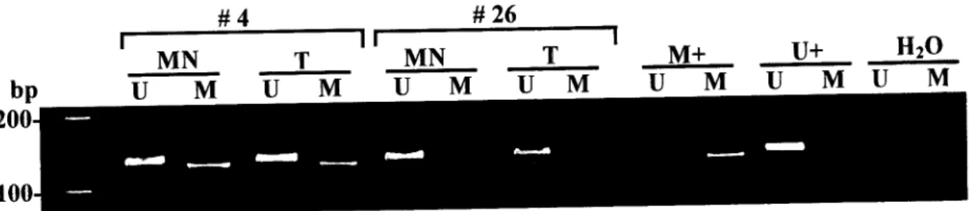
We collected 28 primary resectable prostate tumors of pathological grade and stage amenable to surgical cure [7 T2A (Gleason 5-8), 5 T2B (Gleason 6-7), 15 T3A (Gleason 5-7) and 1 T3B (Gleason 7)] and extracted genomic DNA from tumor, peripheral blood lymphocytes (normal control) and urine sediment (from a simple voided urine obtained preoperatively). The DNA samples were coded and the methylation status of GSTP1 assessed in a blinded manner. Decoding of the results revealed that 22 of 28 (79%) prostate tumors were positive for GSTP1 methylation. In 6 of 22 (27%) cases the corresponding urine sediment DNA was positive for GSTP1 methylation indicating the presence of neoplastic DNA in the urine (Fig. 1, Tumors 1 and 2). There was no case where a urine sediment DNA gave a positive methylation result in the absence of methylation in the corresponding tumor (potential false positive) (Fig. 1, Tumor 3). The 6 tumors with positive urine results were Gleason 5-7 and stages T2A (1), T2B (1) and T3A (4).
Although we only detected GSTP1 methylation in under a third of voided urine samples, we have clearly demonstrated that molecular diagnosis of prostate neoplasia in urine is feasible. Moreover, albeit in a limited study, we observed absolute specificity because we did not find any GSTP1 hypermethylation in the urine DNA from the 6 patients with unmethylated GSTP1 tumor DNA. We detected GSTP1 hypermethylation in a minority of paired urine and this level of sensitivity can likely be improved upon. Goessl et al20 reported a higher percentage of positive cases with a fluorescence based conventional PCR technique. It is possible that prostatic massage and the higher number of cycles used in their study yielded more positive urine DNAs but specificity is known to decrease in MSP, as in other PCR protocols, with increased cycle number.21 Indeed, in the study by Goessl, some urine samples were positive yet the primary tumor was not found to harbor GSTP1 hypermethylation.
Further work needs to focus on understanding factors such as tumor size and localization within the prostate, urine collection techniques for example the potential benefits of a prostatic massage before urine collection, and continuing improvements in molecular technology to increase the detection rate.
Six tumors of 28 did not have GSTP1 hypermethylation preventing assessment of neoplasia in the urine. However, screening for methylation of other loci, such as the Endothelin B receptor (methylated in -70% of prostate tumors)22 or CD44 (methylated in 77% of tumors)23 is likely to further increase the number of primary tumors with methylation (amenable for screening) to allow 100% diagnostic coverage. GSTP1 hypermethylation has not been reported in bladder cancer and is found infrequently in renal tumors.19 Inadvertent detection of a renal cell carcinoma in urine is therefore possible. Even so, GSTP1
hypermethylation is cancer specific, unlike PSA it is not found in normal prostatic tissue or
BPH.
A further consideration is that of our choice of optimal negative controls. For a study of the type presented here, normal age-matched controls would present problems of ethics and interpretation of results. The high frequency of incidental prostate cancer in men over 50 years of age (estimated at 30-50%), the hypothesized early timing of GSTP1
hypermethylation in prostate tumorigenesis, and the ability of MSP to detect 1 cancer cell in a background of 1000 normal cells argues against initial inclusion of a cohort of age-matched men with no evidence of prostate cancer as controls. Whether a positive MSP test arose from a false positive or from asymptomatic prostate cancer would be difficult to ascertain. In our exploratory study, control urine samples from the 6 of 28 (21%) of patients whose prostate cancer did not show GSTP1 hypermethylation were negative for urine methylation.
Carmen Jerónimo - Doctoral Thesis
existing methods and theoretically more specific for neoplasia than serum PSA. Only 80% of the patients in our study of clinically early cancer (Tla mostly) had elevated PSA. Two subgroups of men in whom GSTP1 hypermethylation has clinical utility would be the 20% of men with prostate cancer with a near normal PSA value and men with a high PSA value but negative biopsies. If our results are confirmed in larger studies, GSTP1 hypermethylation could be used to augment PSA and other current diagnostic procedures for detection of prostate cancer in the general population.
This technique could also be employed to identify neoplastic disease in other diagnostic clinical material such as needle biopsies or serum. Similarly, in prostate cancer patients, hypermethylation may be a marker of neoplastic cell burden or minimal residual disease after removal of the primary tumor. Finally, it has previously been shown that nearly all bladder cancers24 and many kidney cancers25 can be detected by molecular analysis of urine raising the possibility of simultaneous molecular screening for three common adult cancer types in one simple voided urine specimen.
Patient 1 Patient 2 Patient 3 Patient 4
Turn Uri Turn Uri Turn Uri Turn Uri MCF-7 NL H20
L T ~ M T T M U M I T M U M Û ~ M U M T T T V Í T J M U M U M
Fig. 1 Methylation specific PCR of GSTP1 in prostate carcinoma and urine DNAs. The presence of a visible PCR product in the methylated lane (M) of the tumor DNA from patients
1, 2, and 4 indicates the presence of methylated alleles of GSTP1. A PCR product is also present in the methylated lane (M) of the urine DNA from patients 1 and 2 indicating the presence of neoplastic cell DNA in the urine. The absence of a visible PCR product in the methylated lane (M) of urine sediment DNA from patient 4 indicates that neoplastic cell DNA is absent or undetectable in the urine. Patient 3's tumor DNA is not methylated and the corresponding urine DNA also had no PCR product in the methylated lane (M) while a product can be clearly seen in the unmethylated lane (U). The PCR product in the unmethylated lane (U) from patient's 1, 2 and 4 tumor DNA most likely arises from normal cell contamination of the tumor specimen. Tumor cell line MCF-7 DNA as a positive control for GSTP1 methylation, normal lymphocyte DNA (NL) as a negative control, a water control for contamination in the PCR reaction (right) and Mspl digested pBR322 as a molecular weight marker (far left) are also shown.
Carmen Jerónimo - Doctoral Thesis
REFERENCES
1. Greenlee, R.T., Murray, T., Bolden, S., and P.A. Wingo, Cancer Statistics 2000. CA
Cancer J. Clin., 50: 7-33, 2000.
2. Andriole, G. L. and Catalona, W. J. The case for aggressive diagnosis and therapy of localized prostate cancer. In: Principles and Practice of Genitourinary Oncology, chapter 46, 457-464. Ed. Raghavan, D. et al. Lippincott-Raven, 1996.
3. Fearon, E.R. and Vogelstein, B. A genetic model for colorectal tumorigenesis. Cell, 61:
759-767, 1990.
4. Sidransky, D. Nucleic acid-based methods for the detection of cancer. Science, 278: 1054-1058, 1997.
5. Isaacs, W.B. and Isaacs, J. T.: Molecular genetics of prostate cancer progression. In: Principles and Practice of Genitourinary oncology. In: Principles and Practice of Genitourinary Oncology, chapter 39, 403-408. Ed. Raghavan, D. et al. Lippincott-Raven,
1996.
6. Cairns, P., Okami, K., Halachami, S., Halachami, N., Esteller, M., Herman, J. G., Jen, J., Isaacs, W. B., Bova, G. S. and Sidransky, D.: Frequent inactivation of PTEN/MMAC1 in primary prostate cancer. Cancer Res, 57: 4997-5000, 1997.
7. Merlo, A., Herman, J.G., Mao, L., Lee, D.J., Gabrielson, E., Burger, P.C., Baylin, S.B., and Sidransky, D. 5'CpG island methylation is associated with transcriptional silencing of the tumour suppressor gene pl6/CDKN2/MTSl in human cancers. Nat. Med., 1: 686-692,
1995.
8. Baylin, S.B., Herman, J.G., Graff, J.R., Vertino, P., and Issa, J.P. Alterations in DNA methylation: a fundamental aspect of neoplasia. Adv. Cancer Res., 72: 141-146, 1998.
9. Jones, P.A. and Laird, P.W. Cancer epigenetics comes of age. Nat. Gen., 21: 163-167, 1999.
10. Lee, W-H., Morton, R. A., Epstein, J. I., Brooks, J. D., Campbell, P. A., Bova, G. S., Hsieh, W-S., Isaacs, W. B. and Nelson, W.G. Cytidine methylation of regulatory sequences near the pi-class glutathione S-transferase gene accompanies human prostatic carcinogenesis. Proc. Natl. Acad. Sci. USA, 91: 11733-11737, 1994.
11. Herman, J.G., Graff, J.R., Myõhãnen, S., Nelkin, B.D. and Baylin, S.B. Methylation-specific PCR: a novel PCR assay for methylation status of CpG islands. Proc. Natl. Acad. Sci. USA, 95: 9821-9826, 1996.
12. Belinsky, S.A., Nikula, K.J., Palmisano, W.A., Michels, R., Saccomano, G., Gabrielson, E., Baylin, S.B., and Herman, J.G. Aberrant methylation of pl6(INK4a) is an early event in lung cancer and a potential biomarker for early diagnosis. Proc. Natl. Acad. Sci. USA,
95: 11891-11896, 1998.
13. Ahrendt, S.A., Chow, J.T., Xu, L-H, Yang, S.C., Eisenberger, CF., Esteller, M., Herman, J.G., Wu, L., Decker, P.A., Jen, J. and Sidransky, D. Molecular detection of tumor cells in bronchoalveolar lavage fluid from patients with early stage lung cancer. J Natl Cancer Inst, 97: 332-339, 1999.
14. Esteller, M., Sanchez-Cespedes, M., Roseli, R., Sidransky, D., Baylin, S.B., and Herman, J.G. Detection of aberrant promoter hypermethylation of tumor suppressor genes in Serum DNA from non-small cell lung cancer patients. Cancer Res, 59: 67-70, 1999
15. Wong, I.H.N., Lo, Y.M.D., Zhang, J., Liew, C-T., Wong, N., Lai, P.B.S., Lau, W.Y. and Johnson, P.J. Detection of aberrant pl6 methylation in the plasma and serum of liver cancer patients. Cancer Res., 59: 71-73, 1999.
Carmen Jerónimo - Doctoral Thesis
17. Silva, J.M., Dominguez, G., Garcia, J.M., Gonzalez, R., Villanueva, M.J., Navarro, F., Provencio, M., San Martin, S., Espana P., and Bonilla, F. Presence of tumor DNA in plasma of breast cancer patients: clinicopathological correlations. Cancer Res. 59: 3251-3256, 1999.
18. Sambrook, J., Fritsch, E.F., and Maniatis, T. Molecular Cloning: A Laboratory Manual, Ed. 2, 9.14-9.23. Cold Spring Harbor, NY: Cold Spring Harbor Laboratory, 1989.
19. Esteller, M., Corn, P.G., Urena, J.M., Gabrielson, E., Baylin, S.B., and Herman, J.G. Inactivation of Glutathione S-Transferase PI gene by promoter hypermethylation in human neoplasia. Cancer Res, 58: 4515-4518, 1999.
20. Goessl, C , Krause, H., Muller, M., Heicappell, R., Schrader, M., Sachsinger, J., and Miller, K. Fluorescent methylation-specific polymerase chain reaction for DNA-based detection of prostate cancer in bodily fluids. Cancer Res., 60: 5941-5945, 2000.
21. Corn, P.G., Smith, D.B., Ruckdeschel, E.S., Douglas, D., Baylin, S.B., and Herman, J.G. E-Cadherin expression is silenced by 5'CpG island methylation in acute leukemia. Clin. Cancer Res. 6: 4243-4248, 2000.
22. Nelson, J.B., Lee, W.H., Nguyen, S.H., Jarrard, D.F., Brooks, J.D., Magnuson, S.R., Opgenorth, T.J., Nelson, W.G., and Bova, G.S. Methylation of the 5' CpG island of the endothelin B receptor gene is common in human prostate cancer. Cancer Res., 57: 35-37,
1997.
23. Lou, W., Krill, D., Dhir, R., Becich, M.J., Dong, J.T., Frierson, H.F. Jr., Isaacs, W.B., Isaacs, J.T., and Gao, A.C. Methylation of the CD44 metastasis suppressor gene in human prostate cancer. Cancer Res., 59: 2329-2331, 1999.
24. Mao, L., Schoenberg, M., Scicchitano, M., Erozan, Y.S., Merlo, A., Schwab, D., and Sidransky, D. Molecular detection of primary bladder cancer by microsatellite analysis. Science, 277: 659-662, 1996.
25. Eisenberger, CF., Schoenberg, M., Enger, C , Hortopan, S., Shah, S., Chow, N-H., Marshall, F., and Sidransky, D. Diagnosis of renal cancer by molecular urinalysis. J. Natl. Cancer Inst., 91: 2028-2032, 1999.
PAPER III: Quantitative GSTP1 Hypermethylation in Bodily Fluids of Prostate Cancer Patients
QUANTITATIVE GSTP1 HYPERMETHYLATION
IN BODILY FLUIDS OF PROSTATE CANCER PATIENTS
Carmen Jerónimo, Henning Usadel, Rui Henrique, Cristina Silva, Jorge Oliveira, Carlos Lopes, and David Sidransky1
Department of Otolaryngology-Head and Neck Surgery [C.J., H.U., D.S.], Head and Neck Cancer Research Division, The Johns Hopkins University School of Medicine, Baltimore, Maryland 21205-2195, USA; and Unit of Molecular Pathology-Department of Pathology [R.H., C.S., C.L.], and Department of Urology [J.O.], Instituto Português de Oncologia de
Francisco Gentil - Centro Regional do Porto, Portugal.
Running Title: DETECTION OF PROSTATE CANCER IN BODILY FLUIDS BY GSTP1
HYPERMETHYLATION
Key words: real-time quantitative MSP, bodily fluids, prostate cancer, early detection, GSTP1 hypermethylation
Footnotes:
'To whom reprint requests should be addressed at the Head and Neck Cancer Research Division, The Johns Hopkins University School of Medicine, 818 Ross Research Building, 720 Rutland Avenue, Baltimore, MD 21205-2195, USA.
C.J. and H.U. are supported by grants from the Fundação para a Ciência e Tecnologia, Portugal (Program PRAXIS XXI - BD 13398/97), and the Dr. Mildred Scheel-Stiftung fur Krebsforschung, Deutsche Krebshilfe, respectively.
This work was supported by a NIH grant UOl CA 84986 and a Virco collaborate research agreement. Under a licensing agreement between The Johns Hopkins University and Virco, Dr. Sidransky is entitled to a share of royalty received by the University on sales or products described in this article. The University and Dr. Sidransky own Virco stock, which is subject to certain restrictions under University policy. Dr. Sidransky is a paid consultant to Virco. The terms of this arrangement are being managed by The Johns Hopkins University in accordance with its conflict of interest policies.